. 2018 Mar;155(3):1118-1127.e1.
doi: 10.1016/j.jtcvs.2017.08.127. Epub 2017 Nov 29.

Angiogenesis precedes cardiomyocyte migration in regenerating mammalian hearts

Affiliations

Angiogenesis precedes cardiomyocyte migration in regenerating mammalian hearts

Arnar B Ingason et al. J Thorac Cardiovasc Surg. 2018 Mar.

Abstract

Objective: Although the mammalian heart's ability to fully regenerate is debated, its potential to extensively repair itself is gaining support. We hypothesized that heart regeneration relies on rapid angiogenesis to support myocardial regrowth and sought to characterize the timeline for angiogenesis and cell proliferation in regeneration.

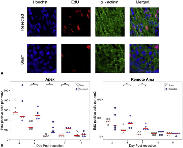
Methods: One-day-old CD-1 mice (P1, N = 60) underwent apical resection or sham surgery. Hearts were explanted at serial time points from 0 to 30 days postresection and analyzed with immunohistochemistry to visualize vessel ingrowth and cardiomyocyte migration into the resected region. Proliferating cells were labeled with 5-ethynyl-2'-deoxyuridine injections 12 hours before explant. 5-Ethynyl-2'-deoxyuridine-positive cells were counted in both the apex and remote areas of the heart. Masson's trichrome was used to assess fibrosis.

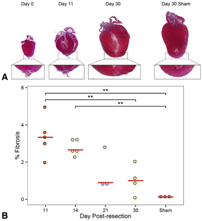
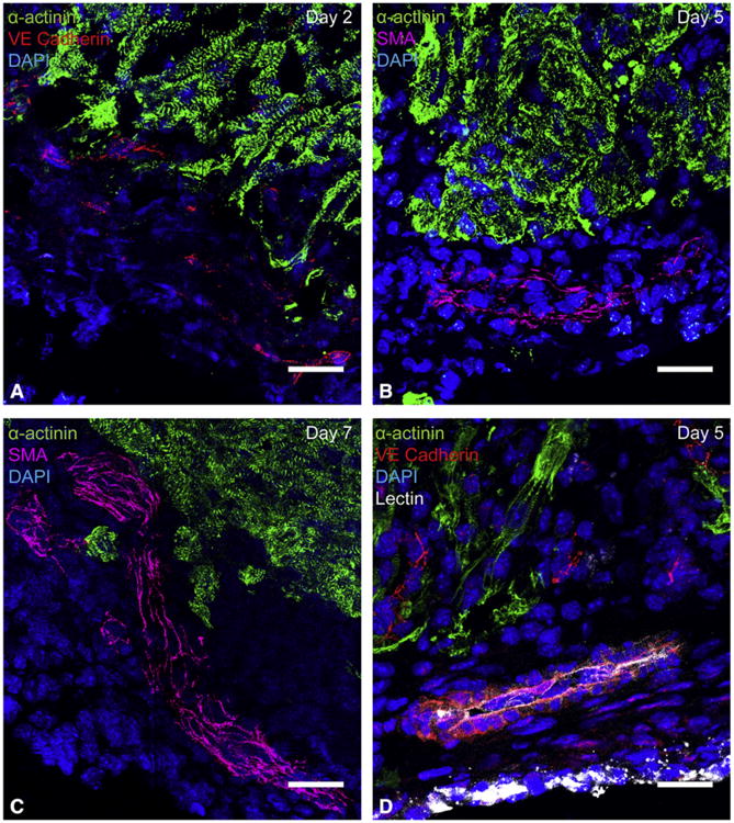
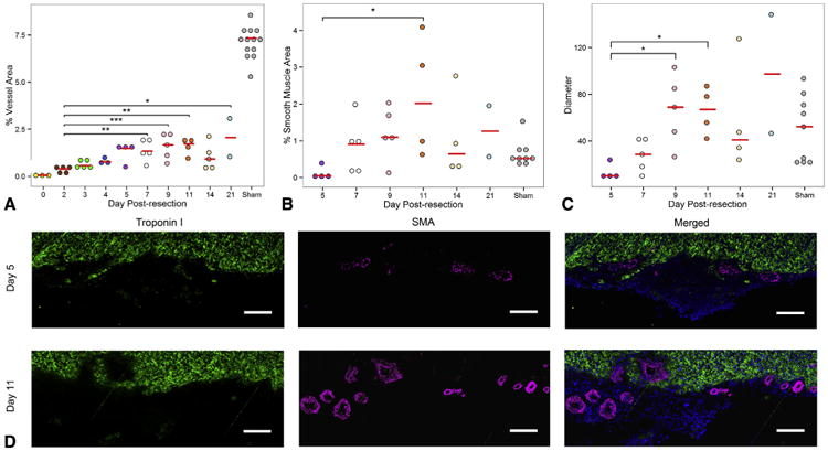
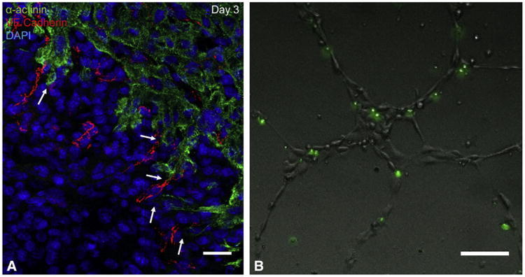
Results: By 30 days postresection, hearts regenerated with minimal fibrosis. Compared with sham surgery, apical resection stimulated a significant increase in proliferation of preexisting cardiomyocytes between 3 and 11 days after injury. Capillary migration into the apical thrombus was detected as early as 2 days postresection, with development of mature arteries by 5 days postresection. New vessels became perfused by 5 days postresection as evidenced by lectin injection. Vessel density and diameter significantly increased within the resected area over 21 days, and vessel ingrowth always preceded cardiomyocyte migration, with coalignment of most migrating cardiomyocytes with ingrowing vessels.

Conclusions: Endothelial cells migrate into the apical thrombus early after resection, develop into functional arteries, and precede cardiomyocyte ingrowth during mammalian heart regeneration. This endogenous neonatal response emphasizes the importance of expeditious angiogenesis required for neomyogenesis.

Keywords: angiogenesis; arteriogenesis; cardiac regeneration; cardiomyocyte migration; vascular biology.


Conflict of interest statement

Conflict of Interest Statement: Authors have nothing to disclose with regard to commercial support.

Figures

Figure 1
Figure 1
The neonatal heart regenerates with minimal fibrosis after apical resection. A, Masson's trichrome staining of hearts at 0, 11, and 30 days postresection. For comparison, sham-operated hearts, harvested 30 days after surgery, are included. A magnified image of each apex is provided below. An apical thrombus quickly formed after resection and became fibrotic by 11 days postresection. By 30 days postresection, the heart had regenerated with minimal fibrosis. B, Quantification of fibrotic area in the apex after apical resection at 11, 14, 21, and 30 days postresection. For comparison, sham-operated hearts harvested 30 days after surgery are included. Values are represented as percentage fibrosis of the apical area. Fibrosis in the apex consistently decreased over time. This suggests that fibrosis is gradually replaced by myocardium as heart regeneration progresses. Red bars represent median value for each timepoint. Asterisks denote statistical significance at **P < .01.
Figure 2
Figure 2
Apical resection prolongs window of cardiomyocyte proliferation. A, Proliferative cardiomyocytes were identified by staining for α-actinin (green), EdU (red), and Hoechst 33342 (blue). The number of EdU-positive cells in the apex 3 days after operation is compared between sham and apical resection groups. EdU staining represents cardiomyocytes that have gone through at least 1 cell cycle of replication. B, EdU measurements comparing the number of proliferating cells in the apex and remote myocardium of resected hearts and sham controls at each time point. Cardiomyocyte proliferation is increased relative to sham controls at 3 days postresection in both the apex and remote area of the heart compared with sham-operated animals and is sustained for over 1 week in the apex. Red bars represent median value for each timepoint. Asterisks denote statistical significance at *P <.05 and **P<.01. EdU, 5-Ethynyl-2-deoxyuridine.
Figure 3
Figure 3
Vessel ingrowth into the apical thrombus precedes cardiomyocyte migration after resection. Histologic analysis of sagittal heart sections demonstrated that vessels enter the apical thrombus early and mature over time. Endothelial cells always extended deeper into the thrombus than cardiomyocytes. A, Migrating endothelial cells in the apical thrombus, visualized by vascular endothelial cadherin staining. Capillary formation was first noted in the apical thrombus at 2 days postresection. B, An artery characterized by a smooth muscle layer in the apical thrombus at 5 days postresection. Arteries in the apical thrombus were first noted at 5 days postresection, but did not consistently appear until 7 days postresection. C, A large ingrowing artery within the apical thrombus at 7 days postresection. Arteries were found within apical thrombus of all hearts treated with apical resection at 7 days postresection and onward. D, A lectin-perfused vessel in the apical thrombus at 5 days postresection. This suggests that ingrowing vessels become functional by 5 days postresection. Green =14; α-actinin, red =14; vascular endothelial cadherin, red, magenta=14; smooth muscle actin, blue =14; DAPI, white =14;lectin. All scale bars represent 25 μm. SMA, Smooth muscle actin; VE, vascular endothelial; DAPI, 4′,6-diamidino-2-phenylindole.
Figure 4
Figure 4
The apical thrombus becomes more vascularized over time to ultimately regrow apical coronary arteries. A, Vascular density of apical thrombus consistently increased from the time of resection to 9 days postresection, after which it plateaued. B, Percentage of smooth muscle cell area in the apical thrombus. C, Maximal vessel diameter in the apical thrombus. For comparison, measurements of apices of sham-operated hearts are provided for all graphs. D, Comparison of arterial formation in the apical thrombus of resected hearts 5 and 11 days postresection. Note the increased arterial density and vessel diameter at 11 days postresection compared with 5 days postresection. Green =14; troponin I, magenta =14; smooth muscle actin, blue =14; DAPI. Scale bar represents 25 μm. Red bars represent median value for each timepoint. Asterisks denote statistical significance at *P<.05, **P<.01, and ***P<.001. SMA, Smooth muscle actin.
Figure 5
Figure 5
Migratory cardiomyocytes in the regenerating apex are in close proximity to endothelial cells. A, Immunohistochemical staining of apical thrombus 3 days postresection. After apical resection, endothelial cells migrate into the apical thrombus ahead of cardiomyocytes. Most migrating cardiomyocytes were observed in close proximity to ingrowing vessels. Note the close proximity of cardiomyocyte processes to migrating vessels in the apical thrombus (arrows). Green =14; α-actinin, red =14; vascular endothelial cadherin, blue =14; DAPI. Scale bars represent 25 μm. B, In vitro co-culturing of murine GFP-positive cardiomyocytes and human umbilical vein endothelial cells. Nearly all cardiomyocytes co-align with the endothelial network. Scale bars represent 25 μm. Together, this suggests that a strong relationship exists between the 2 cell types. VE, Vascular endothelial; DAPI, 4′,6-diamidino-2-phenylindole.

Comment in

Similar articles

Cited by

References

    1. Poss KD, Wilson LG, Keating MT. Heart regeneration in zebrafish. Science. 2002;298:2188–90. - PubMed
    1. Witman N, Murtuza B, Davis B, Arner A, Morrison JI. Recapitulation of developmental cardiogenesis governs the morphological and functional regeneration of adult newt hearts following injury. Dev Biol. 2011;354:67–76. - PubMed
    1. Oberpriller JO, Oberpriller JC. Response of the adult newt ventricle to injury. J Exp Zool. 1974;187:249–59. - PubMed
    1. Tam SKC, Gu W, Mahdavi V, Nadal-Ginard B. Cardiac myocyte terminal differentiation. Ann N Y Acad Sci. 1995;752:72–9. - PubMed
    1. Li FQ, Wang XJ, Capasso JM, Gerdes AM. Rapid transition of cardiac myocytes from hyperplasia to hypertrophy during postnatal development. J Mol Cell Car-diol. 1996;28:1737–46. - PubMed