. 2018 Jun 15;83(12):1012-1023.
doi: 10.1016/j.biopsych.2018.01.003. Epub 2018 Jan 12.

Glutamatergic Ventral Pallidal Neurons Modulate Activity of the Habenula-Tegmental Circuitry and Constrain Reward Seeking

Affiliations

Glutamatergic Ventral Pallidal Neurons Modulate Activity of the Habenula-Tegmental Circuitry and Constrain Reward Seeking

Jessica Tooley et al. Biol Psychiatry. .

Abstract

Background: The ability to appropriately integrate and respond to rewarding and aversive stimuli is essential for survival. The ventral pallidum (VP) plays a critical role in processing both rewarding and aversive stimuli. However, the VP is a heterogeneous structure, and how VP subpopulations integrate into larger reward networks to ultimately modulate these behaviors is not known. We identify a noncanonical population of glutamatergic VP neurons that play a unique role in responding to aversive stimuli and constraining inappropriate reward seeking.

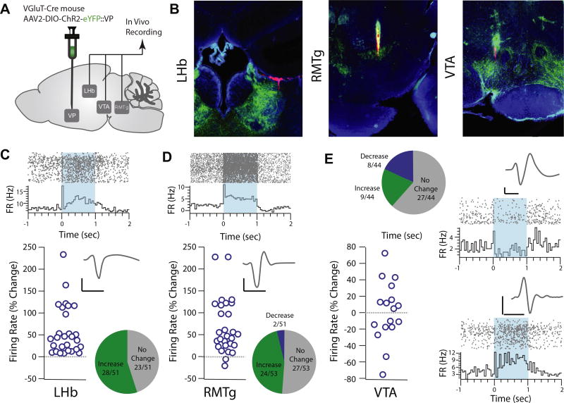
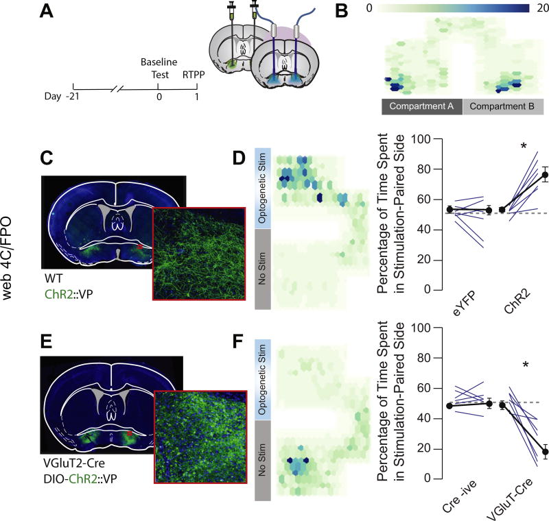
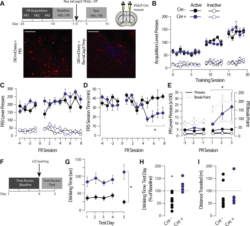
Methods: Using neurochemical, genetic, and electrophysiological approaches, we characterized glutamatergic VP neurons (n = 4-8 mice/group). We performed patch clamp and in vivo electrophysiology recordings in the lateral habenula, rostromedial tegmental nucleus, and ventral tegmental area to determine the effect of glutamatergic VP neuron activation in these target regions (n = 6-10 mice/group). Finally, we selectively optogenetically stimulated glutamatergic VP neurons in a real-time place preference task and ablated these neurons using a virally expressed caspase to determine their necessity for reward seeking.

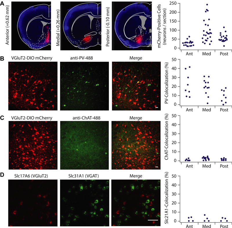
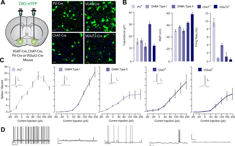
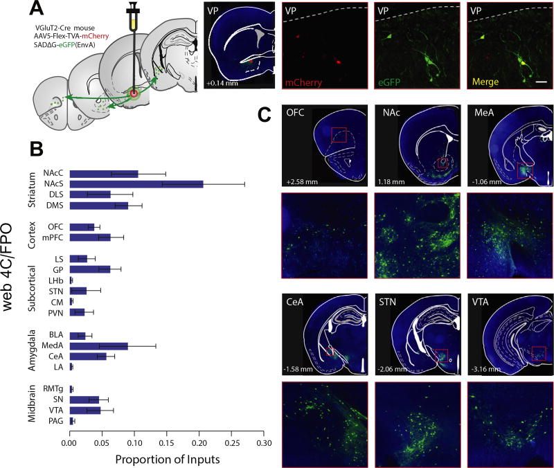
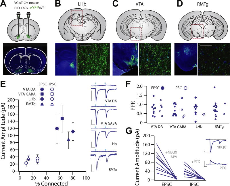
Results: Glutamatergic VP neurons exhibit little overlap with cholinergic or gamma-aminobutyric acidergic markers, the canonical VP subtypes, and exhibit distinct membrane properties. Glutamatergic VP neurons innervate and increase firing activity of the lateral habenula, rostromedial tegmental nucleus, and gamma-aminobutyric acidergic ventral tegmental area neurons. While nonselective optogenetic stimulation of the VP induced a robust place preference, selective activation of glutamatergic VP neurons induced a place avoidance. Viral ablation of glutamatergic VP neurons increased reward responding and abolished taste aversion to sucrose.

Conclusions: Glutamatergic VP neurons constitute a noncanonical subpopulation of VP neurons. These glutamatergic VP neurons increase activity of the lateral habenula, rostromedial tegmental nucleus, and gamma-aminobutyric acidergic ventral tegmental area neurons and adaptively constrain reward seeking.

Keywords: Aversion; Dopamine (DA); Electrophysiology; Gamma-aminobutyric acid (GABA); Rostromedial tegmental nucleus (RMTg); Ventral tegmental area (VTA).


Conflict of interest statement

DISCLOSURES

The authors report no biomedical financial interests or potential conflicts of interest.

Figures

Figure 1
Figure 1
Vesicular glutamate transporter (VGluT2)-positive ventral pallidum (VP) neurons exhibit distinct neurochemical properties from canonical VP subtypes. (A) VGluT2-Cre mice were injected with floxed mCherry in the VP to selectively label VGluT2-positive VP cells. VGluT2-positive VP neurons were expressed through the anterior (Ant) (+0.98 to 0.50 mm anterior–posterior relative to bregma; 27.6 ± 3.4 cells/section), medial (Med) (+0.50 to 0.02 mm anterior–posterior relative to bregma; 97.4 ± 11.8 cells/section), and posterior (Post) (0.02 to −0.46 mm anterior–posterior relative to bregma; 55.2 ± 5.6 cells/section) planes of the VP. To measure the percentage of selectively labeled glutamatergic neurons that colocalize with parvalbumin (PV) and choline acetyltransferase (ChAT), confocal images (20×) were taken of VP sections of VGluT2-Cre mice infected with floxed mCherry and immunostained against PV and ChAT primary antibodies, with the secondary antibody conjugated to Alexa Fluor 488. (B) Colocalization of PV and mCherry was observed in the anterior (22.6 ± 4.6%), medial (19.7 ± 2.5%), and posterior (9.2 ± 2.4%) planes, with highest colocalization occurring in the anterior VP (F2 = 4.178, p = .027). (C) ChAT and mCherry exhibited little coexpression (anterior 7.4 ± 5.0%, medial 3.3 ± 0.5%, posterior 2.89 ± 0.59%; F2 = 1.780, p = .183). (D) Fluorescent images and quantification of in situ hybridization against VGluT2 (Slc17A6, red) and VGAT (Slc31A1, green) revealed low colocalization of VGluT and VGAT cells throughout the VP (anterior 2.0 ± 1.2%, medial 2.3 ± 1.6%, posterior 2.1 ± 1.1%; F2 = 0.100, p = .906). Scale bars: 50 µM. VGAT, vesicular GABA transporter.
Figure 2
Figure 2
Vesicular glutamate transporter (VGluT2)-positive ventral pallidum (VP) neurons exhibit distinct membrane properties from canonical VP subtypes. (A) Experimental schematic. Floxed adeno-associated virus construct encoding enhanced yellow fluorescent protein (eYFP) was injected into the VP of VGAT-Cre, ChAT-Cre, PV-Cre, or VGluT2-Cre mice to selectively label genetically defined cell types in the VP. Using patch clamp electrophysiology, passive membrane properties were measured from parvalbumin (PV)-positive (PV+; n = 11), gamma-aminobutyric acid (GABA) type I (n = 14), and GABA type II (n = 14), choline acetyltransferase (ChAT)-positive (ChAT+; n = 8), and VGluT2-expressing (VGluT2+; n = 7) VP neurons. (B) Summary of average capacitance values (PV+ = 17.6 ± 2.3 pF, GABA I = 18.6 ± 1.7 pF, GABA II = 13.4 ± 2.6 pF, ChAT+ = 32.4 ± 2.1 pF, VGluT+ = 14.1 ± 1.3 pF), resting membrane potential (PV+ = −51.3 ± 1.6 mV, GABA I = −64.5 ± 1.5 mV, GABA II = −51.2 ± 2.8 mV, ChAT+ = −58.1 ± 1.8 mV, VGluT+ = −78.5 ± 2.8 mV), and firing rate (PV+ = 9.77 ± 0.89 Hz, GABA I = 0.47 ± 0.16 Hz, GABA II = 4.78 ± 0.42 Hz, ChAT+ = 2.03 ± 1.00 Hz, VGluT+ = 0.86 ± 0.75 Hz) is shown for each genetically defined population of VP neurons. VGluT+ VP neurons were significantly hyperpolarized, F4 = 27.24 p < .001, Tukey post hoc tests for comparison of VGluT against all other populations < .01. (C) For each genetically identified cell population, the number of action potentials per second in response to successive current injection (−40 to 200 pA) is plotted. Excitability was calculated as area under the curve. There was a significant effect of cell type on excitability (F4 = 25.08, p < .001); excitability was lower in VGluT2+ cells relative to all other subtypes (ChAT: t = 2.83, p = .011; PV: t = 6.62, p < .001; GABA type I: t = 3.57, p = .001; GABA type II: t = 2.20, p = .034). (D) Representative trace demonstrating spontaneous firing rate and waveform of genetically identified VP subpopulations. Scale bars for action potential trace: 10 mV, 20 ms. Scale bars for spontaneous activity trace: 10 mV, 1 s.
Figure 3
Figure 3
Pseudorabies tracing reveals monosynaptic inputs to glutamatergic ventral pallidum (VP) neurons. (A) Experimental schematic. Floxed adeno-associated virus constructs encoding the rabies G protein and envelope protein, followed by the G-deleted pseudorabies virus encoding green fluorescent protein (eGFP), were injected into the VP of VGluT2-Cre mice to map monosynaptic inputs to the VP. A confocal image of example starter cells expressing mCherry and GFP is shown. (B) GFP-positive cells were quantified in striatal, cortical, subcortical, and amygdala subregions and midbrain areas and expressed as a proportion of total GFP-positive cells (see Supplemental Table S1). (C) Representative pictomicrographs (4×) and insets (20×) showing GFP-labeled cells in the orbitofrontal cortex (OFC) (top left), nucleus accumbens core (NAc) and striatum (top center), medial amygdala (MeA) (top right), central amygdala (CeA) (bottom left), subthalamic nucleus (STN) (bottom center), and ventral tegmental area (VTA) (bottom right). Scale bar = 100 µM. BLA, basolateral amygdala; CM, central medial nucleus; DLS, dorsolateral striatum; DMS, dorsomedial striatum; GP, globus pallidus; LA, lateral amygdala; LHb, lateral habenula; LS, lateral septum; mPFC, medial prefrontal cortex; NAcS, nucleus accumbens shell; PAG, periaqueductal gray; PVN, paraventricular thalamus; RMTg, rostromedial tegmentum; SN, substantia nigra.
Figure 4
Figure 4
Glutamatergic ventral pallidum (VP) neurons exhibit functional projections to the lateral habenula (LHb), ventral tegmental area (VTA), and rostromedial tegmental nucleus (RMTg). (A–D) Experimental schematic and widefield images showing floxed channelrhodopsin-2 (ChR2) with enhanced yellow fluorescent protein (eYFP) injection sites to the VP of VGluT2-Cre mice and terminal fields in the LHb (B), VTA (C), and RMTg (D). (E) Patch clamp recordings were made of neurons in the terminal fields of vesicular glutamate transporter–positive VP neurons in the LHb (n = 20, capacitance = 17.63 ± 1.32 pF), RMTg (n = 15, capacitance = 26.83 ± 2.56 pF), and VTA (dopamine [DA] neuron n = 14, capacitance = 55.53 ± 5.097 pF, gamma-aminobutyric acid [GABA] neuron n = 14, capacitance = 19.96 ± 1.64). Recordings made at −70 mV revealed robust inward excitatory postsynaptic currents (EPSCs) (LHb: 80.0% connected, −89.7 ± 20.5 pA; RMTg: 73.3% connected, −98.6 ± 23.4 pA; VTA DA: 64.3% connected, −113.4 ± 43.4 pA; VTA GABA: 57.1% connected, −104.9 ± 40.6 pA), and recordings made at 0 mV revealed infrequent outward inhibitory postsynaptic currents (IPSCs) (LHb: 10% connected, 33.5 ± 19.6 pA; RMTg: 13.3% connected, 32.5 ± 5.5 pA; VTA DA: 42.8% connected, 31.8 ± 7.2 pA; VTA GABA: 35.7% connected, 83.2 ± 44.6 pA). There was no difference between areas with respect to proportion of neurons exhibiting light-evoked currents, and EPSCs were observed more frequently than IPSCs [t test between proportions; VTA DA χ2(1) = 1.25, p = .26; VTA GABA χ2(1) = 1.25, p = .26; LHb χ2(1) = 14.04, p < .001; RMTg χ2(1) = 10.63, p = .001]. (F) There was no significant difference in EPSC or IPSC paired-pulse ratio (PPR) in any region (VTA DA: t4 = 0.6941, p = .5258; VTA GABA: t3 = 0.2195, p = .8403; LHb: t2 = 0.0187, p = .9867; RMTg: t1 = 0.7603, p = .5862) or in PPR between any brain region in terms of EPSC (one-way analysis of variance, EPSC F3 = 0.2552, p = .8572; mean VTA DA = 0.637 ± 0.091, VTA GABA = 0.784 ± 0.139, LHb = 0.691 ± 0.122, RMTg = 0.654 ± 0.116) or IPSC amplitude (one-way analysis of variance, IPSC F3 = 0.2487, p = .8605; VTA DA = 0.794 ± 0.166, VTA GABA = 0.849 ± 0.193, LHb = 0.620 ± 0.190, RMTg = 0.709 ± 0.265). (G) We verified that outward EPSCs were blocked by the combination of alpha-amino-3-hydroxy-5-methyl-4-isoxazole propionic acid and N-methyl-D-aspartate receptor antagonists (6-nitro-7-sulfamobenzoquinoxaline-2-3-dione [NBQX] and D, L-2-amino-5-phosphonovaleric acid [APV] at 100 µM), and outward IPSCs were blocked by the chloride channel blocker picrotoxin (PTX; 50 µM). Scale bars: 20 pA, 20 ms.
Figure 5
Figure 5
Optogenetic activation of glutamatergic ventral pallidum (VP) neurons increases firing rates of lateral habenula (LHb) and rostromedial tegmental nucleus (RMTg) neurons and modulates ventral tegmental area (VTA) firing rates in vivo. (A) Experimental schematic. Floxed channelrhodopsin-2 (ChR2) with enhanced yellow fluorescent protein (eYFP) was injected into the VP of VGluT2-Cre mice, and in vivo recordings were performed in the LHb, RMTg, and VTA. (B) Representative images showing histological verification of recording sites in the LHb (left), RMTg (center), and VTA (right). (C) Representative recording from an isolated LHb unit (top; raster plot and peri-event histogram) aligned to onset of a 1-s optogenetic stimulation of vesicular glutamate transporter–positive VP neurons. Representative recording shows firing rate 1 s before optogenetic onset to 2 s following onset. Unit waveform is shown in inset. In total, 51 units were identified, 28 of which were significantly modulated, and all significantly modulated units exhibited an increased firing rate (45.1% unmodulated, 54.9% increased). The firing rate increased by 56.7 ± 11.8% in significantly modulated units. (D) Representative recording from an isolated unit from the RMTg. In total, 53 units were identified, 26 of which were significantly modulated (50.9% unmodulated, 45.3% increased, 3.8% decreased). Together, the firing rate of the RMTg increased by 61.55 ± 12.5% in significantly modulated units. (E) In the VTA, 44 units were identified; of these, 61.4% were unmodulated, 20.5% increased their firing rate, and 18.2% decreased their firing rate in response to optogenetic activation of vesicular glutamate transporter–positive VP neurons. A representative triphasic waveform and raster plot from a negatively modulated unit (top) and a positively modulated unit (bottom) are shown. Scale bars: 20 µs, 20 mV. FR, firing rate.
Figure 6
Figure 6
Activation of glutamatergic ventral pallidum (VP) neurons induces real-time place aversion. (A) Experimental schematic. Unfloxed channelrhodopsin-2 (ChR2) was injected into the VP of wild-type (WT) mice, floxed ChR2 was injected into the VP of VGluT2-Cre mice, and optic fibers were implanted over the injection site. A baseline place preference task was conducted 24 hours before the real-time place preference (RTPP) task. (B) Representative heat map showing no baseline preference for either chamber of the test apparatus (scale indicates activity counts of tracked position). (C) Widefield image of infection site of unfloxed ChR2-enhanced yellow fluorescent protein (eYFP) in the VP. (D) Nonspecific optogenetic stimulation (stim) of the VP induced an RTPP (baseline 51.1 ± 1.4%, stim 77.1 ± 4.9%; t6 = 6.17, p < .001), while stimulation with the control fluorophore had no effect (baseline 49.7 ± 1.7%, stim 47.0 ± 5.1%; t6 = 1.036, p = .34). (E) Widefield image showing infection site of floxed ChR2-eYFP in the VP of VGluT2-Cre mice. (F) Selective optogenetic activation of glutamatergic VP neurons induced a real-time place aversion (baseline 49.9 ± 2.8%, stim 23.1 ± 6.03%; t6 = 6.05, p < .001), while Cre-negative littermates showed no response to light stimulation (baseline 53.1 ± 2.5%, stim 52.8 ± 3.1%; t5 = 0.11, p = .914).
Figure 7
Figure 7
Genetically encoded, caspase-mediated ablation of glutamatergic ventral pallidum (VP) neurons increases responding for sucrose and impairs sucrose taste aversion learning. (A) Experimental schematic. Mice were trained on an operant task to lever press for a sucrose pellet at fixed ratio (FR) 1 schedule. When performance stabilized on FR 1 responding, five sessions each of FR 2 and 3 schedules were completed before baseline operant testing (consisting of FR 5 and progressive ratio [PR] test sessions). VGluT2-Cre mice or Cre-negative control mice were then injected bilaterally with Cre-dependent viral caspase, and testing was resumed. Representative pictomicrographs showing floxed mCherry expression in the VP of unlesioned (left) and taCasp-lesioned (right) mice are shown. (B) There was no effect of genotype on the initial acquisition of the FR responding task (Fgenotype = 0.196, p = .662); lever presses increased as a function to FR schedule (Fsession = 314.117, p < .001), and there was a significant difference between active and inactive lever presses (Flever = 281.999, p < .001). (C) There was no effect of lesion on number of FR 5 lever presses (Cre− = pre 197.83 ± 9.5, post 264.13 ± 12.4 presses/session; Cre+ = pre 183.78 ± 10.65, post 214.53 ± 10.8 presses/session; Fsession × genotype = 2.612, p = .120). (D) Following lesion, the amount of time to earn 30 sucrose rewards decreased in VGluT2-Cre mice (Cre− = pre 36.09 ± 1.9, post 39.36 ± 2.54 min/session; Cre+ = pre 36.12 ± 2.6, post 22.04 ± 2.31 min/session; Fsession × genotype = 64.193, p = .003). (E) Following lesion, the number of lever presses in the PR task increased in VGluT2-Cre mice (Cre− = pre 372.11 ± 28.14, post 443.39 ± 32.9 presses/session; Cre+ = pre 496.24 ± 48.87, post 1656.62 ± 444.88 presses/session; Fsession × genotype = 5.198, p = .004), as did the breakpoint (Cre− = pre 14.47 ± 0.38, post 15.0 ± 0.41, Cre+ = pre 15.2 ± 0.3, post 19.0 ± 0.42, Fsession × genotype = 5.09, p = .001). (F) Schematic of sucrose aversion task. Lesioned mice were given free access to sucrose solution over a 4-day baseline. Sucrose solution was then paired with lithium chloride (LiCl), and mice were retested the following day. (G, H) Absolute time spent drinking sucrose was higher in VGluT2-Cre-lesioned mice (84.8 ± 8.4 s/session) than in wild-type control mice (40.3 ± 4.6 s/session). Wild-type mice decreased their sucrose consumption following LiCl pairing (50.3 ± 11.1% decrease from baseline; t7 paired t test = 2.844, p = .0249), whereas VGluT2-Cre-lesioned mice did not decrease their sucrose consumption in response to LiCl pairing (31.4 ± 9.4% nonsignificant change; t5 paired t test = 1.9024, p = .115). There was a significant effect of genotype and day by genotype by day interaction (Fgenotype = 39.034, p < .001; Fgenotype × test day = 9.603, p = .009. (I) There was no difference in spontaneous locomotor activity between lesioned WT (82.7 ± 20.8 m) and VGLuT2-Cre (77.7 ± 17.5 m) mice in an open field arena task (t12 = 0.184, p = .858). Scale bar = 50 µM.

Comment in

  • The Control of Reward Seeking.
    Darcq E, Kieffer BL. Darcq E, et al. Biol Psychiatry. 2018 Jun 15;83(12):981-983. doi: 10.1016/j.biopsych.2018.04.003. Biol Psychiatry. 2018. PMID: 29804588 No abstract available.

References

    1. Haber SN, Groenewegen HJ, Grove EA, Nauta WJ. Efferent connections of the ventral pallidum: Evidence of a dual striato pallidofugal pathway. J Comp Neurol. 1985;235:322–335. - PubMed
    1. Dichter GS, Damiano CA, Allen JA. Reward circuitry dysfunction in psychiatric and neurodevelopmental disorders and genetic syndromes: Animal models and clinical findings. J Neurodev Disord. 2012;4:19. - PMC - PubMed
    1. Nusslock R, Alloy LB. Reward processing and mood-related symptoms: An RDoC and translational neuroscience perspective. J Affect Disord. 2017;216:3–16. - PMC - PubMed
    1. Feil J, Sheppard D, Fitzgerald PB, Yucel M, Lubman DI, Bradshaw JL. Addiction, compulsive drug seeking, and the role of frontostriatal mechanisms in regulating inhibitory control. Neurosci Biobehav Rev. 2010;35:248–275. - PubMed
    1. O’Donnell P, Lavin A, Enquist LW, Grace AA, Card JP. Interconnected parallel circuits between rat nucleus accumbens and thalamus revealed by retrograde transynaptic transport of pseudorabies virus. J Neurosci. 1997;17:2143–2167. - PMC - PubMed

Publication types

MeSH terms