

Structural biology of the separase-securin complex with crucial roles in chromosome segregation
- PMID: 29452922
- PMCID: PMC5915870
- DOI: 10.1016/j.sbi.2018.01.012
Structural biology of the separase-securin complex with crucial roles in chromosome segregation
Abstract
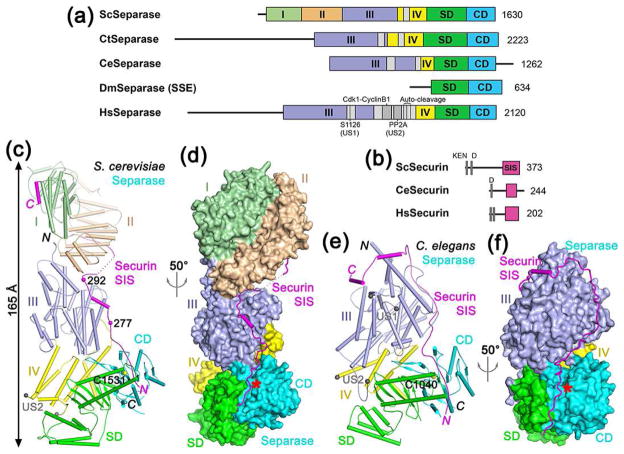
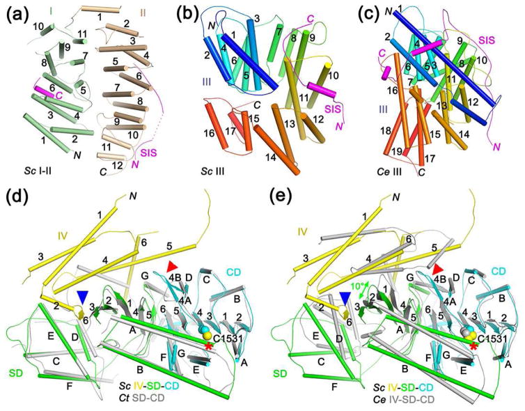
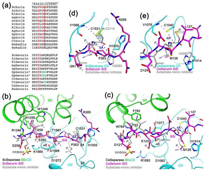
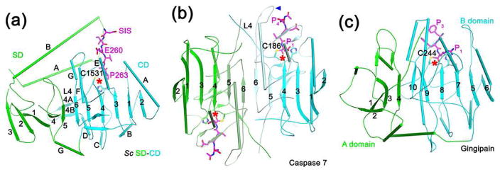
The cysteine protease separase opens the cohesin ring by cleaving its kleisin subunit and is a pivotal cell cycle factor for the transition from metaphase to anaphase. It is inhibited by forming a complex with the chaperone securin, and in vertebrates, also by the Cdk1-cyclin B1 complex. Separase is activated upon the destruction of securin or cyclin B1 by the proteasome, after ubiquitination by the anaphase-promoting complex/cyclosome (APC/C). Here we review recent structures of the active protease segment of Chaetomium thermophilum separase in complex with a substrate-mimic inhibitor and full-length Saccharomyces cerevisiae and Caenorhabditis elegans separase in complex with securin. These structures define the mechanism for substrate recognition and catalysis by separase, and show that securin has extensive contacts with separase, consistent with its chaperone function. They confirm that securin inhibits separase by binding as a pseudo substrate.
Copyright © 2018 Elsevier Ltd. All rights reserved.
Figures
Similar articles
-
Structure and Function of the Separase-Securin Complex.Subcell Biochem. 2021;96:217-232. doi: 10.1007/978-3-030-58971-4_4. Subcell Biochem. 2021. PMID: 33252730 Free PMC article. Review.
-
Structural basis of human separase regulation by securin and CDK1-cyclin B1.Nature. 2021 Aug;596(7870):138-142. doi: 10.1038/s41586-021-03764-0. Epub 2021 Jul 21. Nature. 2021. PMID: 34290405 Free PMC article.
-
Structural basis of cohesin cleavage by separase.Nature. 2016 Apr 7;532(7597):131-4. doi: 10.1038/nature17402. Epub 2016 Mar 30. Nature. 2016. PMID: 27027290 Free PMC article.
-
Human chromosome segregation involves multi-layered regulation of separase by the peptidyl-prolyl-isomerase Pin1.Mol Cell. 2015 May 7;58(3):495-506. doi: 10.1016/j.molcel.2015.03.025. Epub 2015 Apr 23. Mol Cell. 2015. PMID: 25921067
-
How do so few control so many?Cell. 2005 Mar 25;120(6):739-46. doi: 10.1016/j.cell.2005.03.006. Cell. 2005. PMID: 15797376 Review.
Cited by
-
The Interplay of Cohesin and RNA Processing Factors: The Impact of Their Alterations on Genome Stability.Int J Mol Sci. 2022 Apr 1;23(7):3939. doi: 10.3390/ijms23073939. Int J Mol Sci. 2022. PMID: 35409298 Free PMC article. Review.
-
Cystathionine β-synthase is required for oocyte quality by ensuring proper meiotic spindle assembly.Cell Prolif. 2022 Dec;55(12):e13322. doi: 10.1111/cpr.13322. Epub 2022 Aug 19. Cell Prolif. 2022. PMID: 36053797 Free PMC article.
-
Structure and Function of the Separase-Securin Complex.Subcell Biochem. 2021;96:217-232. doi: 10.1007/978-3-030-58971-4_4. Subcell Biochem. 2021. PMID: 33252730 Free PMC article. Review.
-
The Functions and Mechanisms of the Cohesin Complex in Regulating the Fate Determinations of Stem Cells.Research (Wash D C). 2025 Jul 10;8:0757. doi: 10.34133/research.0757. eCollection 2025. Research (Wash D C). 2025. PMID: 40642059 Free PMC article. Review.
-
The Involvement of Ubiquitination Machinery in Cell Cycle Regulation and Cancer Progression.Int J Mol Sci. 2021 May 27;22(11):5754. doi: 10.3390/ijms22115754. Int J Mol Sci. 2021. PMID: 34072267 Free PMC article. Review.
References
-
- Uhlmann F, Wernic D, Poupart MA, Koonin EV, Nasmyth K. Cleavage of cohesin by the CD clan protease separin triggers anaphase in yeast. Cell. 2000;103:375–386. - PubMed
-
- Nasmyth K. Segregating sister genomes: the molecular biology of chromosome separation. Science. 2002;297:559–565. - PubMed
-
- Uhlmann F. Separase regulation during mitosis. Biochem Soc Trans. 2003;70:243–251. - PubMed
-
- Yanagida M. Cell cycle mechanisms of sister chromatid separation; role of Cut1/separin and Cut2/securin. Genes to Cells. 2000;5:1–8. - PubMed
-
- Kamenz J, Hauf S. Time to split up: dynamics of chromosome separation. Trends Cell Biol. 2017;27:42–54. A recent review on chromosome segregation and separase function. - PubMed
Publication types
MeSH terms
Substances
Grants and funding
LinkOut - more resources
Full Text Sources
Other Literature Sources
Molecular Biology Databases
Miscellaneous
