. 2018 Mar 28;38(13):3177-3189.
doi: 10.1523/JNEUROSCI.3368-17.2018. Epub 2018 Feb 16.

Codeficiency of Lysosomal Mucolipins 3 and 1 in Cochlear Hair Cells Diminishes Outer Hair Cell Longevity and Accelerates Age-Related Hearing Loss

Affiliations

Codeficiency of Lysosomal Mucolipins 3 and 1 in Cochlear Hair Cells Diminishes Outer Hair Cell Longevity and Accelerates Age-Related Hearing Loss

Teerawat Wiwatpanit et al. J Neurosci. .

Abstract

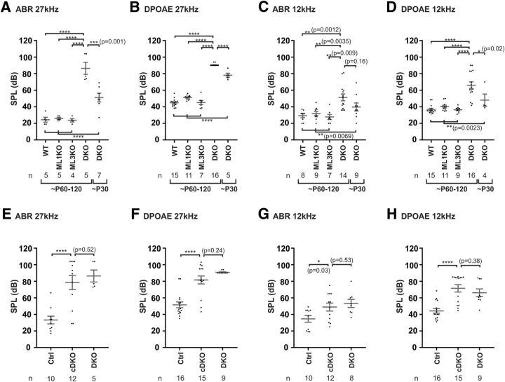
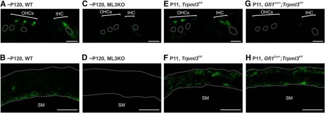
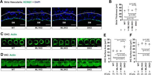
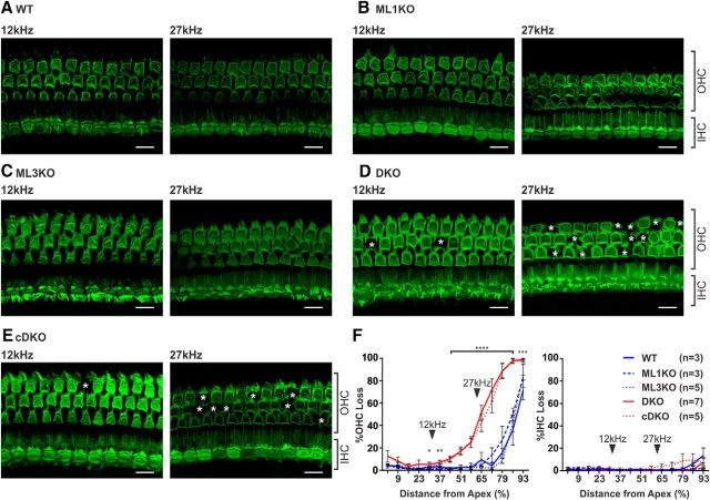
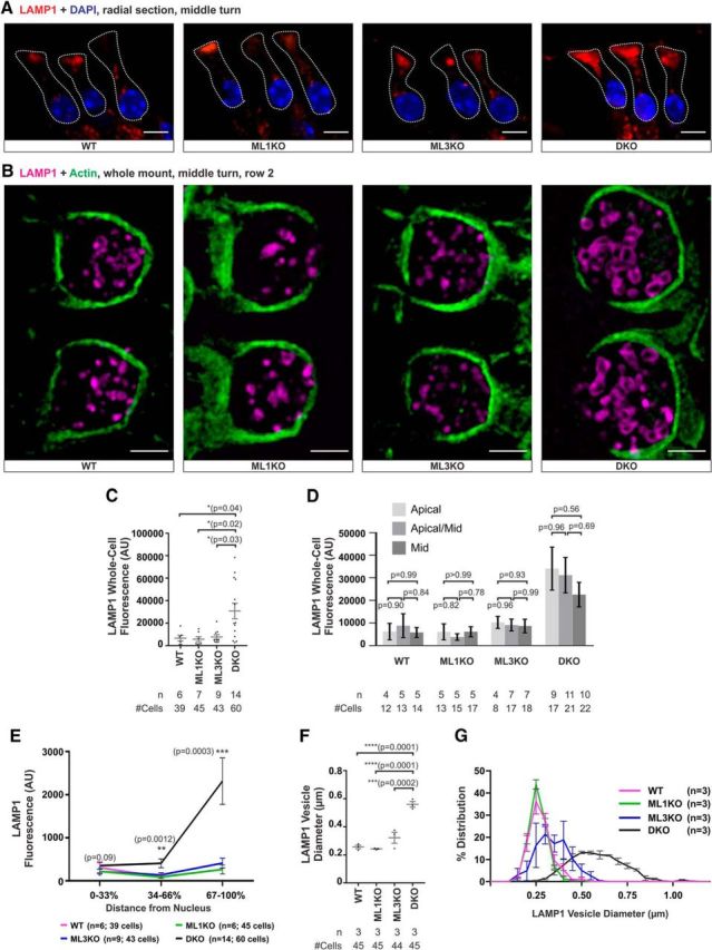
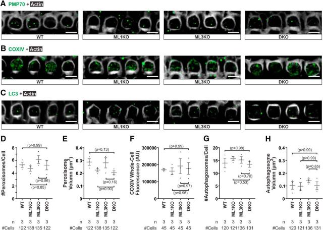
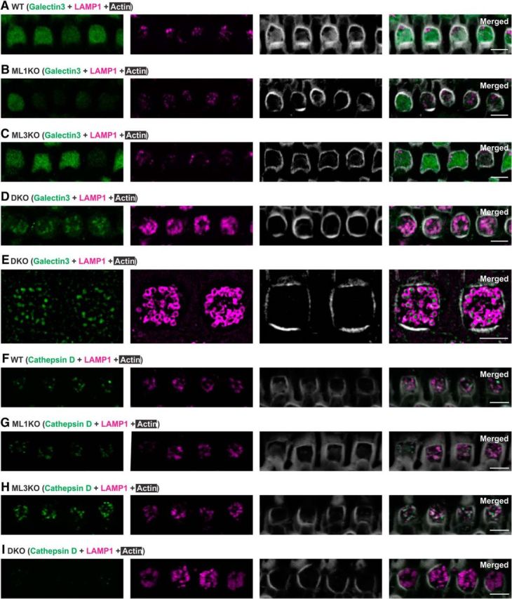
Acquired hearing loss is the predominant neurodegenerative condition associated with aging in humans. Although mutations on several genes are known to cause congenital deafness in newborns, few genes have been implicated in age-related hearing loss (ARHL), perhaps because its cause is likely polygenic. Here, we generated mice lacking lysosomal calcium channel mucolipins 3 and 1 and discovered that both male and female mice suffered a polygenic form of hearing loss. Whereas mucolipin 1 is ubiquitously expressed in all cells, mucolipin 3 is expressed in a small subset of cochlear cells, hair cells (HCs) and marginal cells of the stria vascularis, and very few other cell types. Mice lacking both mucolipins 3 and 1, but not either one alone, experienced hearing loss as early as at 1 month of age. The severity of hearing impairment progressed from high to low frequencies and increased with age. Early onset of ARHL in these mice was accompanied by outer HC (OHC) loss. Adult mice conditionally lacking mucolipins in HCs exhibited comparable auditory phenotypes, thereby revealing that the reason for OHC loss is mucolipin codeficiency in the HCs and not in the stria vascularis. Furthermore, we observed that OHCs lacking mucolipins contained abnormally enlarged lysosomes aggregated at the apical region of the cell, whereas other organelles appeared normal. We also demonstrated that these aberrant lysosomes in OHCs lost their membrane integrity through lysosomal membrane permeabilization, a known cause of cellular toxicity that explains why and how OHCs die, leading to premature ARHL.SIGNIFICANCE STATEMENT Presbycusis, or age-related hearing loss (ARHL), is a common characteristic of aging in mammals. Although many genes have been identified to cause deafness from birth in both humans and mice, only a few are known to associate with progressive ARHL, the most prevalent form of deafness. We have found that mice lacking two lysosomal channels, mucolipins 3 and 1, suffer accelerated ARHL due to auditory outer hair cell degeneration, the most common cause of hearing loss and neurodegenerative condition in humans. Lysosomes lacking mucolipins undergo organelle membrane permeabilization and promote cytotoxicity with age, revealing a novel mechanism of outer hair cell degeneration and ARHL. These results underscore the importance of lysosomes in hair cell survival and the maintenance of hearing.

Keywords: ARHL; TRPML; hearing; lysosome; mucolipin; presbycusis.


Figures

Figure 1.
Figure 1.
Coabsence of mucolipins 3 and 1 in HCs, but not either alone, causes age-related hearing loss progressing from high to low frequencies. A–H, Hearing thresholds at 27 and 12 kHz determined using ABRs (A, C, E, G) and DPOAEs (B, D, F, H) in adult (∼P60–P120) and juvenile (∼P30) mice. A, B, Adult mice lacking both mucolipins 3 and 1 (DKO), but not either alone (ML1KO or ML3KO), have raised ABR (A) and DPOAE (B) thresholds at 27 kHz compared with WT mice. Juvenile DKO mice (∼P30) exhibit somewhat smaller threshold shifts at 27 kHz for both ABR and DPOAE. C, D, Adult DKO mice have a mild hearing loss at 12 kHz indicated by both ABR (C) and DPOAE (D) threshold shifts. At 12 kHz, juvenile DKO mice have intermediate ABR and DPOAE threshold shifts. E–H, Hearing tests from mice in which mucolipins 3 and 1 are conditionally absent in HCs only (cDKO) indicate that adult cDKO mice (∼P120) have raised hearing thresholds at both 27 (E, F) and 12 (G, H) kHz. Age-matched (P120) data from adult DKO mice from (A–D) were selected and replotted (E–H) to statistically compare the ABR (E, G) and DPOAE (F, H) thresholds between mice lacking mucolipins in all cells versus mice lacking mucolipins in HCs only. *p < 0.05; **p < 0.01; ***p < 0.001; ****p < 0.0001.
Figure 2.
Figure 2.
Mucolipin 3 is localized to HCs and the stria vascularis in both adult and neonatal cochleae. Immunohistochemistry of the mucolipin 3 carboxyl terminus (TRPML3-CT) on adult (∼P120) (A–D) and neonatal (P11) (E–H) cochleae. A, TRPML3-CT labels vesicles in both IHCs and OHCs as well as cells of stria vascularis (B) in WT cochlea. C, D, In ML3KO tissues, there is no TRPML3-CT immunoreactivity detected above the background signals in either HCs (C) or stria vascularis (D). E, F, Neonatal Trpml3F/F HCs express mucolipin 3 in a similar pattern as adult HCs; however, the levels of TRPML3-CT immunoreactivity in P11 OHCs and IHCs are comparable (E). Mucolipin 3 appears more dispersed in the P11 stria vascularis (F). G, H, There is no TRPML3-CT immunoreactivity detected in GfiCre/+;Trpml3F/F HCs (G), although its signal is detected in the stria vascularis (H). Scale bars: A, C, E, G, 10 μm; B, D, F, H, 20 μm. For clarity, HC nuclei and strial boarder were outlined in top panels. SM, Scala media.
Figure 3.
Figure 3.
There is an anatomical defect in OHCs but not in the stria vascularis from cochleae lacking both mucolipin 3 and 1. A, Immunoreactivity of potassium channel KCNQ1 in the stria vascularis shows that there is no mislocalization of this channel in DKO cochlea compared with WT, ML1KO, and ML3KO animals. SM, Scala media. B, Quantification of strial thickness indicates that there is no strial degeneration in DKO cochlea. C, D, Actin labeling reveals that OHCs (C), but not IHCs (D), from DKO cochlea are larger than those of control cochleae. E, F, Quantification of HC apical surface area, measured just under the cuticular plate of HCs from middle turn, confirms that DKO OHCs are larger than the controls. Samples were from 4- to 4.5-month-old animals. Scale bars: A, 20 μm; C, D, 5 μm. ****p ≤ 0.0001.
Figure 4.
Figure 4.
Age-related hearing loss in animals lacking both mucolipin 3 and 1 is accompanied by OHC death. A–E, Actin labeling shows HCs from a whole-mount surface preparation of the adult cochlea (∼P120). Cochlear positions transmitting sound at 12 and 27 kHz were calculated using a mouse frequency-place map (Müller et al., 2005). There is no HC loss at 12 and 27 kHz positions in WT (A), ML1KO (B), and ML3KO (C) cochleae. However, OHC loss can be observed in DKO (D) and cDKO (E) cochleae (asterisks). The extent of OHC loss is more severe at the 27 kHz position. No IHC loss was observed at either position. F, Cytocochleograms showing %OHC and %IHC loss along the cochlear spiral for all genotypes (A–E). *p = 0.014. **p = 0.009. ***p = 0.0008. ****p ≤ 0.0001. Arrowheads indicate positions corresponding to 12 and 27 kHz. Scale bars, 10 μm.
Figure 5.
Figure 5.
OHCs lacking mucolipins 3 and 1 possess pathologically enlarged lysosomes. A, Immunoreactivity of LAMP1 (a lysosomal membrane protein) in adult (∼P120) OHCs reveals apical accumulation of lysosomes in DKO OHCs but not in those from WT, ML1KO, and ML3KO control cochleae. B, Super-resolution structured illumination microscopy shows that DKO OHCs, but not the controls, possess enlarged lysosomes as indicated by LAMP1 immunoreactivity. B, Images were OHCs from row 2. C, Quantification of total whole-cell LAMP1 fluorescence from A reveals a significant increase in LAMP1 in DKO OHCs but not in control cochleae. D, LAMP1 whole-cell intensity values used in C are replotted separately based on cochlear position (apical, apical/middle, and middle). We observed no difference in OHC LAMP1 intensity between cochlear turns within each genotype. E, LAMP1 intensity values used in C are replotted, binning the apical-basal axis of OHCs in three segments based on the percentage distance from the nucleus. F, By measuring LAMP1 vesicle diameter from images taken from confocal and structured illumination microscopy, we found that lysosomal diameter of DKO OHCs is ∼2.18-fold larger than that from control OHCs. G, A frequency distribution plot of LAMP1 vesicle diameter in all genotypes. Scale bars: A, 5 μm; B, 2 μm. *p < 0.05; **p < 0.01; ***p < 0.001; ****p ≤ 0.0001.
Figure 6.
Figure 6.
Enlarged lysosomes in OHCs lacking mucolipin 3 and 1 do not cause defects in lysosomal degradation of damaged organelles. A–C, Whole-mount cochleae from WT, ML1KO, ML3KO, and DKO adult (∼P120) animals using different organelle markers: peroxisome (PMP70, A), mitochondria (COXIV, B), and autophagosome (LC3, C). There is no apparent defect in distributions and numbers of peroxisomes (A), mitochondria (B), and autophagosomes (C). Representative images were reconstructed from a projection of 8 confocal optical sections spanning 0.88 μm under the cuticular plate. D, E, The number of peroxisomes per OHC and peroxisomal volume, indicated by PMP70 immunoreactivity, from DKO OHCs are similar to those from controls. F, Whole-cell fluorescence of COXIV did not differ between DKO and control OHCs. G, H, The number of autophagosomes per OHC and autophagosomal volume, indicated by LC3 immunoreactivity, from DKO OHCs are similar to controls. Scale bars, 5 μm.
Figure 7.
Figure 7.
OHCs lacking mucolipin 3 and 1 exhibit lysosomal membrane permeabilization. A–D, Immunoreactivity of galectin 3, a membrane-impermeable cytosolic lectin with high affinity for the luminal lysosomal glycocalyx (Aits et al., 2015), and LAMP1 reveal that lysosomal membranes in DKO OHCs are permeabilized. Galectin 3 is cytosolic in WT (A), ML1KO (B), and ML3KO (C) OHCs, and its expression level varied. D, In contrast, galectin 3 immunoreactivity has a vesicular pattern that is associated with LAMP1 signal in DKO OHCs. E, Super-resolution–structured illumination microscopy shows that galectin 3 is localized to lysosomal lumen. F–I, Immunoreactivity of cathepsin D and LAMP1 reveals that, upon lysosomal permeabilization, DKO lysosomes contain very low levels of cathepsin D. Cathepsin D has a vesicular expression pattern strongly associated with lysosomes in WT (F), ML1KO (G), and ML3KO (H) OHCs. I, In contrast, cathepsin D levels are low or undetectable in DKO lysosomes. Samples were from adult (∼P120) animals. Scale bars: A–D, F–I, 5 μm; E, 2 μm.

Similar articles

Cited by

References

    1. Aits S, Kricker J, Liu B, Ellegaard AM, Hämälistö S, Tvingsholm S, Corcelle-Termeau E, Høgh S, Farkas T, Holm Jonassen A, Gromova I, Mortensen M, Jäättelä M (2015) Sensitive detection of lysosomal membrane permeabilization by lysosomal galectin puncta assay. Autophagy 11:1408–1424. 10.1080/15548627.2015.1063871 - DOI - PMC - PubMed
    1. Bach G. (2001) Mucolipidosis type IV. Mol Genet Metab 73:197–203. 10.1006/mgme.2001.3195 - DOI - PubMed
    1. Bach G, Webb MB, Bargal R, Zeigler M, Ekstein J (2005) The frequency of mucolipidosis type IV in the Ashkenazi Jewish population and the identification of 3 novel MCOLN1 mutations. Hum Mutat 26:591. 10.1002/humu.9385 - DOI - PubMed
    1. Boya P, Kroemer G (2008) Lysosomal membrane permeabilization in cell death. Oncogene 27:6434–6451. 10.1038/onc.2008.310 - DOI - PubMed
    1. Brunk UT, Dalen H, Roberg K, Hellquist HB (1997) Photo-oxidative disruption of lysosomal membranes causes apoptosis of cultured human fibroblasts. Free Radic Biol Med 23:616–626. 10.1016/S0891-5849(97)00007-5 - DOI - PubMed

Publication types

Substances