. 2018 Apr 16;84(9):e02810-17.
doi: 10.1128/AEM.02810-17. Print 2018 May 1.

Molecular and Functional Study of a Branching Sucrase-Like Glucansucrase Reveals an Evolutionary Intermediate between Two Subfamilies of the GH70 Enzymes

Affiliations

Molecular and Functional Study of a Branching Sucrase-Like Glucansucrase Reveals an Evolutionary Intermediate between Two Subfamilies of the GH70 Enzymes

Minghui Yan et al. Appl Environ Microbiol. .

Abstract

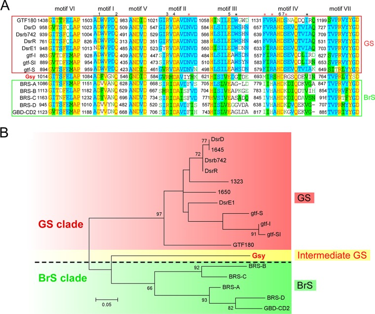
Glucansucrases (GSs) in glycoside hydrolase family 70 (GH70) catalyze the synthesis of α-glucans from sucrose, a reaction that is widely seen in lactic acid bacteria (LAB). These enzymes have been implicated in many aspects of microbial life. Products of GSs have great commercial value as food supplements and medical materials; therefore, these enzymes have attracted much attention from both science and industry. Certain issues concerning the origin and evolution of GSs are still to be addressed, although an increasing number of GH70 enzymes have been characterized. This study describes a GS enzyme with the appearance of a branching sucrase (BrS). Structural analysis indicated that this GS enzyme produced a type of glucan composed of an α-(1→6) glucosidic backbone and α-(1→4) branches, as well as a considerable amount of α-(1→3) branches, distinguishing it from the GSs identified so far. Moreover, sequence-based analysis of the catalytic core of this enzyme suggested that it might be an evolutionary intermediate between the BrS and GS subgroups. These results provide an evolutionary link between these subgroups of GH70 enzymes and shed new light on the origination of GSs.IMPORTANCE GH70 GSs catalyze the synthesis of α-glucans from sucrose, a reaction that is widely seen in LAB. Products of these enzymes have great commercial value as food supplements and medical materials. Moreover, these enzymes have attracted much attention from scientists because they have potential in tailored synthesis of α-glucans with desired structures and properties. Although more and more GSs have been characterized, the origin and evolution of these enzymes have not been well addressed. This study describes a GS with the appearance of a BrS (i.e., high levels of similarity to BrSs in sequence analysis). Further analysis indicated that this enzyme synthesized a type of insoluble glucan composed of an α-(1→6) glucosidic backbone and many α-(1→4)- and α-(1→3)-linked branches, the linkage composition of which has rarely been reported in the literature. This BrS-like GS enzyme might be an evolutionary intermediate between BrS and GS enzymes.

Keywords: GH70 enzymes; Leuconostoc mesenteroides; branching sucrase; glucansucrase; insoluble glucan; intermediate.


Figures

FIG 1
FIG 1
De novo polymerization activity of the recombinant Gsy. (A) Morphology of E. coli strains carrying pET28a(+)-gsy (left), compared to those carrying the empty vector (right). (B) SDS-PAGE analyses. (Left) Total protein extracts of E. coli cells were analyzed by SDS-PAGE, followed by staining with Coomassie brilliant blue (CBB). Note that a differentially expressed protein band was apparent in samples induced with IPTG. (Right) An equivalent gel was subjected to an in situ polymerization assay. At the site similar to the differential band in the CBB-stained gel, a polysaccharide-active band of ∼170 kDa was obvious.
FIG 2
FIG 2
Effects of pH and temperature on the activity of the purified Gsy. The effect of pH was assayed in 50 mM sodium acetate (pH 3.5 to 7.5), and the effect of temperature (20°C to 40°C) was assayed in 50 mM sodium acetate (pH 5.4). Reactions were carried out for 15 min. The enzyme activity was reflected by the amount of fructose released. The highest activity was set as 100%, and the other values were calculated accordingly (relative activity). Shown are the average values of three independent experiments, and the error bars represent the standard errors (n = 3).
FIG 3
FIG 3
FTIR spectrum of the glucan synthesized by purified Gsy. The glucan sample was prepared as described in Materials and Methods. The FTIR spectrum was obtained using a Nicolet Nexus 470 spectrometer.
FIG 4
FIG 4
1H NMR spectrum of the glucan produced by purified Gsy. The 1H NMR spectrum was recorded at 500 MHz in D2O at 30°C. 1H chemical shifts are given using sodium 2,2,3,3-tetradeutero-3-trimethylsilylpropanoate (1H = 0 ppm) as an internal reference. The 1H NMR spectrum was obtained with a Varian Mercury Plus 400 spectrometer (Varian, USA).
FIG 5
FIG 5
Sequence-based analysis of motifs I to VII of the catalytic core of different GH70 enzymes. (A) Alignment of motifs I to VII from different GH70 enzymes. Sequence alignment was performed with Align X (Vector NTI, Invitrogen), and motifs I to VII are indicated above the sequences. GSs are shown in the upper lines enclosed in the red box, while BrSs are shown in the lower lines enclosed in the green box; Gsy is shown between the two subgroups. The numbers 1 to 7 indicate the highly conserved sites of GH70 enzymes, and the asterisks indicate the sites conserved exclusively in BrSs, as identified by Vuillemin et al. (16). The black asterisks indicate the conserved sites shared by Gsy, and the red asterisks indicate the sites at which Gsy showed a pattern distinct from that of the BrSs identified so far. (B) Phylogenetic analysis of GH70 enzymes, as indicated. Phylogenetic analysis was performed using MEGA 6.0, and the evolutionary tree was constructed based on the sequences of the core motifs shown in panel A. The scale bar represents a 5% difference in amino acid sequences. To illustrate the peculiar characteristics of Gsy, it is marked in red font to emphasize its enzyme function as a GS enzyme, but it is shaded yellow to indicate its appearance as a BrS enzyme as well as a GS enzyme. The dashed line indicates the separation of the GS and BrS clades.
FIG 6
FIG 6
Phylogenetic analysis of the four subgroups of GH70 enzymes and GH13 proteins. Alignment was performed with the GH70 and GH13 enzymes listed in Table S1 in the supplemental material. Overall, the GH70 enzymes were obtained by a BLAST analysis using the Gsy protein (GenBank accession number ANJ45894.1) as the query sequence. Notably, all of the characterized GH70 enzymes (http://www.cazy.org/GH70_characterized.html) were included in the alignment. Members of GH13 in the alignment were chosen according to previous studies (7, 19). The evolutionary tree is based on alignment of the full sequences of these enzymes. The biochemically characterized GSs and GTFB-like and GTFC-like 4,6-α-GTs of GH70, as well as members of GH13, are included. The evolutionary history was inferred using the maximum likelihood method, based on the JTT matrix-based model. The sequences are labeled with their GenBank accession numbers. The bar represents a genetic distance of 0.3 substitution per position. The BrS and GS subgroups of GH70 are indicated in green and red fonts, respectively. Gsy is shown in red font shaded with yellow, and its homologs that have not been biochemically characterized are shown in gray.

References

    1. Puanglek S, Kimura S, Enomoto-Rogers Y, Kabe T, Yoshida M, Wada M, Iwata T. 2016. In vitro synthesis of linear α-1,3-glucan and chemical modification to ester derivatives exhibiting outstanding thermal properties. Sci Rep 6:30479. doi:10.1038/srep30479. - DOI - PMC - PubMed
    1. Freitas F, Alves VD, Reis MA. 2011. Advances in bacterial exopolysaccharides: from production to biotechnological applications. Trends Biotechnol 29:388–398. doi:10.1016/j.tibtech.2011.03.008. - DOI - PubMed
    1. Monsan P, Bozonnet S, Albenne C, Joucla G, Willemot RM, Remaud-Siméon M. 2001. Homopolysaccharides from lactic acid bacteria. Int Dairy J 11:675–685. doi:10.1016/S0958-6946(01)00113-3. - DOI
    1. Janeček Š, Svensson B, MacGregor EA. 2014. α-Amylase: an enzyme specificity found in various families of glycoside hydrolases. Cell Mol Life Sci 71:1149–1170. doi:10.1007/s00018-013-1388-z. - DOI - PMC - PubMed
    1. Moulis C, André I, Remaud-Siméon M. 2016. GH13 amylosucrases and GH70 branching sucrases, atypical enzymes in their respective families. Cell Mol Life Sci 73:2661–2679. doi:10.1007/s00018-016-2244-8. - DOI - PMC - PubMed

Publication types

MeSH terms

LinkOut - more resources