

AR Expression in Breast Cancer CTCs Associates with Bone Metastases
- PMID: 29453314
- PMCID: PMC5882540
- DOI: 10.1158/1541-7786.MCR-17-0480
AR Expression in Breast Cancer CTCs Associates with Bone Metastases
Abstract
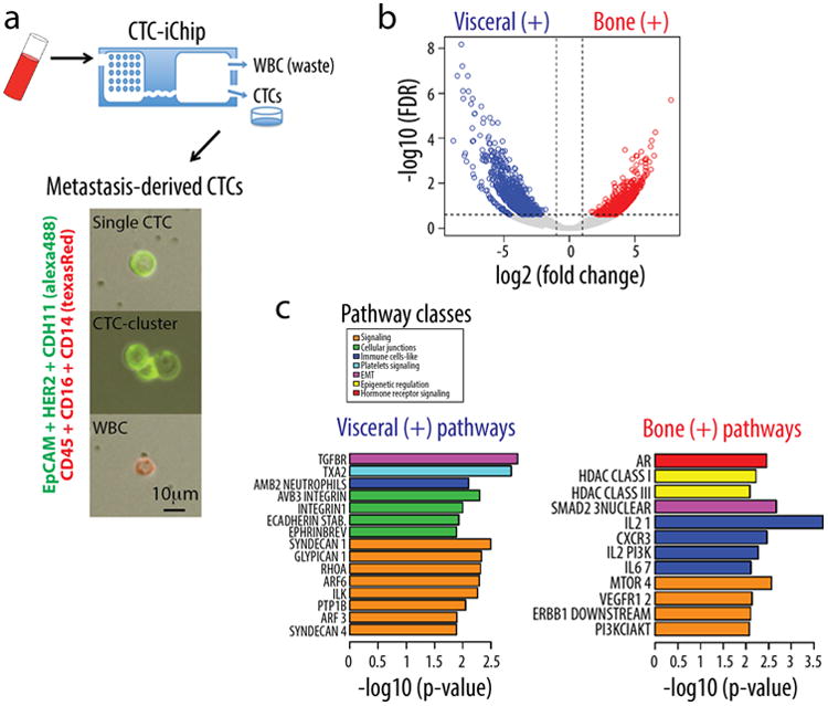
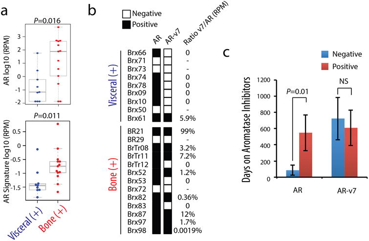
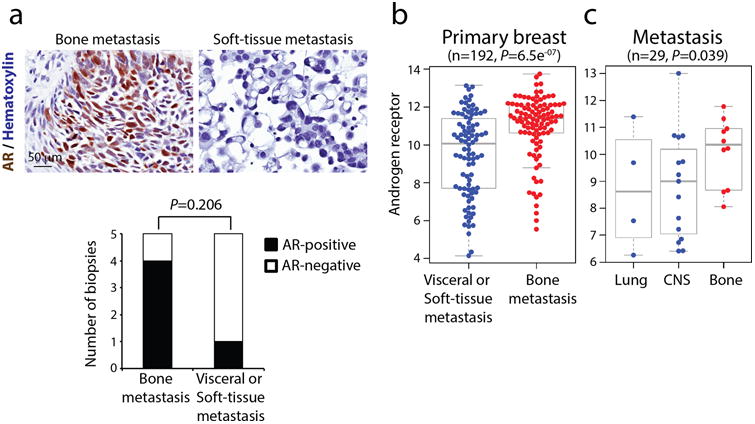
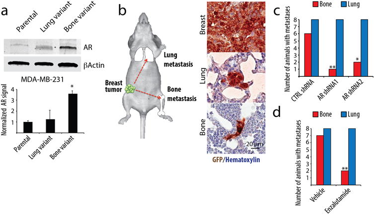
Molecular drivers underlying bone metastases in human cancer are not well understood, in part due to constraints in bone tissue sampling. Here, RNA sequencing was performed of circulating tumor cells (CTC) isolated from blood samples of women with metastatic estrogen receptor (ER)+ breast cancer, comparing cases with progression in bone versus visceral organs. Among the activated cellular pathways in CTCs from bone-predominant breast cancer is androgen receptor (AR) signaling. AR gene expression is evident, as is its constitutively active splice variant AR-v7. AR expression within CTCs is correlated with the duration of treatment with aromatase inhibitors, suggesting that it contributes to acquired resistance to endocrine therapy. In an established breast cancer xenograft model, a bone-tropic derivative displays increased AR expression, whose genetic or pharmacologic suppression reduces metastases to bone but not to lungs. Together, these observations identify AR signaling in CTCs from women with bone-predominant ER+ breast cancer, and provide a rationale for testing androgen inhibitors in this subset of patients.Implications: This study highlights a role for the AR in breast cancer bone metastasis, and suggests that therapeutic targeting of the AR may benefit patients with metastatic breast cancer. Mol Cancer Res; 16(4); 720-7. ©2018 AACR.
©2018 American Association for Cancer Research.
Conflict of interest statement
Conflict of Interest: The authors declare no potential conflict of interest.
Figures
Similar articles
-
Detecting androgen receptor (AR), AR variant 7 (AR-V7), prostate-specific membrane antigen (PSMA), and prostate-specific antigen (PSA) gene expression in CTCs and plasma exosome-derived cfRNA in patients with metastatic castration-resistant prostate cancer (mCRPC) by integrating the VTX-1 CTC isolation system with the QIAGEN AdnaTest.BMC Cancer. 2024 Apr 16;24(1):482. doi: 10.1186/s12885-024-12139-3. BMC Cancer. 2024. PMID: 38627648 Free PMC article.
-
Expression pattern of androgen receptors, AR-V7 and AR-567es, in circulating tumor cells and paired plasma-derived extracellular vesicles in metastatic castration resistant prostate cancer.Analyst. 2019 Nov 4;144(22):6671-6680. doi: 10.1039/c9an00999j. Analyst. 2019. PMID: 31596277
-
AR-V7 in circulating tumor cells cluster as a predictive biomarker of abiraterone acetate and enzalutamide treatment in castration-resistant prostate cancer patients.Prostate. 2018 Jun;78(8):576-582. doi: 10.1002/pros.23501. Epub 2018 Mar 5. Prostate. 2018. PMID: 29508425
-
Androgen receptor variant-driven prostate cancer: clinical implications and therapeutic targeting.Prostate Cancer Prostatic Dis. 2016 Sep;19(3):231-41. doi: 10.1038/pcan.2016.17. Epub 2016 May 17. Prostate Cancer Prostatic Dis. 2016. PMID: 27184811 Free PMC article. Review.
-
CTC-derived AR-V7 detection as a prognostic and predictive biomarker in advanced prostate cancer.Expert Rev Mol Diagn. 2018 Feb;18(2):155-163. doi: 10.1080/14737159.2018.1427068. Epub 2018 Jan 16. Expert Rev Mol Diagn. 2018. PMID: 29319382 Free PMC article. Review.
Cited by
-
Combinatorial Power of cfDNA, CTCs and EVs in Oncology.Diagnostics (Basel). 2022 Mar 31;12(4):870. doi: 10.3390/diagnostics12040870. Diagnostics (Basel). 2022. PMID: 35453918 Free PMC article. Review.
-
Transcriptomic profiling of single circulating tumor cells provides insight into human metastatic gastric cancer.Commun Biol. 2022 Jan 11;5(1):20. doi: 10.1038/s42003-021-02937-x. Commun Biol. 2022. PMID: 35017627 Free PMC article.
-
Differential Expression of the Androgen Receptor, Splice Variants and Relaxin 2 in Renal Cancer.Life (Basel). 2021 Jul 22;11(8):731. doi: 10.3390/life11080731. Life (Basel). 2021. PMID: 34440475 Free PMC article.
-
Future perspectives from lung cancer pre-clinical models: new treatments are coming?Transl Lung Cancer Res. 2020 Dec;9(6):2629-2644. doi: 10.21037/tlcr-20-189. Transl Lung Cancer Res. 2020. PMID: 33489823 Free PMC article. Review.
-
The Diversity of Liquid Biopsies and Their Potential in Breast Cancer Management.Cancers (Basel). 2023 Nov 17;15(22):5463. doi: 10.3390/cancers15225463. Cancers (Basel). 2023. PMID: 38001722 Free PMC article. Review.
References
-
- Kang Y, Siegel PM, Shu W, Drobnjak M, Kakonen SM, Cordon-Cardo C, et al. A multigenic program mediating breast cancer metastasis to bone. Cancer cell. 2003;3(6):537–49. - PubMed
Publication types
MeSH terms
Substances
Grants and funding
LinkOut - more resources
Full Text Sources
Other Literature Sources
Medical
Molecular Biology Databases
Research Materials
