. 2018 Feb 16;8(1):3184.
doi: 10.1038/s41598-018-21596-3.

A role for tau in learning, memory and synaptic plasticity

Affiliations

A role for tau in learning, memory and synaptic plasticity

Fabrizio Biundo et al. Sci Rep. .

Abstract

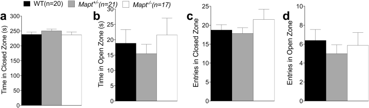
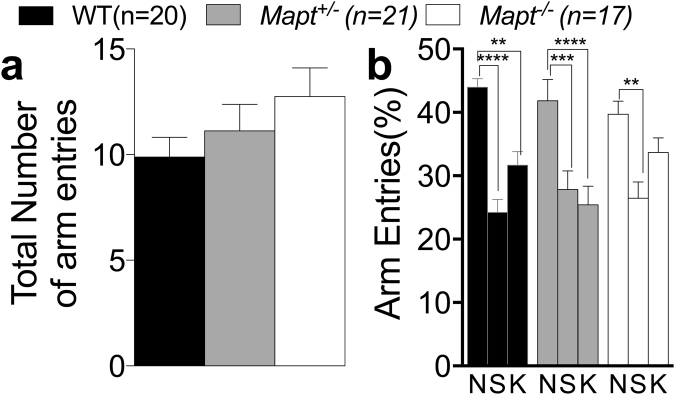
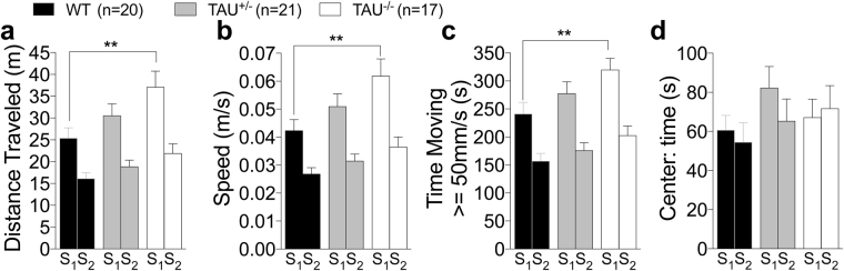
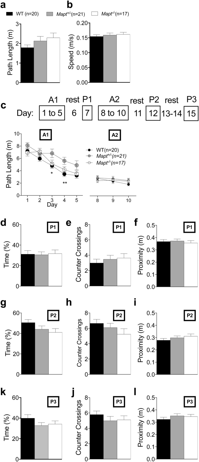
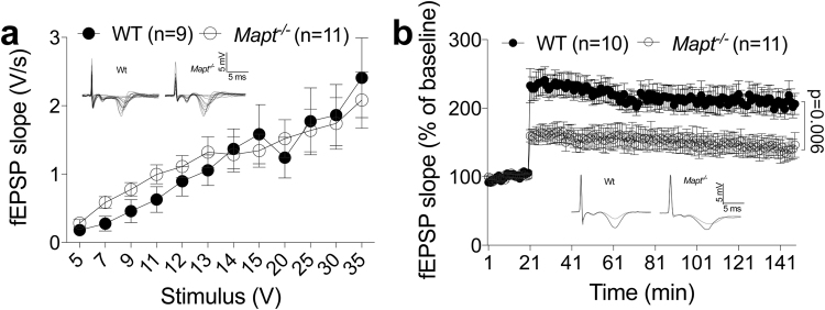
Tau plays a pivotal role in the pathogenesis of neurodegenerative disorders: mutations in the gene encoding for tau (MAPT) are linked to Fronto-temporal Dementia (FTD) and hyper-phosphorylated aggregates of tau forming neurofibrillary tangles (NFTs) that constitute a pathological hallmark of Alzheimer disease (AD) and FTD. Accordingly, tau is a favored therapeutic target for the treatment of these diseases. Given the criticality of tau to dementia's pathogenesis and therapy, it is important to understand the physiological function of tau in the central nervous system. Analysis of Mapt knock out (Mapt-/-) mice has yielded inconsistent results. Some studies have shown that tau deletion does not alter memory while others have described synaptic plasticity and memory alterations in Mapt-/- mice. To help clarifying these contrasting results, we analyzed a distinct Mapt-/- model on a B6129PF3/J genetic background. We found that tau deletion leads to aging-dependent short-term memory deficits, hyperactivity and synaptic plasticity defects. In contrast, Mapt+/- mice only showed a mild short memory deficit in the novel object recognition task. Thus, while tau is important for normal neuronal functions underlying learning and memory, partial reduction of tau expression may have fractional deleterious effects.


Conflict of interest statement

The authors declare no competing interests.

Figures

Figure 1
Figure 1
The elevated zero maze revealed no anxiety deficit in both Mapt+/− and Mapt−/− mice at the age of six months. The amount of time spent in the open and closed zones and the number of entries into the open and closed zones of the elevated zero maze during the 5-min testing period were analyzed (a–d). One-way ANOVA revealed no significant effect of genotype for all the measures.
Figure 2
Figure 2
The two trial Y-maze task showed a mild deficit of short-term spatial recognition memory in Mapt−/− mice at the age of 6 months. Mice were analyzed at 6 months of age. (a) Total number of arm entries during trial 2. (b) Percentage of entries into the novel (N), start (S) and known (K) arms. Data are expressed as means ± S.E.M.: **P < 0.01; ***P < 0.001, ****P < 0.0001 (Dunnett’s multiple comparisons test).
Figure 3
Figure 3
Increased locomotor activity of Mapt−/− mice at the age of 7 months in the open field test. (a) Mean distance traveled: Mapt−/− mice traveled significantly longer distances than WT animals during the first session. (b) Mean speed: Mapt−/− mice traveled at a significantly higher speed than littermate control mice during the first session. (c) Time spent traveling at speed greater than 50 mm/s. Mapt−/− mice spent more time moving > =50 mm/s than WT mice during the first session. (d) Mean time spent in the center of the open field is not significantly different among the three genotypes. Data are expressed as means ± S.E.M.: **P < 0.01, Tukey’s multiple comparisons test.
Figure 4
Figure 4
Novel object recognition test shows that 7-month-old Mapt−/− mice present a mild short-term memory deficit. Data are expressed as means ± S.E.M. (a) Time spent during the exploration of two familiar objects. (b) Time spent exploring the familiar and novel objects. While WT mice explore more the novel object (***P = 0.0002, Sidak’s multiple comparisons test), Mapt+/− and Mapt−/− mice spent comparable amount of time exploring the two objects. Discriminatory ratio and discriminatory index are shown in (c) and (d).
Figure 5
Figure 5
The visible platform and the Morris water maze tests show no visual, motivational spatial learning and retention memory deficit in 10-month-old Mapt−/− mice. Visual task: path length swam to reach the visible platform (a) and mean speed with which mice swam to reach the visible platform (b) revealed no significant effect of genotype (one-way ANOVA). (c) The schematic illustration of the Morris water maze task is represented above the mean path lengths for the A1 and A2 sessions. (df) Time spent in the target quadrant during the P1, P2 and P3 trials; (gh) Counter crossings in the target platform during the P1, P2 and P3 trials; (kl) Proximity to the target platform during the P1, P2 and P3 trials. Data are expressed as means ± S.E.M, *P < 0.05, **P < 0.01, Tukey’s multiple comparisons test.
Figure 6
Figure 6
The elevated zero maze revealed no anxiety deficit in both Mapt+/− and Mapt−/− mice at the age of twelve months. The amount of time spent in the open and closed zones and the number of entries into the open and closed zones of the elevated zero maze during the 5-min testing period were analyzed (a–d). One-way ANOVA revealed no significant effect of genotype for all the measures.
Figure 7
Figure 7
Short-term spatial recognition memory deficits of Mapt−/− mice in the two trial Y-maze task exacerbate with aging. Mice were analyzed at 12 months of age. (a) Total number of arm entries during trial 2. (b) Percentage of entries into the novel (N), start (S) and known (K) arms. Data are expressed as means ± S.E.M; *P < 0.05, **P < 0.01, ****P < 0.0001, Dunnett’s multiple comparisons test.
Figure 8
Figure 8
Increased locomotor activity of Mapt−/− mice at the age of 11 months in the open field test. (a) Mean distance traveled: Mapt−/− mice traveled significantly longer distances than WT animals during the first session. (b) Mean speed: Mapt−/− mice traveled at a significantly higher speed than littermate control mice during the first session. (c) Time spent traveling at speed greater than 50 mm/s. Mapt−/− mice spent more time moving > =50 mm/s than WT mice during the first session. (d) Mean time spent in the center of the open field is not significantly different among the three genotypes. Data are expressed as means ± S.E.M: **P < 0.01, ***P < 0.001, Tukey’s multiple comparisons test.
Figure 9
Figure 9
Mapt−/− mice exhibited a dysfunction in working memory at the age of 16 months in the 6-arm radial water maze. Visual task: path length swam to reach the visible platform (a) and mean speed with which mice swam to reach the visible platform (b) revealed no significant effect of genotype (one-way ANOVA). (c) average number of errors in finding the platform across four trials. Data are expressed as means ± S.E.M: *P < 0.05, ***P < 0.001, ****P < 0.0001, Dunnett’s multiple comparisons test.
Figure 10
Figure 10
Fear conditioning paradigm conducted at the age of 18 months show contextual memory deficit for Mapt−/−. (a) Percentage of freezing recorded during the stage preceding the administration of the US and CS (baseline). (b) Percentages of freezing during the contextual test 24 h after the administration of the stimuli. (c) Time course of the percentage of freezing in 1-min bins during the contextual test. Data are expressed as means ± S.E.M: *P < 0.05, Tukey’s multiple comparisons test.
Figure 11
Figure 11
Impaired LTP in old Mapt−/− mice. (a) Input/output curve: field potentials were recorded from 400 μm hippocampal slices at increasing values of voltage intensity stimulation. Data are expressed as means ± S.E.M. (b) Deletion of the Mapt gene significantly reduces LTP evoked with theta-burst tetanus at Schaeffer collateral CA3–CA1 synapses. Representative traces 1 min before (thin) and 120 min after (thick) θ-burst stimulation are shown.
Figure 12
Figure 12
Tau protein is reduced in Mapt+/− brains and absent in Mapt−/− brains. Western blot analysis of 5 brain homogenates from 5 20-month-old mice for each genotype (Mapt+/+, i.e. wild type, Mapt+/− and Mapt−/−) rains for with either the anti-APP antibody Y188, which recognize a C-terminal epitope of APP (top panel), of the anti-tau monoclonal antibody DA9 (bottom panel). The data show similar levels of APP and the APP metabolite αCTF in Mapt+/+, Mapt+/− and Mapt−/− brains. Yet, levels of tau are highest in Mapt+/+ samples, reduced in Mapt+/− homogenates and ablated in Mapt−/− brains. As indicated in the Methods section, the brain samples used for Western blot analysis were prepared from brains of animals perfused under anesthesia with PBS followed by a 4% formaldide solution. This explains why the APP and tau bands appear smeared rather than sharp bands. For this reason, a precise quantitation of signals has not been performed. Nevertheless, it appears clear that the levels of tau are genotype-related and that levels of APP and APP-CTF are, on the contrary, similar in all animals.

References

    1. Spillantini MG, Goedert M. Tau protein pathology in neurodegenerative diseases. Trends Neurosci. 1998;21:428–433. doi: 10.1016/S0166-2236(98)01337-X. - DOI - PubMed
    1. Goedert M, Crowther RA, Spillantini MG. Tau mutations cause frontotemporal dementias. Neuron. 1998;21:955–958. doi: 10.1016/S0896-6273(00)80615-7. - DOI - PubMed
    1. Grundke-Iqbal I, et al. Abnormal phosphorylation of the microtubule-associated protein tau (tau) in Alzheimer cytoskeletal pathology. Proc Natl Acad Sci USA. 1986;83:4913–4917. doi: 10.1073/pnas.83.13.4913. - DOI - PMC - PubMed
    1. Mandelkow EM, Mandelkow E. Biochemistry and cell biology of tau protein in neurofibrillary degeneration. Cold Spring Harb Perspect Med. 2012;2:a006247. doi: 10.1101/cshperspect.a006247. - DOI - PMC - PubMed
    1. Hutton M, et al. Association of missense and 5’-splice-site mutations in tau with the inherited dementia FTDP-17. Nature. 1998;393:702–705. doi: 10.1038/31508. - DOI - PubMed

LinkOut - more resources