. 2018 Feb 16;9(1):699.
doi: 10.1038/s41467-018-03164-5.

Physical basis of amyloid fibril polymorphism

Affiliations

Physical basis of amyloid fibril polymorphism

William Close et al. Nat Commun. .

Abstract

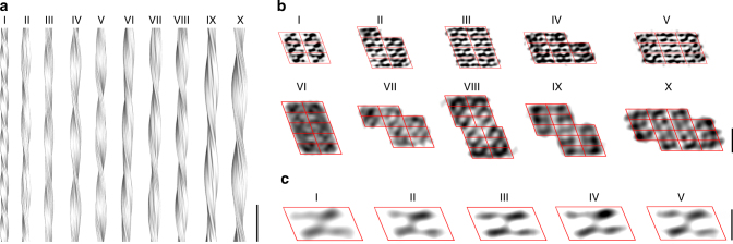
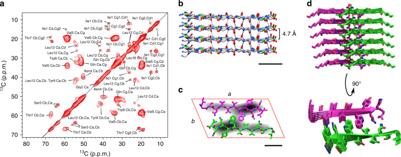
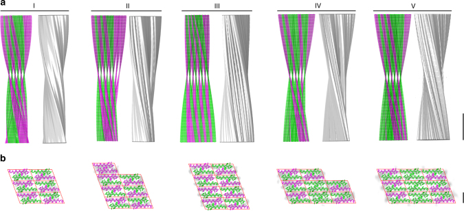
Polymorphism is a key feature of amyloid fibril structures but it remains challenging to explain these variations for a particular sample. Here, we report electron cryomicroscopy-based reconstructions from different fibril morphologies formed by a peptide fragment from an amyloidogenic immunoglobulin light chain. The observed fibril morphologies vary in the number and cross-sectional arrangement of a structurally conserved building block. A comparison with the theoretically possible constellations reveals the experimentally observed spectrum of fibril morphologies to be governed by opposing sets of forces that primarily arise from the β-sheet twist, as well as peptide-peptide interactions within the fibril cross-section. Our results provide a framework for rationalizing and predicting the structure and polymorphism of cross-β fibrils, and suggest that a small number of physical parameters control the observed fibril architectures.


Conflict of interest statement

The authors declare no competing financial interests.

Figures

Fig. 1
Fig. 1
Structural polymorphism of AL1 peptide fibrils. a Side view of reconstructions of fibril morphologies I–X (scale bar, 50 nm). b Cross-sectional slices of the reconstructed densities superimposed with a lattice of parallelograms (red). Reconstructions of morphologies I–V were filtered to 10 Å and morphologies VI–X to a resolution corresponding to their FSC values at 0.143 (scale bar, 5 nm). c Aligned and averaged parallelograms of morphologies I–V sharing a quasi-twofold symmetry. The data from morphology I were included as published (scale bar, 2 nm)
Fig. 2
Fig. 2
Protomer conformation revealed by ss-NMR. a The 2D 13C–13C proton-driven spin diffusion spectrum shows the cross-peaks between aliphatic carbon atoms. b Backbone model of the cross-β sheet implementing Φ/Ψ angles as predicted by the program TALOS+ (scale bar, 1 nm). c Averaged parallelograms of morphologies I–V (gray) superimposed with a peptide dimer (green, magenta) as obtained by NMR. The placement of the peptides followed the considerations described previously for morphology I8 (scale bar, 1 nm). d Side view and top view of a six-layer stack illustrating the structure of a PF. Side-chain conformations were not determined by experiments and represent only conformations that are compatible with the packing shown here
Fig. 3
Fig. 3
Peptide assembly of fibril morphologies I to V. a Side-by-side presentation of the experimentally obtained density and of the peptide assembly of fibril morphologies I to V (scale bar, 10 nm). b Peptide cross-sections showing an exposed N terminus (magenta) and those not exposed (green) within one peptide layer, superimposed with reconstructions (gray) and filtered to 10 Å. Side-chain conformations were not determined by experiments and represent only conformations that are compatible with the packing shown here. The 3D map of morphology I was included as published (scale bar, 2 nm)
Fig. 4
Fig. 4
Mathematical analysis of the observed and theorized fibril morphologies. a Graphical representation of the definition of the parameters nl, ns, and ne for a given fibril cross-section. b Plot of E for all theorized fibril morphologies or cross-sections (blue) vs. n as obtained by the even fit (gray columns). Hence, only white columns have predictive power for the even fit. Red symbols: experimentally observed fibril morphologies. c Close-up of b. Black horizontal division markers show the 1% cutoff of the most stable morphologies for each n value

References

    1. Riek R, Eisenberg DS. The activities of amyloids from a structural perspective. Nature. 2016;539:227–235. doi: 10.1038/nature20416. - DOI - PubMed
    1. Sipe JD, et al. Amyloid fibril proteins and amyloidosis: Chemical identification and clinical classification International Society of Amyloidosis 2016 Nomenclature Guidelines. Amyloid. 2016;23:209–213. doi: 10.1080/13506129.2016.1257986. - DOI - PubMed
    1. Chiti F, Dobson CM. Protein misfolding, functional amyloid, and human disease. Annu. Rev. Biochem. 2006;75:333–366. doi: 10.1146/annurev.biochem.75.101304.123901. - DOI - PubMed
    1. Cherny I, Gazit E. Amyloids: not only pathological agents but also ordered nanomaterials. Angew. Chem. Int. Ed.Engl. 2008;47:4062–4069. doi: 10.1002/anie.200703133. - DOI - PubMed
    1. Sunde M, et al. Common core structure of amyloid fibrils by synchrotron x-ray diffraction. J. Mol. Biol. 1997;273:729–739. doi: 10.1006/jmbi.1997.1348. - DOI - PubMed

Publication types

MeSH terms