. 2018 Feb 16;9(1):701.
doi: 10.1038/s41467-018-03147-6.

Host defense against oral microbiota by bone-damaging T cells

Affiliations

Host defense against oral microbiota by bone-damaging T cells

Masayuki Tsukasaki et al. Nat Commun. .

Abstract

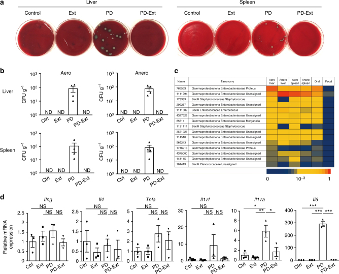
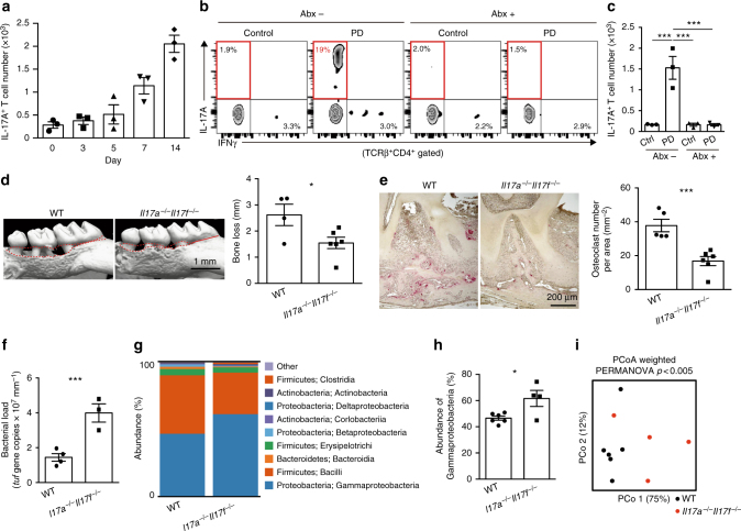
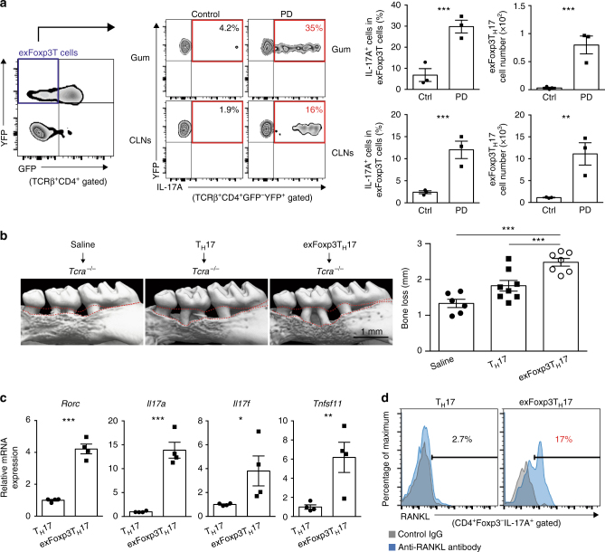
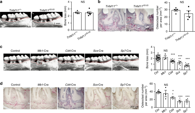
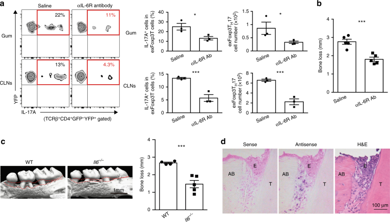
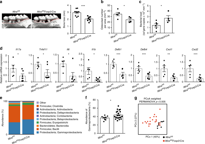
The immune system evolved to efficiently eradicate invading bacteria and terminate inflammation through balancing inflammatory and regulatory T-cell responses. In autoimmune arthritis, pathogenic TH17 cells induce bone destruction and autoimmune inflammation. However, whether a beneficial function of T-cell-induced bone damage exists is unclear. Here, we show that bone-damaging T cells have a critical function in the eradication of bacteria in a mouse model of periodontitis, which is the most common infectious disease. Bacterial invasion leads to the generation of specialized TH17 cells that protect against bacteria by evoking mucosal immune responses as well as inducing bone damage, the latter of which also inhibits infection by removing the tooth. Thus, bone-damaging T cells, which may have developed to stop local infection by inducing tooth loss, function as a double-edged sword by protecting against pathogens while also inducing skeletal tissue degradation.


Conflict of interest statement

K.O. declares that the Department of Osteoimmunology, Graduate School of Medicine and Faculty of Medicine, The University of Tokyo in which he works is an endowmentdepartment, supported with an unrestricted grant from Chugai Pharmaceutical Co.,751 LTD., AYUMI Pharmaceutical Corporation and Noevir Co., Ltd. The remaining authors declare no competing financial interests.

Figures

Fig. 1
Fig. 1
Suppression of oral microbial invasion and local inflammation by tooth extraction. a Bacterial colony formation in the culture of the liver and spleen cells from mice subjected to an experimental periodontitis model (PD). This formation was abrogated by tooth extraction (PD-Ext). Colony formation was not observed in the control (Ctrl) or tooth-extracted (Ext) group. Representative pictures of more than three independent experiments are shown. b Colony-forming units (CFUs) in aerobic (Aero) and anaerobic (Anero) cultures of liver and spleen cells from mice in Ctrl (n = 5), Ext (n = 4), PD (n = 4), or PD-Ext (n = 4) groups, pooled from two independent experiments. c 16S sequence analysis of tissue cultures, ligature (Oral), or fecal samples (Fecal) collected from a mouse in the PD group. The top fifteen frequently detected bacterial species in the aerobic liver culture are listed on the left and the sequence frequency (represented by the bottom bar) in each sample is shown. Representative data of more than three independent experiments is presented. d Quantitative RT-PCR analysis of inflammatory cytokines in the periodontal tissues collected from mice in the Ctrl, Ext, PD, or PD-Ext groups (n = 3). The data were obtained from duplicated experiments. All samples were collected at day 42. All data are shown as the mean ± s.e.m. Statistical analyses were performed using ANOVA with Tukey’s multiple-comparison test. *P < 0.05; **P < 0.01; ***P < 0.005; ND, not detected; NS, not significant
Fig. 2
Fig. 2
TH17 cells protect against the invasion of oral microbiota and induce bone damage. a Number of IL-17A+CD4+TCRβ+ cells in the periodontal tissues at various time points after the ligature placement (n = 3). Representative data of two independent experiments is presented. b, c Effects of an antibiotic cocktail (Abx; ampicillin 1 mg ml−1, streptomycin 5 mg ml−1, and colistin 1 mg ml−1 in drinking water) on the accumulation of TH17 cells. Mice were treated with Abx from 1 week before the ligature placement. The frequency (b) and number (c) of IL-17A+CD4+TCRβ+ cells in the periodontal tissues were analyzed 7 days after the ligature placement (n = 3). PD: periodontitis. d Micro-CT analysis of periodontitis-induced bone loss in the wild-type (n = 4) or Il17a−/−Il17f−/− mice (n = 6), pooled from three independent experiments. The upper red dotted line indicates the cementoenamel junction and the lower red dotted line indicates the alveolar bone crest in the left panel. e Osteoclast number in the maxilla of the wild-type (n = 5) or Il17a−/−Il17f−/− mice (n = 6), pooled from two independent experiments. f Total bacterial load in ligatures collected from wild-type (n = 4) or Il17a−/−Il17f−/− mice (n = 3). Representative data of two independent experiments is presented. g Oral bacterial composition (major phylum; class) of the wild-type (n = 6) or Il17a−/−Il17f–/–mice (n = 4). h Abundance of γ-proteobacteria in the oral bacteria of the wild-type (n = 6) or Il17a−/−Il17f−/−mice (n = 4). i Differences in the bacterial composition between the wild-type (n = 6) and Il17a−/−Il17f−/−mice (n = 4). Principal coordinate analysis (PCoA) and permutational ANOVA (PERMANOVA) comparisons of the weighted UniFrac distances are shown. PCo1: principal coordinate 1; PCo2: principal coordinate 2. The data were pooled from two independent experiments (gi). All data are shown as the mean ± s.e.m. Statistical analyses were performed using ANOVA with Tukey’s multiple-comparison test (c), Student’s t-test (df and h) or PERMANOVA of the weighted UniFrac distances (i). *P < 0.05; ***P < 0.005
Fig. 3
Fig. 3
A crucial role for exFoxp3TH17 cells in the bone destruction during oral infection. a Frequency and number of exFoxp3TH17 cells in the periodontal tissues (Gum) and the cervical lymph nodes (CLNs) in control or periodontitis-induced mice 7 days after the ligature placement (n = 3). Representative data of more than three independent experiments is presented. PD: periodontitis. b Effects of adoptive transfer of TH17 cells (n = 8) or exFoxp3TH17 cells (n = 7) on periodontitis-induced bone loss compared to the saline group (n = 6). The data were pooled from more than three independent experiments. The upper red dotted line indicates the cementoenamel junction and the lower red dotted line indicates the alveolar bone crest in the left panel. All data are shown as the mean ± s.e.m. c Quantitative RT-PCR analysis of Rorc, Il17a, Il17f, and Tnfsf11 transcripts in TH17 cells or exFoxp3TH17 cells (n = 4). The data were pooled from two independent experiments. d FACS profiles of RANKL expression in TH17 cells or exFoxp3TH17 cells. Representative data of more than three independent experiments is shown. Statistical analyses were performed using Student’s t-test (a, c), ANOVA with Tukey’s multiple-comparison test (b). *P < 0.05; **P < 0.01; ***P < 0.005
Fig. 4
Fig. 4
Osteoblasts and periodontal ligament cells are the major source of RANKL in periodontitis. a Micro-CT analysis of periodontitis-induced bone loss in Tnfsf11+/+ mice (n = 4) or Tnfsf11ΔS/ΔS mice (n = 6). The upper red dotted line indicates the cementoenamel junction and the lower red dotted line indicates the alveolar bone crest. b Histological analysis of periodontitis-induced osteoclast development in Tnfsf11+/+ mice (n = 5) or Tnfsf11ΔS/ΔS mice (n = 4). c Micro-CT analysis of periodontitis-induced bone loss in mice in which RANKL was specifically deleted in B cells (Mb1-Cre) (n = 3), T cells (Cd4-Cre) (n = 17), periodontal ligament cells (Scx-Cre) (n = 9) or osteoblastic cells (Sp7-Cre) (n = 12) compared to control mice (n = 31). The upper red dotted line indicates the cementoenamel junction and the lower red dotted line indicates the alveolar bone crest in the left panel. d Osteoclast number in the maxilla of mice in which RANKL was specifically deleted in B cells (Mb1-Cre) (n = 3), T cells (Cd4-Cre) (n = 5), periodontal ligament cells (Scx-Cre) (n = 4) or osteoblastic cells (Sp7-Cre) (n = 4) compared to control mice (n = 7) evaluated by TRAP staining. The data were pooled from more than three independent experiments (c, d). All data are shown as the mean ± s.e.m. Statistical analyses were performed using Student’s t-test (a, b), ANOVA with Dunnett’s multiple-comparison test (c, d). *P < 0.05; ***P < 0.005; NS, not significant
Fig. 5
Fig. 5
IL-6 facilitates the generation of exFoxp3TH17 cells during periodontal infection. a Frequency and number of exFoxp3TH17 cells in periodontal tissues (Gum) and cervical lymph nodes (CLNs) in periodontitis-induced mice treated with saline or anti-IL-6R antibody (Ab) 7 days after the ligature placement (n = 3). Two milligrams of anti-IL-6R Ab (MR16-1) were injected intraperitoneally after 2 days of the ligature placement. Representative data of two independent experiments is shown. b Periodontitis-induced bone loss in control mice or anti-IL-6R Ab-treated mice (n = 5), pooled from two independent experiments. c Micro-CT analysis of periodontitis-induced bone loss in wild-type mice (n = 4) or Il6−/− mice (n = 5). The upper red dotted line indicates the cementoenamel junction and the lower red dotted line indicates the alveolar bone crest. d In situ hybridization of Il6 mRNA in periodontitis-induced wild-type mice 3 days after the ligature placement. Representative data of more than three independent experiments is shown. H&E, haematoxylin and eosin stain; E, epithelium; T, tooth; AB, alveolar bone. All data are shown as the mean ± s.e.m. Statistical analyses were performed using Student’s t-test. *P < 0.05; ***P < 0.005
Fig. 6
Fig. 6
Induction of anti-bacterial response and bone damage by exFoxp3TH17 cells. a Micro-CT analysis of periodontitis-induced bone loss in Il6raflox/flox mice (n = 9) or Il6raflox/flox Foxp3-Cre mice (n = 8). The upper red dotted line indicates the cementoenamel junction and the lower red dotted line indicates the alveolar bone crest. b Number of osteoclasts in the maxilla of periodontitis-induced Il6raflox/flox mice (n = 5) or Il6raflox/flox Foxp3-Cre mice (n = 4), pooled from two independent experiments. c Total bacterial load determined by analyzing the tuf gene copy number in ligatures collected from Il6raflox/flox mice (n = 6) or Il6raflox/flox Foxp3-Cre mice (n = 4). d Quantitative RT-PCR analysis of Il17a, Tnfsf11, Il6, Il1b, Defb1, Defb4, Cxcl1, and Cxcl2 transcripts in the periodontal tissues collected from periodontitis-induced Il6raflox/flox mice (n = 5) or Il6raflox/flox Foxp3-Cre mice (n = 6). The data were obtained from duplicated experiments. e Bacterial composition (major phylum; class) of DNA collected from ligatures of Il6raflox/flox mice (n = 8) or Il6raflox/flox Foxp3-Cre mice (n = 20). f Abundance of γ-proteobacteria in the bacterial DNA collected from the ligatures of Il6raflox/flox mice (n = 8) or Il6raflox/flox Foxp3-Cre mice (n = 20). g Differences in the bacterial communities between Il6raflox/flox mice (n = 8) and Il6raflox/flox Foxp3-cre mice (n = 20). PCo1: principal coordinate 1; PCo2: principal coordinate 2. The data were pooled from more than three independent experiments (eg). PERMANOVA comparisons of the weighted UniFrac distances are shown. All data are shown as the mean ± s.e.m. Statistical analyses were performed using Student’s t-test (ad and f) or PERMANOVA of the weighted UniFrac distances (g). *P < 0.05; **P < 0.01; ***P < 0.005

Similar articles

Cited by

References

    1. Round JL, Mazmanian SK. The gut microbiota shapes intestinal immune responses during health and disease. Nat. Rev. Immunol. 2009;9:313–323. doi: 10.1038/nri2515. - DOI - PMC - PubMed
    1. Bosshardt DD, Lang NP. The junctional epithelium: from health to disease. J. Dent. Res. 2005;84:9–20. doi: 10.1177/154405910508400102. - DOI - PubMed
    1. Hajishengallis G. Immunomicrobial pathogenesis of periodontitis: keystones, pathobionts, and host response. Trends Immunol. 2014;35:3–11. doi: 10.1016/j.it.2013.09.001. - DOI - PMC - PubMed
    1. Miller WD. The human mouth as a focus of infection. Lancet. 1891;138:340–342. doi: 10.1016/S0140-6736(02)01387-9. - DOI
    1. O’Reilly PG, Claffey NM. A history of oral sepsis as a cause of disease. Periodontol. 2000. 2000;23:13–18. doi: 10.1034/j.1600-0757.2000.2230102.x. - DOI - PubMed

Publication types