. 2018 Feb 16;9(1):700.
doi: 10.1038/s41467-018-03019-z.

Loss of microRNA-128 promotes cardiomyocyte proliferation and heart regeneration

Affiliations

Loss of microRNA-128 promotes cardiomyocyte proliferation and heart regeneration

Wei Huang et al. Nat Commun. .

Abstract

The goal of replenishing the cardiomyocyte (CM) population using regenerative therapies following myocardial infarction (MI) is hampered by the limited regeneration capacity of adult CMs, partially due to their withdrawal from the cell cycle. Here, we show that microRNA-128 (miR-128) is upregulated in CMs during the postnatal switch from proliferation to terminal differentiation. In neonatal mice, cardiac-specific overexpression of miR-128 impairs CM proliferation and cardiac function, while miR-128 deletion extends proliferation of postnatal CMs by enhancing expression of the chromatin modifier SUZ12, which suppresses p27 (cyclin-dependent kinase inhibitor) expression and activates the positive cell cycle regulators Cyclin E and CDK2. Furthermore, deletion of miR-128 promotes cell cycle re-entry of adult CMs, thereby reducing the levels of fibrosis, and attenuating cardiac dysfunction in response to MI. These results suggest that miR-128 serves as a critical regulator of endogenous CM proliferation, and might be a novel therapeutic target for heart repair.


Conflict of interest statement

The authors declare no competing financial interests.

Figures

Fig. 1
Fig. 1
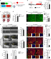
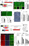
MiR-128 increases as the heart progresses from neonate to adult. a Evaluation of wild-type mouse cardiomyocyte (CM) proliferative activity using Ki67 immunostaining at postnatal day 1 (P1), P7, and P28. Scale bars, 25 µm. b Percentage of CM Ki67+/cTnT+ in P1, P7, and P28 hearts (n = 5 mice for each time point, ~600 CMs/heart). c Wheat germ agglutinin (WGA) staining of P1, P7, and P28 hearts. Scale bars, 25 µm. d Quantification of CM size in P1, P7, and P28 hearts stained with WGA (n = 5 mice for each time point, ~250 CMs/heart). e Evaluation of miR-128 expression level during heart development using qPCR analysis, including embryonic day 14.5 (E14.5), E19.5, P1, P3, P7, P14, and P28 hearts (n = 5). f qPCR analysis of miR-128 expression in neonatal (P1) and adult (P28) CMs (n = 5). g Comparison of miR-128 expression by qPCR in cardiac fibroblast (CF) and CMs (n = 5). Statistical significance was calculated using ANOVA in b, d and Student’s t-test in f, g. Data are represented as means ± SEM. *P < 0.05
Fig. 2
Fig. 2
Overexpression of miR-128 in cardiomyocytes impairs cardiac homeostasis. a Schematic showing the generation of mice that overexpress CM-specific miR-128 after doxycycline (Dox) withdrawal. Control mice were miR-128TetRE mice, and miR-128OE mice were α-MHC-tTA; miR-128TetRE mice. b Schematic of experimental design for CM-specific overexpression of miR-128 at P1 (left panel). Right panel shows the qPCR analysis of miR-128 expression in Ctrl and miR-128OE mice (n = 5). c Gross morphology (upper panel) and Masson trichrome staining (lower panel) of hearts at P1. Scale bars, 0.25 cm (upper panel); 1 mm (lower panel). d Heart weight (HW) to body weight (BW) ratio of P1 mice (n = 8). e Wheat germ agglutinin (WGA) staining of P1 neonatal hearts. Scale bars, 25 µm. f Quantification of CM size as determined by WGA staining (n = 8 mice, ~200 CMs/heart). g Heart function analyzed by echocardiography in P1 mice as measured by left ventricular end-diastolic diameter (LVDd), LV end-systolic diameter (LVDs), ejection fraction (EF), and fraction shortening (FS) (n = 6). h Evaluation of CM proliferative activity by Ki67 immunostaining in Ctrl and miR-128OE hearts. Scale bars, 25 µm (yellow); 10 µm (white). i Quantification data of CM proliferative activity by Ki67 staining in Ctrl and miR-128OE hearts (n = 6 mice, ~800 CMs/heart). Statistical significance was calculated using Student’s t-test. Data are represented as means ± SEM. *P < 0.05
Fig. 3
Fig. 3
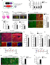
Cardiac miR-128 deletion promotes postnatal CM proliferation in vivo. a Schematic diagram depicting the generation of cardiac-specific miR-128 knockout (miR-128−/−) mice. Control mice were miR-128fl/fl mice, and miR-128−/− mice were Nkx2.5Cre; miR-128fl/fl mice. b The expression level of miR-128 during heart development (n = 6) analyzed by qPCR, including embryonic day 10.5 (E10.5), E14.5, postnatal day 7 (P7), and P28. c Masson trichrome staining of mouse hearts at P7. Scale bars, 2.0 mm. d Comparison of cardiac function between Ctrl and miR-128−/− hearts analyzed by echocardiography at P7, and measured by EF and FS (n = 6). e Measurement of HW to BW ratio in Ctrl and miR-128−/− mice (n = 6). f Evaluation of CM size in P7 Ctrl and miR-128−/− hearts assessed by WGA and cardiac troponin T (cTnT) staining (n = 5 mice, ~250 CMs/heart). Scale bars, 50 µm (yellow); 10 µm (white). g Assessment of CM proliferative activity and sarcomere structure in P7 hearts by immunofluorescence of cTnT and Ki67. Arrows indicate Ki67-positive CMs with sarcomere disassembly. Scale bars, 500 µm (yellow); 25 µm (white). hj Quantification of Ki67+ CMs, sarcomere disassembled CMs and Ki67+ disassembled CMs (n = 4860 CMs pooled from six mice). k CM apoptosis analyzed by TUNEL staining. l Schematic diagram depicting the protocol for EdU intraperitoneal (i.p.) injection at P7 mice to label proliferating CMs in vivo. m Analysis of CM proliferation by EdU incorporation assay in Ctrl and miR-128−/− hearts at P14 (n = 6 mice, ~250 CMs/heart). Scale bars, 50 µm. n Schematic diagram depicting the protocol for EdU intraperitoneal (i.p.) injection at P14 to label proliferating CMs in vivo. o Comparison of EdU+ CMs in Ctrl and miR-128−/− hearts at P21 (n = 6 mice, ~200 CMs/heart). Statistical significance was calculated using Student’s t-test. Data are represented as means ± SEM. *P < 0.05
Fig. 4
Fig. 4
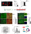
MiR-128 deletion is associated with activation of cell cycle-related genes. a The predicted conserved target site of miR-128 in the 3′UTR of Suz12 from different species. b, c Western blot analysis of SUZ12 expression in mouse hearts at P1, P7, and P28 (n = 5). d, e Western blot analysis of SUZ12 expression in neonatal CMs treated with either vehicle (Ctrl), miR-128 mimic (miR-128) or miR-128 inhibitor (Anti-miR-128) (n = 3). f Luciferase reporter assay for wild-type (WT) and mutant Suz12 3′UTR (Mut) in cells treated with either vehicle (Ctrl) or miR-128 mimic (miR-128) (n = 3). g Western blot assay for cell cycle-related protein expression in control (miR-128fl/fl), and miR-128−/− (Nkx2.5Cre; miR-128fl/fl) hearts at P7 (n = 5). h Quantification data of p27 mRNA levels in Ctrl and miR-128−/− hearts by qPCR (n = 5). i Comparison of SUZ12, EZH2, and H3K27me3 enrichment on the p27 promoter by ChIP-qPCR (n = 5). Statistical significance was calculated using ANOVA in c, e and Student’s t-test in f, h, and i. Data are represented as means ± SEM. *P < 0.05. NS designates not significant
Fig. 5
Fig. 5
MiR-128 regulates CM proliferation through targeting Suz12. a In vitro, evaluation of CM proliferation by immunofluorescence staining of Ki67 in miR-128−/− (Nkx2.5Cre; miR-128fl/fl) neonatal CMs transfected with either scrambled control siRNA (si-Ctrl) or Suz12 siRNA (si-Suz12). Cells are counter-stained with DAPI to visualize nuclei and with antibody to cTnT to identify CMs. Scale bars, 40 µm. b Expression of Suz12 in miR-128−/− neonatal CMs transfected with either a scrambled control siRNA (si-Ctrl) or Suz12 siRNA (si-Suz12) (n = 5). c Quantification of CM proliferation by Ki67 immunostaining (n = 12 samples, ~150 CMs/sample). d Schematic diagram depicting the protocol for siRNA and EdU intraperitoneal (i.p.) injection for P1 mice. e CM size analysis by WGA and cTnT staining in si-Ctrl and si-Suz12 treated miR-128−/− hearts at P7 (n = 5 mice, ~300 CMs/heart). Scale bars, 50 µm (yellow); 10 µm (white). f Comparison of EdU+ CMs in si-Ctrl and si-Suz12-treated miR-128−/− hearts at P7 (n = 5 mice, ~400 CMs/heart).). Scale bars, 50 µm (yellow), 20 µm (white). g, h Western blot analysis of cell cycle-related genes in si-Ctrl and si-Suz12 treated miR-128−/− hearts at P7 (n = 3). i Proposed model by which miR-128 deletion promotes CM proliferation through coordinating the expression of cell cycle-related genes. Statistical significance was calculated using Student’s t-test in b, c, e, f, and h. Data are represented as means ± SEM. *P < 0.05
Fig. 6
Fig. 6
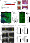
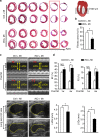
Overexpression of miR-128 inhibits neonatal cardiac regeneration. a Schematic diagram depicting the timing of apex resection (AR) and EdU labeling for P1 mice. Control (Ctrl) mice were miR-128TetRE mice, and miR-128 overexpressing mice (miR-128OE) were α-MHC-tTA; miR-128TetRE mice. b Masson trichrome staining of Ctrl and miR-128OE hearts at day 21 after AR. Scale bars, 0.25 cm (red); 200 µm (black). c Evaluation of CM proliferation by EdU incorporation. Scale bars, 50 µm. d Quantification of EdU+ CMs in P1 hearts at day 21 post AR (n = 5 mice, ~250 CMs/heart). e Staining of mouse hearts with WGA at day 21 after AR (n = 5 mice, ~200 CMs/heart). Scale bars, 25 µm. f, g Heart function analyzed by echocardiography and quantification of LVDd, LVDs, EF, and FS (n = 6) 21 days post AR. Statistical significance was calculated using Student’s t-test in d, e, and g. Data are represented as means ± SEM. *P < 0.05
Fig. 7
Fig. 7
MiR-128 deletion promotes proliferation of adult CMs. a Schematic diagram depicting the protocol of tamoxifen (TAM)-inducible miR-128 deletion (iKO) in adult hearts (P28). Control (Ctrl) mice were miR-128fl/fl mice, and iKO mice were α-MHCMerCreMer; miR-128fl/fl mice. b WGA staining in adult control and iKO hearts. Scale bars, 50 µm. c Measurement of HW to BW ratio in control and iKO hearts (n = 6). d Quantification of CM size determined by WGA staining (n = 6 mice, ~150 CMs/heart). e Evaluation of CM proliferation by EdU incorporation (n = 1130 CMs pooled from five mice). Scale bars, 25 µm. f Representative images of isolated adult CMs in control and iKO hearts. Scale bars, 100 µm. g Comparison of CM number in hearts from control and iKO mice. Approximately 2000 CMs were counted per sample, three independent samples per group. h Schematic diagram for the TAM-inducible dual-lineage tracing protocol for the mouse models. Control (Ctrl-tdTomato) mice were α-MHCMerCreMer; RosatdTomato. The iKO-tdTomato mice were αMHCMerCreMer; miR-128fl/fl; RosatdTomato. i qPCR analysis of miR-128 expression in Ctrl-tdTomato and iKO-tdTomato hearts (n = 5). j Analysis of in vivo sarcomere structure of hears from Ctrl-tdTomato and iKO-tdTomato mice by immunofluorescence staining of cTnT (n = 5). Scale bars, 10 µm. k Expression of sarcomere genes, fetal genes, and genes associated with cell proliferation analyzed by qPCR in adult hearts from Ctrl-tdTomato and iKO-tdTomato mice (n = 6). Statistical significance was calculated using Student’s t-test in b, d, e, g, i, and k. Data are represented as means ± SEM. *P < 0.05. NS, not significant
Fig. 8
Fig. 8
MiR-128 deletion promotes adult cardiomyocyte proliferation after MI. a Schematic of the experimental design for assessing adult (12-weeks-old) cardiac regeneration following MI in TAM-inducible miR-128 knockout (iKO) mice. Control (Ctrl) mice were miR-128fl/fl mice, and iKO mice were α-MHCMerCreMer; miR-128fl/fl mice. b Western blot analysis of cell cycle-related gene expression in infarcted hearts at day 7 after TAM administration (n = 3). c Immunostaining of Aurora B in infarcted hearts at day 14 after TAM administration (n = 5 mice, ~400 CMs/heart). Scale bars, 250 pixel. d Evaluation of CM proliferation by EdU incorporation assay. Scale bars, 50 µm. e Quantification of EdU+ CMs in the Ctrl and iKO hearts at 1 week (1 W) and 4 weeks (4 W) post MI (n = 5 mice, ~250 CMs/heart). Statistical significance was calculated using Student’s t-test in c, e. Data are represented as means ± SEM. *P < 0.05
Fig. 9
Fig. 9
MiR-128 deletion promotes adult cardiac regeneration after MI. a Representative images of Masson trichrome-stained heart section at 28 days after MI. Serial sectioning was performed at 500 µm intervals. The most significant difference between two groups is highlighted by the red box. Scale bars, 1 mm. b Measurement of fibrotic areas in heart sections following MI in control and iKO mice analyzed by Masson trichrome staining. (n = 8). c, d Heart function analyzed by echocardiography and quantified by LVDd, LVDs, EF, and FS (n = 6). Control (Ctrl) mice were miR-128fl/fl mice, iKO mice were α-MHCMerCreMer; miR-128fl/fl mice. Statistical significance was calculated using Student’s t-test in b, d. Data are represented as means ± SEM. *P < 0.05. NS, not significant
Fig. 10
Fig. 10
Loss of miR-128 activates endogenous cardiac regeneration. A schematic diagram proposing that loss of miR-128 activates cardiac regeneration by promoting cardiomyocyte proliferation, while the necrotic tissue of wild-type heart is replaced by myofibroblasts with fibrous scars in response to MI

Similar articles

Cited by

References

    1. Li M, Izpisua Belmonte JC. Mending a faltering heart. Circ. Res. 2016;118:344–351. doi: 10.1161/CIRCRESAHA.115.306820. - DOI - PubMed
    1. Mummery C, et al. Differentiation of human embryonic stem cells to cardiomyocytes: role of coculture with visceral endoderm-like cells. Circulation. 2003;107:2733–2740. doi: 10.1161/01.CIR.0000068356.38592.68. - DOI - PubMed
    1. Zhang J, et al. Functional cardiomyocytes derived from human induced pluripotent stem cells. Circ. Res. 2009;104:e30–e41. doi: 10.1161/CIRCRESAHA.108.192237. - DOI - PMC - PubMed
    1. Kikuchi K. Dedifferentiation, transdifferentiation, and proliferation: Mechanisms underlying cardiac muscle regeneration in zebrafish. Curr. Pathobiol. Rep. 2015;3:81–88. doi: 10.1007/s40139-015-0063-5. - DOI - PMC - PubMed
    1. Foglia MJ, Poss KD. Building and re-building the heart by cardiomyocyte proliferation. Development. 2016;143:729–740. doi: 10.1242/dev.132910. - DOI - PMC - PubMed

Publication types

MeSH terms