. 2018 Apr;38(3):225-233.
doi: 10.1007/s10875-018-0476-0. Epub 2018 Feb 17.

Low Serum IgE Is a Sensitive and Specific Marker for Common Variable Immunodeficiency (CVID)

Affiliations

Low Serum IgE Is a Sensitive and Specific Marker for Common Variable Immunodeficiency (CVID)

Monica G Lawrence et al. J Clin Immunol. 2018 Apr.

Abstract

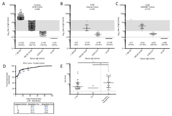
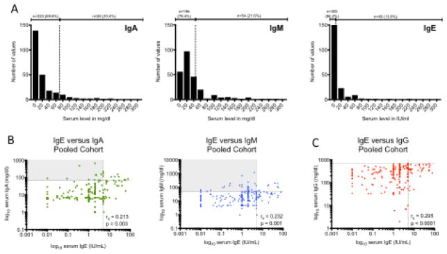
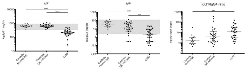
Although small prior studies have suggested that IgE can be low in common variable immunodeficiency (CVID), the workup for patients with recurrent infections and suspected hypogammaglobulinemia does not include the routine measurement of serum IgE. We sought to test the hypothesis that low/undetectable serum IgE is characteristic of CVID by comparing the frequency of low/undetectable serum IgE in healthy controls and patients with CVID. We measured total serum IgE in a large multi-center cohort of patients with CVID (n = 354) and compared this to large population-based cohorts of children and adults. We further compared IgE levels in patients with CVID to those with other forms of humoral immunodeficiency, and in a subset, measured levels of allergen-specific serum IgE and IgG subclasses. Lastly, we evaluated for the presence of IgE in commercially available immunoglobulin replacement therapy (IgRT) products. An undetectable serum IgE (< 2 IU/ml) occurs in only 3.3% (95% CI, 1.9-5.7%) of the general population. In contrast, an undetectable IgE occurs in 75.6% (95% CI, 65.6-85.7%) of patients with CVID. Conversely, a high IgE (> 180 IU/ml) is very uncommon in CVID (0.3% of patients). IgE is > 2 IU/ml in 91.2% of patients with secondary hypogammaglobulinemia, and thus, an IgE < LLOD is suggestive of a primary humoral immunodeficiency. Allergen-specific IgE is not detectable in 96.5% of patients with CVID. Sufficient quantities of IgE to change the total serum IgE are not contained in IgRT. The IgG1/IgG4 ratio is increased in subjects with low IgE, regardless of whether they are controls or have CVID. These findings support the routine measurement of serum IgE in the workup of patients with hypogammaglobulinemia.

Keywords: Common variable immunodeficiency; IgE; IgE deficiency; secondary hypogammaglobulinemia.


Conflict of interest statement

DISCLOSURE OF CONFLICTS OF INTEREST

John Routes received independent contractor fees from CSL Behring. The rest of the authors declare that they have no relevant conflicts of interest.

Figures

Fig. 1
Fig. 1
Serum IgE in controls and hypogammaglobulinemia. Serum total IgE levels were measured in A) 964 controls from the OLIN population-based cohort in Northern Sweden; B) an internal cohort of 181 patients with CVID; and C) a multi-center USIDNET cohort of 173 patients with CVID. Shaded region represents normal range for IgE based on UVA lab. D) ROC curve characteristics based on pooled internal and USIDNET cohort (n=354). E) Serum IgE levels in patients with primary hypogammaglobulinemia (CVID, n=62 or XLA, n=9) compared to those with secondary hypogammaglobulinemia (n=34). Horizontal line is drawn at median and error bars show interquartile range (IQR). **** p<0.0001 using Kruskal-Wallis test with Dunn’s correction for multiple comparisons
Fig. 2
Fig. 2
IgE compared to other serum Ig isotypes in patients with CVID. A) Distribution of serum IgA, IgM and IgE in CVID. Vertical dotted line represents LLN. B) Correlation of serum IgE with serum IgA and IgM. C) Correlation of serum IgE with serum IgG. Horizontal dotted lines represent LLN; vertical dotted lines represent IgE cutoff of 5 IU/ml. Rs = Spearman r
Fig. 3
Fig. 3
Serum Ig subclasses in controls with normal IgE (n=15), controls with IgE

References

    1. Seppanen M, Aghamohammadi A, Rezaei N. Is there a need to redefine the diagnostic criteria for common variable immunodeficiency? Expert review of clinical immunology. 2014;10(1):1–5. doi: 10.1586/1744666X.2014.870478. - DOI - PubMed
    1. Knight AK, Cunningham-Rundles C. Inflammatory and autoimmune complications of common variable immune deficiency. Autoimmunity reviews. 2006;5(2):156–9. doi: 10.1016/j.autrev.2005.10.002. - DOI - PubMed
    1. Bonilla FA, Barlan I, Chapel H, Costa-Carvalho BT, Cunningham-Rundles C, de la Morena MT, et al. International Consensus Document (ICON): Common Variable Immunodeficiency Disorders. J Allergy Clin Immunol Pract. 2016;4(1):38–59. doi: 10.1016/j.jaip.2015.07.025. - DOI - PMC - PubMed
    1. Chapel H, Lucas M, Lee M, Bjorkander J, Webster D, Grimbacher B, et al. Common variable immunodeficiency disorders: division into distinct clinical phenotypes. Blood. 2008;112(2):277–86. doi: 10.1182/blood-2007-11-124545. - DOI - PubMed
    1. Chapel H, Cunningham-Rundles C. Update in understanding common variable immunodeficiency disorders (CVIDs) and the management of patients with these conditions. British journal of haematology. 2009;145(6):709–27. doi: 10.1111/j.1365-2141.2009.07669.x. - DOI - PMC - PubMed

Publication types

MeSH terms