. 2018 Feb 17;36(8):2118-2127.
doi: 10.1002/jor.23872. Online ahead of print.

Inflammatory and degenerative phases resulting from anterior cruciate rupture in a non-invasive murine model of post-traumatic osteoarthritis

Affiliations

Inflammatory and degenerative phases resulting from anterior cruciate rupture in a non-invasive murine model of post-traumatic osteoarthritis

Sophie J Gilbert et al. J Orthop Res. .

Abstract

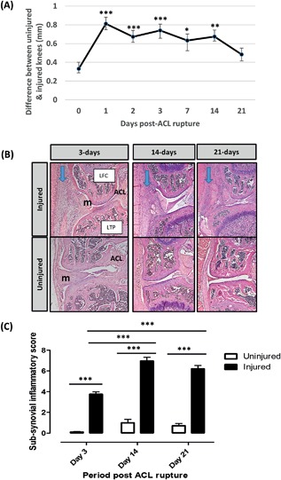
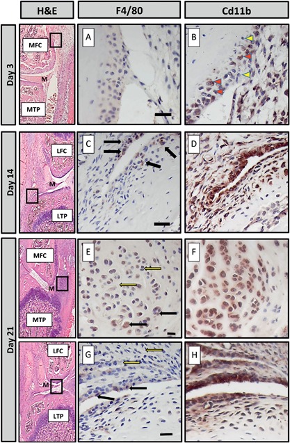
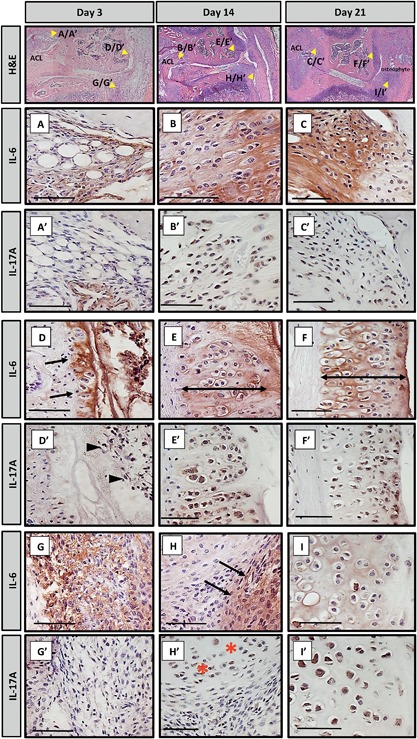
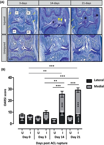
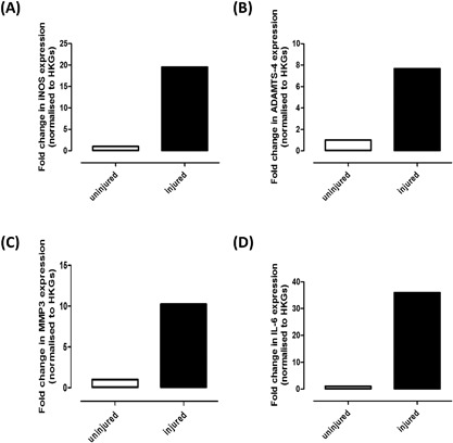
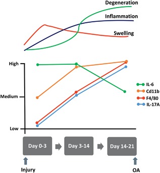
Joint injury is the predominant risk factor for post-traumatic osteoarthritis development (PTOA). Several non-invasive mouse models mimicking human PTOA investigate molecular mechanisms of disease development; none have characterized the inflammatory response to this acute traumatic injury. Our aim was to characterize the early inflammatory phase and later degenerative component in our in vivo non-invasive murine model of PTOA induced by anterior cruciate ligament (ACL) rupture. Right knees of 12-week-old C57Bl6 mice were placed in flexion at a 30° offset position and subjected to a single compressive load (12N, 1.4 mm/s) to induce ACL rupture with no obvious damage to surrounding tissues. Tissue was harvested 4 h post-injury and on days 3, 14, and 21; contralateral left knees served as controls. Histological, immunohistochemical, and gene analyzes were performed to evaluate inflammatory and degenerative changes. Immunohistochemistry revealed time-dependent expression of mature (F4/80 positive) and inflammatory (CD11b positive) macrophage populations within the sub-synovial infiltrate, developing osteophytes, and inflammation surrounding the ACL in response to injury. Up-regulation of genes encoding acute pro-inflammatory markers, inducible nitric oxide synthase, interleukin-6 and interleukin-17, and the matrix degrading enzymes, ADAMTS-4 and MMP3 was detected in femoral cartilage, concomitant with extensive cartilage damage and bone remodelling over 21-days post-injury. Our non-invasive model describes pathologically distinct phases of the disease, increasing our understanding of inflammatory episodes, the tissues/cells producing inflammatory mediators and the early molecular changes in the joint, thereby defining the early phenotype of PTOA. This knowledge will guide appropriate interventions to delay or arrest disease progression following joint injury. © 2018 The Authors. Journal of Orthopaedic Research® Published by Wiley Periodicals, Inc. on behalf of the Orthopaedic Research Society. J Orthop Res 9999:1-10, 2018.

Keywords: degeneration; inflammation; mechanical load; non-invasive mouse model; post-traumatic osteoarthritis.


Figures

Figure 1
Figure 1
Knee joint swelling was measured by digital callipers and expressed as the difference in size between the uninjured and injured leg. *p < 0.05, **p < 0.01, ***p < 0.001 GLM ANOVA versus day‐0 (A). Coronal sections from uninjured and injured legs over the 21‐day time course were stained with Haematoxylin and Eosin (B). Cellular infiltrate in the sub‐synovial lining is indicated (arrows). Whole joints were scored for the presence of sub‐synovial inflammation (C). LTP, lateral tibial plateau; LFC, lateral femoral condyle; m, meniscus. *** GLM ANOVA (p < 0.001).
Figure 2
Figure 2
The localization of F4/80 and CD11b in consecutive sections taken from injured knees of mice culled at 3, 14, and 21‐days post‐ACL rupture was determined by immunohistochemistry. Haematoxylin and Eosin (H&E) stained sections indicate where magnified images are derived from. At day‐3 (A and B), staining for F4/80 was very weak whereas CD11b was found in a large number of cells within the inflammatory infiltrate (solid arrowheads); some cells remained negative (white arrowheads). At day‐14 (C and D) and ‐21 (E and G), a limited number of F4/80+ cells were located within the synovial infiltrate (black arrows); many cells remained negative (white arrows). Cells within the developing osteophytes (E) were also positive. CD11b cells were located throughout the developing osteophyte (F) and synovial infiltrate. LTP, lateral tibial plateau, LFC, lateral femoral condyle; MTP, medial tibial plateau; MFC, medial femoral condyle, m, meniscus. Scale bar = 20 μM.
Figure 3
Figure 3
Consecutive sections were taken from injured knees of mice culled at 3, 14, and 21‐days post‐ACL rupture and stained for IL‐6 and IL‐17A by immunohistochemistry. Haematoxylin and Eosin (H&E) stained sections indicate where magnified images are derived from. Medial compartment inflammation is shown. Strong staining for IL‐6 was observed within the extracellular matrix of the ACL ligament at day‐3 (A), day‐14 (B), and day‐21 (C). In contrast, at day‐3, no staining for IL‐17A was found within the ligament apart from surrounding a blood vessel (A’). At day‐14 (B’) and day‐21 (C’), IL‐17A was detected within the cells of the ligaments. At day‐3 the periosteum of the femoral condyle (arrows, D) was positive for IL‐6 but negative for IL‐17A (D’); intracellular staining in the inflammatory infiltrate nearby was, however, present (arrowheads D’). At day‐14 and ‐21, IL‐6 was observed within the matrix surrounding the hypertrophic chondrocytes of the developing osteophytes (double headed arrow, E and F, respectively). Intracellular staining for IL‐17A was seen on consecutive sections (E’ and F’, respectively). At day‐3 there was abundant staining for IL‐6 within the matrix surrounding the synovial infiltrate (G); consecutive sections were negative for IL‐17 (G’). At day‐14, IL‐6 was still found within this region (H); IL‐17A was absent but present within the nearby meniscus (H’, *). At day‐21, the matrix surrounding hypertrophic chondrocytes within the meniscus were positive for IL‐6 (I) and strong staining for IL‐17A was found within these cells (I’). ACL, anterior cruciate ligament. Scale bar = 50 μM.
Figure 4
Figure 4
Toluidine blue stained coronal sections taken from knees at 3, 14, and 21‐days post‐ACL rupture (A). Injured knees are shown in the top panel and uninjured knees in the bottom panel. All four quadrants of the knee joint are indicated: Lateral and medial tibial plateaus (LTP, MTP, respectively), lateral and medial femoral condyle (LFC, MFC, respectively). Synovial thickening and increased cellular infiltrate (arrows), new matrix deposition within ACLs and collateral ligaments (*), significant medial compartment cartilage loss (arrowheads), bone remodelling (arrows) and osteophyte formation (arrowheads) are shown. Scale bar = 500 μM. Sections were scored for degeneration using the OARSI score (B–F). GLM ANOVA comparing the effect of time (days), uninjured (U) versus injured (I), medial versus lateral, femoral condyle vs tibial plateaus; *p < 0.05, **p < 0.01, ***p < 0.001. Total score increased with time (B) and at day‐14 and ‐21, the medial compartment OARSI score in the injured leg was significantly different to the uninjured leg ***p < 0.001; in addition, at day‐21, the lateral compartment OARSI score in the injured leg was significantly different to the uninjured leg ***p < 0.001. OARSI scores for the femoral condyle and tibial plateaus (C) and sub‐parameters, osteoarthritic changes (D), proteoglycan depletion (E), and subchondral bone (D). At day‐14 and ‐21, the tibial compartment OARSI score in the injured leg was significantly different to the uninjured leg ***p < 0.001; at day‐21, the femoral compartment OARSI score in the injured leg was also significantly different to the uninjured leg ***p < 0.001.
Figure 5
Figure 5
RNA pooled from the femoral condyles of nine uninjured and nine injured knees was analyzed by quantitative PCR to determine the relative expression of (A) iNOS, (B) ADAMTS‐4, (C) MMP‐3, and (D) IL‐6. Data are presented as fold change relative to uninjured knees calculated using the ΔΔCT method with 18S and β‐actin as housekeeping genes (HKGs).
Figure 6
Figure 6
Timeline of changes that occur in inflammatory markers over the 21‐days post‐ACL rupture in our mouse model of PTOA.

References

    1. Brown TD, Johnston RC, Saltzman CL, et al. 2006. Posttraumatic osteoarthritis: a first estimate of incidence, prevalence, and burden of disease. J Orthop Trauma 20:739–744. - PubMed
    1. Little CB, Hunter DJ. 2013. Post‐traumatic osteoarthritis: from mouse models to clinical trials. Nat Rev Rheumatol 9:485–497. - PubMed
    1. Lohmander LS, Englund PM, Dahl LL, et al. 2007. The long‐term consequence of anterior cruciate ligament and meniscus injuries: osteoarthritis. Am J Sports Med 35:1756–1769. - PubMed
    1. Oiestad BE, Engebretsen L, Storheim K, et al. 2009. Knee osteoarthritis after anterior cruciate ligament injury: a systematic review. Am J Sports Med 37:1434–1443. - PubMed
    1. Blaker CL, Clarke EC, Little CB. 2017. Using mouse models to investigate the pathophysiology, treatment, and prevention of post‐traumatic osteoarthritis. J Orthop Res 35:424–439. - PubMed

LinkOut - more resources