. 2018 May:163:67-75.
doi: 10.1016/j.biomaterials.2018.01.035. Epub 2018 Feb 6.

STINGel: Controlled release of a cyclic dinucleotide for enhanced cancer immunotherapy

Affiliations

STINGel: Controlled release of a cyclic dinucleotide for enhanced cancer immunotherapy

David G Leach et al. Biomaterials. 2018 May.

Abstract

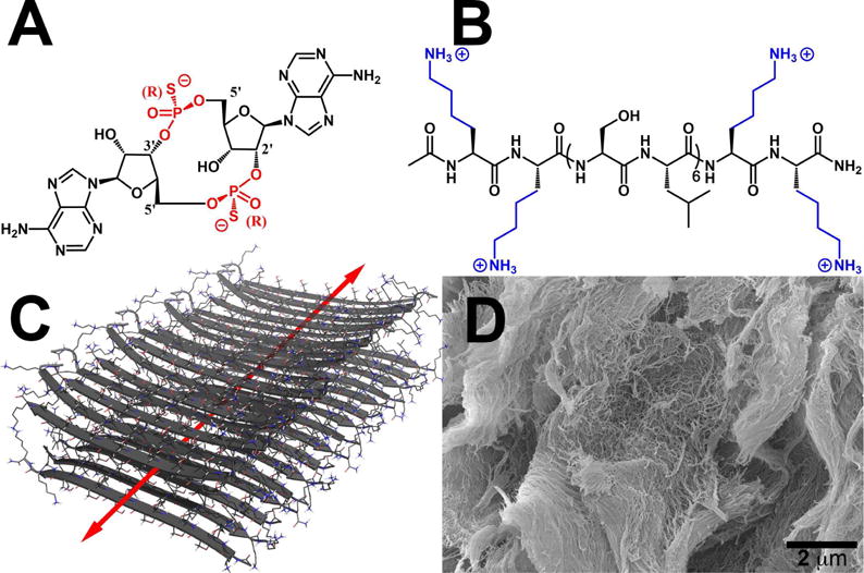
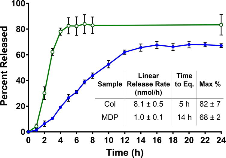
Recent advancements in the field of immunotherapy have yielded encouraging results for the treatment of advanced cancers. Cyclic dinucleotides (CDNs) are a powerful new class of immunotherapy drugs known as STING (Stimulator of Interferon Genes) agonists, currently in clinical trials. However, previous studies of CDNs in murine cancer models have required multiple injections, and improve survival only in relatively nonaggressive tumor models. Therefore, we sought to improve the efficacy of CDN immunotherapy by developing a novel biomaterial we call "STINGel." STINGel is an injectable peptide hydrogel that localizes and provides controlled release of CDN delivery, showing an 8-fold slower release rate compared to a standard collagen hydrogel. The carrier hydrogel is a positively charged, MultiDomain Peptide (MDP) which self-assembles to form a nanofibrous matrix and is easily delivered by syringe. The highly localized delivery of CDN from this nanostructured biomaterial affects the local histological response in a subcutaneous model, and dramatically improves overall survival in a challenging murine model of head and neck cancer compared to CDN alone or CDN delivered from a collagen hydrogel. This study demonstrates the feasibility of biomaterial-based immunotherapy platforms like STINGel as strategies for increasing the efficacy of CDN immunotherapies.

Keywords: Extended drug release; Immunotherapy; Intratumoral injection; Peptide hydrogel; STINGel.


Figures

Fig. 1
Fig. 1
Chemical structures of (A) ML RR-S2 CDA synthetic STING agonist (CDN), (B) K2(SL)6K2 multidomain peptide (MDP), showing charge-pair complementarity of positive lysine termini and negative thiophosphate linkages. (C) Model of anti-parallel β-sheet nanofiber formed by the MDP in solution. The red arrow indicates the axis of the nanofiber and orientation of hydrogen bonding. (D) Scanning Electron Microscopy image of the MDP gel showing a wide field image of the self-assembled nanofibers.
Fig. 2
Fig. 2
CDN drug release kinetics profiles of MDP hydrogels (blue, closed circles) compared to collagen control hydrogels (green, open circles). Samples are 30 μL gels in 96 well plates, loaded with 910 μM CDN and placed under 200 μL HBSS. Absorbance was measured at 259 nm and converted to total percent released for 24 h to monitor release rate and time until equilibrium. Values represent the mean and standard deviation in all plots (n = 3).
Fig. 3
Fig. 3
Cell viability in unloaded and loaded MDP hydrogel. MOC2-E6E7 cells were seeded at a density of approximately 35,000 cells within 70 μL of gel under 200 μL of media (changed every two days) and processed under Live/Dead viability assays (green- live cells; red- dead cells; blue- nuclei). (A) Unloaded hydrogel control showing cell viability over time from small clumps into large spreading masses throughout the peptide hydrogel. (B) CDN dose response assays with images shown from day 3 time point, at which time cells had either died or survived past the initial stage of exposure to CDN. All scale bars are 50 μm, and z-stacks are 100 μm in thickness.
Fig. 4
Fig. 4
Live/dead viability assay quantification used to assess CDN toxicity to MOC2-E6E7 cells. The graph shows the number of viable cells per mm3 of hydrogel over days 1–7 post seeding with cells, testing increasing concentrations of CDN loaded into the MDP hydrogel. The formula image 
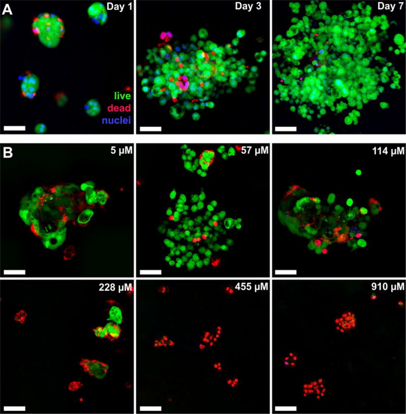
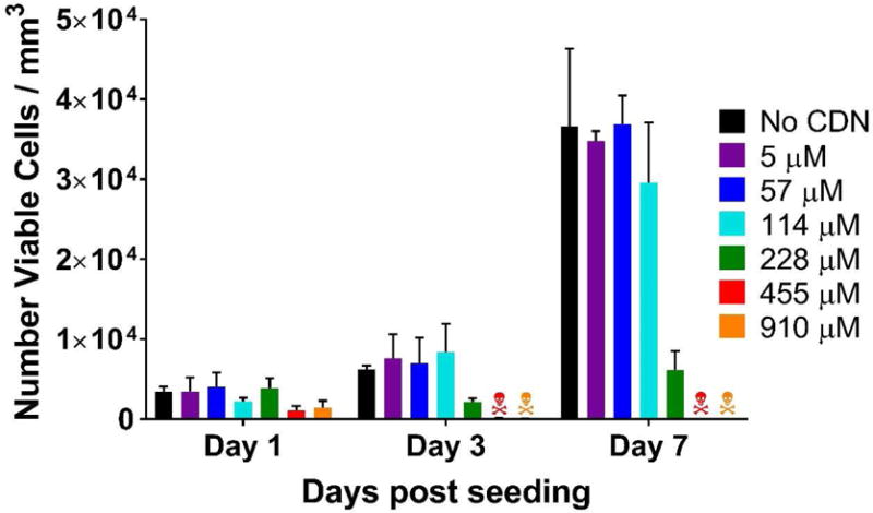
symbols refers to >99% cell death. Values represent the mean and standard deviation in all plots (n = 3).
Fig. 5
Fig. 5
Masson’s trichrome stained MDP hydrogel implants unloaded and loaded with CDN, injected subcutaneously in the dorsal flank of mice. Time point shown is 3 days post injection, at which time hydrogel implant was removed and processed for histology. Scale bars in panels A and D = 1 mm; scale bars in panels B, C, E, and F = 0.1 mm. (A-C) MDP unloaded control implant at 4× magnification showing even infiltration of cells, with boxes drawn around chosen areas whose 40X counterparts are shown in panels B and C, respectively. (D-F) MDP implant loaded with 910 μM CDN (STINGel) at 4× magnification showing uneven infiltration of cells across the implant. Boxes drawn around chosen areas in panel D again have 40X counterparts shown in panels E and F, respectively.
Fig. 6
Fig. 6
Tumor growth curves in controls and STINGel treated animals (n=10 per treatment group). (A) Median primary tumor growth for each group, showing significantly smaller median tumor size in CDN treated groups and a complete delayed growth in STINGel (MDP+CDN) C57BL/6 mice. (B–G) Individual tumor size growth data for tumor bearing mice in each group (number of tumor bearing mice above each plot), showing a clear improvement in progressive tumor free survival for the STINGel treated mice relative to controls and collagen+CDN. (B) HBSS, (C) CDN-alone, (D) MDP gel, (E) collagen gel, (F) collagen+CDN, (G) STINGel.
Fig. 7
Fig. 7
(A) Survival of the different experimental groups based on euthanasia timepoints resulting from excessive tumor burden. The total experimental period was 140 days post-tumor cell inoculation. The 3(IJ) on the x-axis refers to timepoint for intratumoral injection, and 105(RC) refers to timepoint for survivor rechallenge. Whereas 60% of the STINGel-treated C57BL/6 mice survived until the endpoint of the study, nearly all control group (HBSS, MDP gel, and collagen gel) mice were euthanized prior to reaching the endpoint due to excessive tumor burden. Only 10% of CDN alone and collagen+CDN treated mice survived (lines overlaid on plot). *p < 0.0282 vs. CDN, **p < 0.0064 vs. MDP gel, #p < 0.0498 vs. Collagen + CDN. (B) Representative image of STINGel treated mouse that maintained tumor clearance at day 37. (C) Representative image of CDN-only treated mouse growing tumor at day 37.

References

    1. Langer R. New methods of drug delivery. Science. 1990;249(4976):1527–1533. - PubMed
    1. Koshy ST, Ferrante TC, Lewin SA, Mooney DJ. Injectable, porous, and cell-responsive gelatin cryogels. Biomaterials. 2014;35(8):2477–2487. - PMC - PubMed
    1. Bae KH, Wang LS, Kurisawa M. Injectable biodegradable hydrogels: Progress and challenges. J Mater Chem B. 2013;1(40):5371–5388. - PubMed
    1. Langer R, Vacanti JP. Tissue Engineering. Science. 1993;260(5110):920–926. - PubMed
    1. Silva EA, Mooney DJ. Effects of VEGF temporal and spatial presentation on angiogenesis. Biomaterials. 2010;31(6):1235–1241. - PMC - PubMed

Publication types

MeSH terms