

Development and Course of Scars in the Comparison of Age-Related Macular Degeneration Treatments Trials
- PMID: 29454660
- PMCID: PMC6015772
- DOI: 10.1016/j.ophtha.2018.01.004
Development and Course of Scars in the Comparison of Age-Related Macular Degeneration Treatments Trials
Abstract
Purpose: To describe risk factors for scar formation and changes to fibrotic scar through 5 years in the Comparison of Age-related Macular Degeneration Treatments Trials (CATT).
Design: Multicenter, prospective cohort study.
Participants: A total of 1061 subjects in CATT.
Methods: Color photographic and fluorescein angiographic images from baseline and 1, 2, and 5 years were evaluated. Incidence of scar formation was estimated with Kaplan-Meier curves. Risk factors were assessed with Cox regression models.
Main outcome measures: Scar formation, fibrotic scar area, and macular atrophy associated with fibrotic scar ("atrophy").
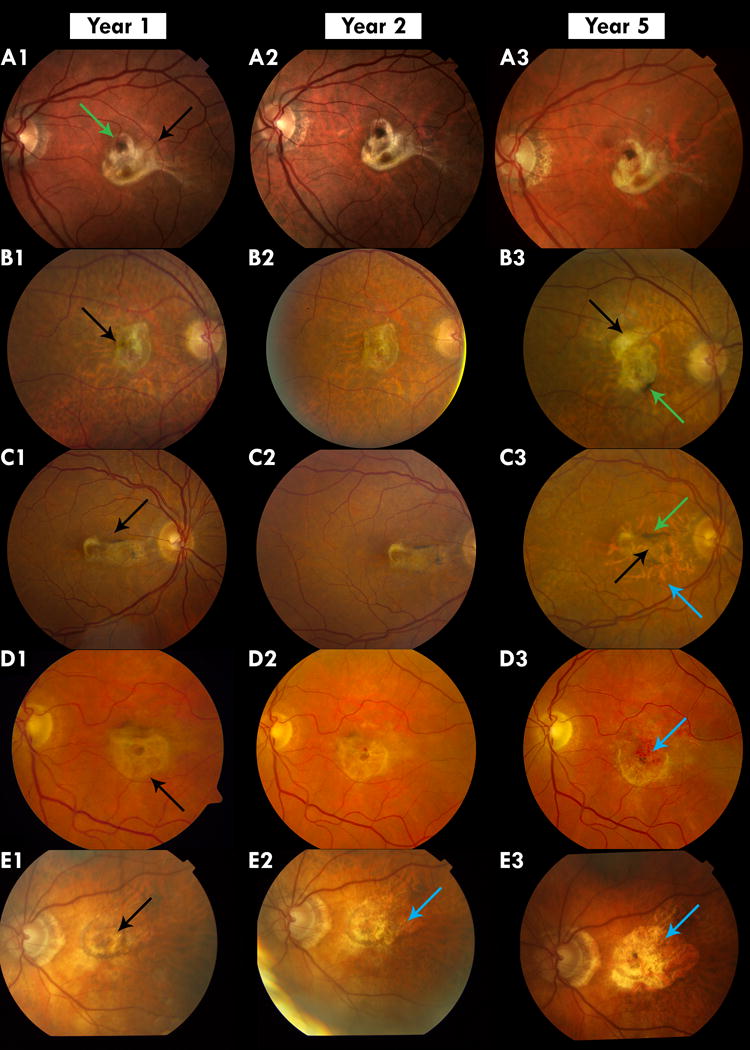
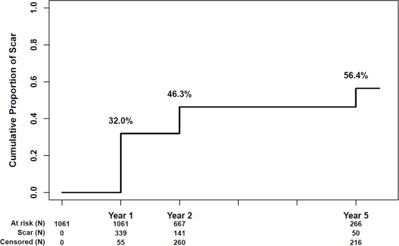
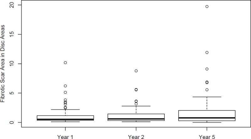
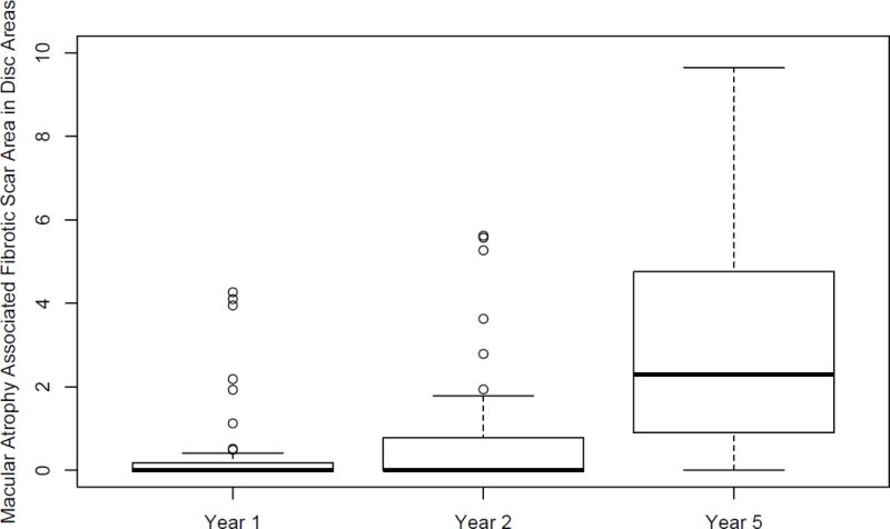
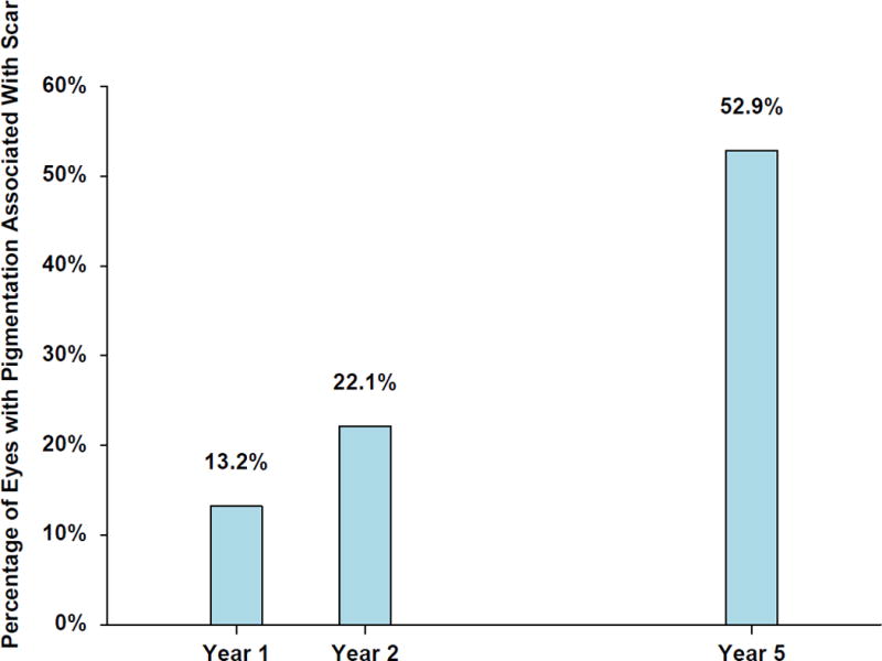
Results: Cumulative proportion of eyes with scar was 32%, 46%, and 56% at years 1, 2, and 5, respectively. Baseline factors associated with increased risk (adjusted hazards ratio [aHR] and 95% confidence interval [CI]) were classic choroidal neovascularization (CNV) (aHR, 4.49; 95% CI, 3.34-6.04) versus occult, hemorrhage >1 disc area (DA) (aHR, 2.28; 95% CI, 1.49-3.47) versus no hemorrhage, retinal thickness >212 μm (aHR, 2.58; 95% CI, 1.69-3.94) versus <120 μm, subretinal tissue complex thickness >275 μm (aHR, 2.64; 95% CI, 1.81-3.84) versus ≤75 μm, subretinal fluid thickness >25 μm (aHR, 1.31; 95% CI, 0.97-1.75) versus no fluid, visual acuity (VA) in fellow eye 20/20 (aHR, 1.72; 95% CI, 1.25-2.36) versus 20/50 or worse, retinal pigment epithelium elevation absence (aHR, 1.71; 95% CI, 1.21-2.41), and subretinal hyperreflective material (aHR, 1.72; 95% CI, 1.25-2.36). Among 68 eyes that developed fibrotic scar at year 1, VA decreased by a mean of additional 13 letters between years 1 and 5. Mean scar area was 1.2, 1.2, and 1.9 DA at 1, 2, and 5 years, respectively. Atrophy was present in 18%, 24%, and 54% of these eyes at years 1, 2, and 5, respectively; the mean areas were 1.6, 2.0, and 3.1 DA, respectively. Atrophy replaced fibrotic scar in 8 eyes at year 5. There was no significant correlation between scar growth and atrophy growth. The rate of growth for both was similar between the clinical trial and observation periods.
Conclusions: Several morphologic features, including classic CNV and large hemorrhage, are associated with scar formation. Rate of new scar formation declined after 2 years. Most fibrotic scars and accompanying macular atrophy expanded over time, reducing VA.
Trial registration: ClinicalTrials.gov NCT00593450.
Copyright © 2018 American Academy of Ophthalmology. All rights reserved.
Conflict of interest statement
Figures
References
-
- Rofagha S, Bhisitkul RB, Boyer DS, Sadda SR, Zhang K, SEVEN-UP Study Group Seven-year outcomes in ranibizumab-treated patients in ANCHOR, MARINA, and HORIZON: a multicenter cohort study (SEVEN-UP) Ophthalmology. 2013;120:2292–9. - PubMed
-
- Gillies MC, Campain A, Barthelmes D, et al. Long-Term Outcomes of Treatment of Neovascular Age-Related Macular Degeneration: Data from an Observational Study. Ophthalmology. 2015;122:1837–45. - PubMed
-
- Peden MC, Suñer IJ, Hammer ME, Grizzard WS. Long-term outcomes in eyes receiving fixed-interval dosing of anti-vascular endothelial growth factor agents for wet age-related macular degeneration. Ophthalmology. 2015;122:803–8. - PubMed
Publication types
MeSH terms
Substances
Associated data
Grants and funding
LinkOut - more resources
Full Text Sources
Other Literature Sources
Medical
