. 2018 Jun;154(8):2209-2221.e14.
doi: 10.1053/j.gastro.2018.02.015. Epub 2018 Feb 15.

P300 Acetyltransferase Mediates Stiffness-Induced Activation of Hepatic Stellate Cells Into Tumor-Promoting Myofibroblasts

Affiliations

P300 Acetyltransferase Mediates Stiffness-Induced Activation of Hepatic Stellate Cells Into Tumor-Promoting Myofibroblasts

Changwei Dou et al. Gastroenterology. 2018 Jun.

Abstract

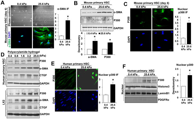
Background & aims: Hepatic stellate cells (HSCs) contribute to desmoplasia and stiffness of liver metastases by differentiating into matrix-producing myofibroblasts. We investigated whether stiffness due to the presence of tumors increases activation of HSCs into myofibroblasts and their tumor-promoting effects, as well as the role of E1A binding protein p300, a histone acetyltransferase that regulates transcription, in these processes.

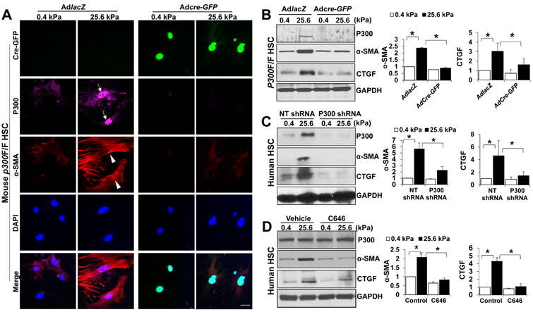
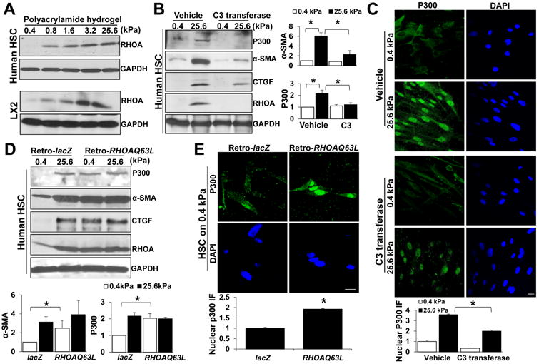
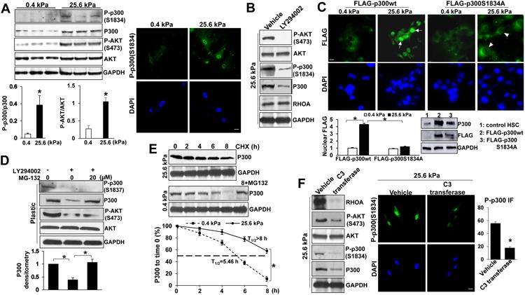
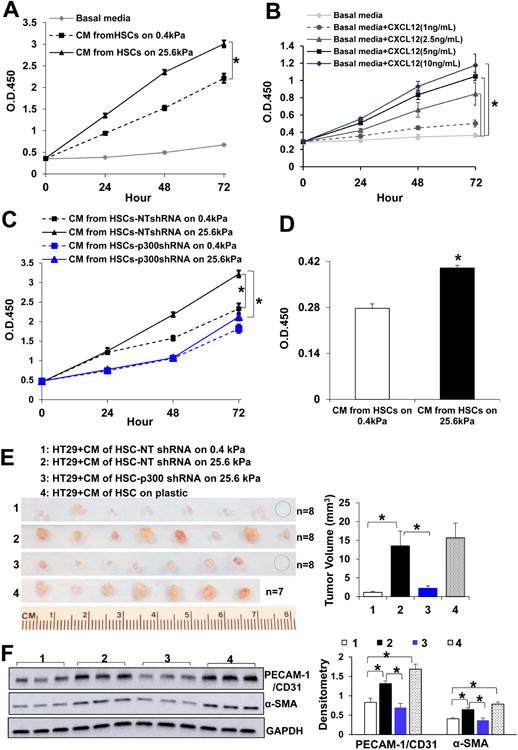
Methods: HSCs were isolated from liver tissues of patients, mice in which the p300 gene was flanked by 2 loxP sites (p300F/F mice), and p300+/+ mice (controls). The HSCs were placed on polyacrylamide gels with precisely defined stiffness, and their activation (differentiation into myofibroblasts) was assessed by immunofluorescence and immunoblot analyses for alpha-smooth muscle actin. In HSCs from mice, the p300 gene was disrupted by cre recombinase. In human HSCs, levels of p300 were knocked down with small hairpin RNAs or a mutant form of p300 that is not phosphorylated by AKT (p300S1834A) was overexpressed. Human HSCs were also cultured with inhibitors of p300 (C646), PI3K signaling to AKT (LY294002), or RHOA (C3 transferase) and effects on stiffness-induced activation were measured. RNA sequencing and chromatin immunoprecipitation-quantitative polymerase chain reaction were used to identify HSC genes that changed expression levels in response to stiffness. We measured effects of HSC-conditioned media on proliferation of HT29 colon cancer cells and growth of tumors following subcutaneous injection of these cells into mice. MC38 colon cancer cells were injected into portal veins of p300F/Fcre and control mice, and liver metastases were measured. p300F/Fcre and control mice were given intraperitoneal injections of CCl4 to induce liver fibrosis. Liver tissues were collected and analyzed by immunofluorescence, immunoblot, and histology.

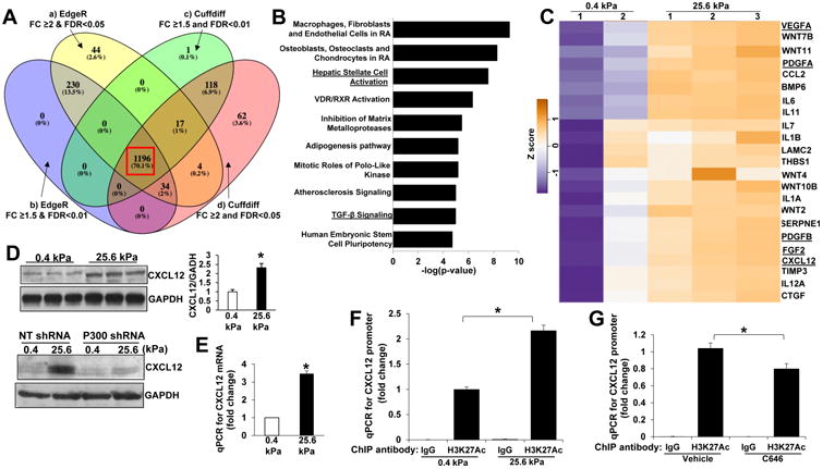
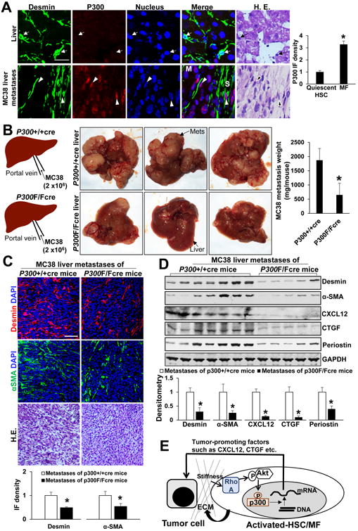
Results: Substrate stiffness was sufficient to activate HSCs, leading to nuclear accumulation of p300. Disrupting p300 level or activity blocked stiffness-induced activation of HSCs. In HSCs, substrate stiffness activated AKT signaling via RHOA to induce phosphorylation of p300 at serine 1834; this caused p300 to translocate to the nucleus, where it up-regulated transcription of genes that increase activation of HSCs and metastasis, including CXCL12. MC38 cells, injected into portal veins, formed fewer metastases in livers of p300F/Fcre mice than control mice. Expression of p300 was increased in livers of mice following injection of CCl4; HSC activation and collagen deposition were reduced in livers of p300F/Fcre mice compared with control mice.

Conclusions: In studies of mice, we found liver stiffness to activate HSC differentiation into myofibroblasts, which required nuclear accumulation of p300. p300 increases HSC expression of genes that promote metastasis.

Keywords: Chromatin Remodeling; Epigenetic Modification; Mechanotransduction; Tumor Progression.


Conflict of interest statement

Conflict of Interest: All authors have declared that no conflict of interest exists

Figures

Fig. 1
Fig. 1. Substrate stiffness promotes p300 nuclear accumulation and MF activation of HSCs
A and B. HSCs isolated from mice were plated on a 0.4 or 25.6 kPa hydrogel and subjected to phase contrast microscopy, IF and WB with quantitative data shown. 25.6 kPa stiffness promoted HSC activation and p300 protein level. *, P<0.05 by t-test, n>20 cells per group for IF and n=3 repeats for WB. Bar, 20 μm. C. P300 IF showed that 25.6 kPa stiffness induced p300 nuclear accumulation as compared to 0.4 kPa. *, P<0.05 by t-test, n>20 cells per group. Bar, 20 μm. D. Primary human HSCs and LX2 cells plated on polyacrylamide hydrogels were collected for WB. E. and F. IF and subcellular fractionation assay were used to analyze stiffness-induced p300 nuclear accumulation. Cell nuclei were counterstained with DAPI. *, P<0.05 by t-test, n>40 cells per group for IF and n=3 for WB. Bar, 20 μm.
Fig. 2
Fig. 2. Stiffness induces HSC activation by a p300-dependent mechanism
A P300F/F HSCs seeded on a 0.4 or 25.6 kPa gel were transduced with adenoviruses encoding lacZ (AdLacZ) or cre-GFP (Adcre-GFP) for IF. Stiffness-mediated upregulation of α-SMA and p300 was inhibited by p300 gene deletion (rows 2 and 3). Cell nuclei were counterstained by DAPI. Bar, 20 μm. B. WB revealed that stiffness-mediated HSC activation was inhibited by p300 gene deletion. *, P<0.05 by ANOVA, n=3. C. Primary human HSCs on 0.4 or 25.6 kPa were transduced with lentiviruses encoding NT shRNA or p300 shRNA. Stiffness-mediated HSC activation was inhibited by p300 knockdown. *, P<0.05 by ANOVA, n=3. D. Stiffness-mediated HSC activation was suppressed by p300 inhibitor C646. *, P<0.05 by ANOVA, n=3.
Fig. 3
Fig. 3. P300 nuclear accumulation by stiffness is mediated by RHOA
A Primary human HSCs and LX2 cells on hydrogels were collected for WB. RHOA protein was increased by stiffness. B and C. Primary human HSCs on 0.4 or 25.6 kPa were incubated with vehicle or RHOA inhibitor C3 transferase. Stiffness-mediated p300 upregulation and nuclear accumulation and HSC activation was inhibited by C3 transferase. *, P<0.05 by ANOVA, n=3 for WB and n>40 cells per group for IF. Bar, 20 μm. D. and E. Primary human HSCs transduced with retroviruses encoding lacZ (Retro-lacZ) or RHOAQ63L (Retro-RHOAQ63L) were seeded on 0.4 or 25.6 kPa for WB and IF. RHOAQ63L overexpression led to HSC activation, p300 upregulation and nuclear accumulation in HSCs on 0.4 kPa. *, P<0.05 by ANOVA, n=3 repeats for WB and n>40 cells per group for IF. Bar, 20 μm.
Fig. 4
Fig. 4. Stiffness induces p300 phosphorylation at S1834 by activating RHOA-AKT pathway
A WB revealed that stiffness induced p300 phosphorylation at S1834 and AKT phosphorylation at S473. The ratio of p-p300 to total p300, the ratio of p-AKT to total AKT, and IF for p-p300(S1834) are shown. *, P<0.05 by t-test, n=3. B. AKT inhibitor LY294002 reduced p-AKT(S473), p-p300(S1834) and total p300 level of HSCs. Data represent multiple repeats with similar results. C. Stiffness induced nuclear accumulation of FLAG-p300wt but not FLAG-p300S1834A mutant. FLAG-tagged p300 fusion proteins were detected by IF and WB for FLAG. *, P<0.05 by ANOVA, n=25 cells per group. Bar, 20 μm. D. LY294002 reduced p300 protein level with its effect reversed by proteasome inhibitor MG-132. *, P<0.05 by ANOVA, n=3 repeats. E. P300 protein stability in HSCs was analyzed by WB in the presence of cycloheximide (CHX). The half-life (T1/2) of p300 in HSCs on 0.4 kPa was 5.46 hours and >8.0 hours in HSCs on 25.6 kPa. *, P<0.05 by ANOVA, n=3. F. RHOA inhibitor C3 transferase reduced p-AKT, p-p300 as well as total p300 level of HSCs. *, P<0.05, by t-test, n>20 cells per group. Bar, 20 μm.
Fig. 5
Fig. 5. Stiffness epigenetically promotes gene transcription of HSCs
A HSCs on 0.4 or 25.6 kPa were subjected to RNA-seq. 1196 genes were identified as stiffness targets by 4 bioinformatic analyzing approaches (4 colors). B. Ingenuity pathway analysis of the 1196 genes. C. A panel of tumor-promoting factors transcriptionally turned on by stiffness is shown by a heatmap. D. Stiffness promoted HSCs to produce CXCL12 protein through HSC p300. *, P<0.05 by t-test, n=3. E. Stiffness increased CXCL12 mRNA level as revealed by qPCR after reverse transcription. *, P<0.05 by t-test, n=6. F. Histone 3/DNA complexes were pulled down by control IgG or anti-histone H3-acetyl K27 for ChIP-qPCR and qPCR was performed for CXCL12 promoter with primer pair 1. Stiffness increased H3K27AC on CXCL12 promoter. *, P<0.05 by ANOVA, n=3. G. C646 reduced H3K27AC on CXCL12 promoter of HSCs. *, P<0.05 by ANOVA, n=3.
Fig. 6
Fig. 6. Stiffness potentiates tumor-promoting effects of HSCs through HSC p300
A HSC-conditioned media (CMs) were used as stimulants for HT29 proliferation assay. HSC 25.6 kPa CM promoted HT29 proliferation as compared to 0.4 kPa CM. *, p<0.05 by ANOVA, n=5. B. Recombinant CXCL12 dose-dependently promoted HT29 proliferation. *, p<0.05 by ANOVA, n=5. C. The effect of HSC-CM on HT29 proliferation was suppressed by p300 knockdown in HSCs. *, p<0.05 by ANOVA, n=5. D. Serum-starved HT29 cells stimulated with HSC-CM at 37°C for 2 hours were plated in a 96-well plate (30,000 cells/well) and cultured in serum-free DMEM for additional 24 hours. MTS assay revealed that in vitro stimulation with HSC-CM accelerated cell proliferation. *, p<0.05 by t-test, n=6. E. Serum-starved HT29 were pretreated with CM at 37°C for 2 hours and resuspended in CM, and HT29/CM were coinjected into SCID mice subcutaneously. Tumor nodules were isolated and quantitated at day 7. *, P<0.05 by ANOVA, n=7 or 8 per group. F. Tumor lysates were subjected to WB for α-SMA and PECAM-1/CD31. *, P<0.05 by ANOVA, n=3.
Fig. 7
Fig. 7. P300 inactivation in activated-HSC/MFs suppresses liver metastasis in mice
A MC38 colorectal cancer cells were implanted into SCID mice by portal vein injection. Desmin IF labeled both quiescent HSCs and activated-HSC/MFs of liver metastases. P300 IF in activated-HSC/MFs of liver metastases was more than 3 times stronger than it in quiescent HSCs (red channel). *, p<0.05 by t-test, n=12, 10. M: metastasis; S: stroma. Bar, 50 μm. B. P300F/F mice were bred with collgen1A1-cre transgenic mice and age-matched male p300F/Fcre and p300+/+cre mice received MC38 portal vein injection. P300F/Fcre mice developed fewer MC38 liver metastases as compared to matched p300+/+cre mice. *, p<0.05 by t-test, n=7, 7. Mets: metastases. C. and D. IF and WB revealed that MC38 liver metastases of p300F/Fcre mice contained reduced levels of desmin, α-SMA, CXCL12, CTGF, and periostin, as compared to those of p300+/+cre mice. *, p<0.05 by t-test, n=7, 7 for IF and n=7, 5 for WB. Bar: 100 μm. E. A schematic illustration of “an amplification loop” for liver metastatic growth.

Comment in

Similar articles

Cited by

References

    1. Vidal-Vanaclocha F. The prometastatic microenvironment of the liver. Cancer Microenviron. 2008;1:113–29. - PMC - PubMed
    1. Kang N, Gores GJ, Shah VH. Hepatic stellate cells: partners in crime for liver metastases? Hepatology. 2011;54:707–13. - PMC - PubMed
    1. Liu C, Billadeau DD, Abdelhakim H, et al. IQGAP1 suppresses TbetaRII-mediated myofibroblastic activation and metastatic growth in liver. J Clin Invest. 2013;123:1138–56. - PMC - PubMed
    1. Kang N, Shah VH, Urrutia R. Membrane-to-Nucleus Signals and Epigenetic Mechanisms for Myofibroblastic Activation and Desmoplastic Stroma: Potential Therapeutic Targets for Liver Metastasis? Mol Cancer Res. 2015;13:604–612. - PMC - PubMed
    1. Nielsen SR, Quaranta V, Linford A, et al. Macrophage-secreted granulin supports pancreatic cancer metastasis by inducing liver fibrosis. Nat Cell Biol. 2016;18:549–60. - PMC - PubMed

Publication types

MeSH terms

Substances