. 2018 May;141(5):1711-1725.e9.
doi: 10.1016/j.jaci.2018.02.003. Epub 2018 Feb 15.

Mechanism for initiation of food allergy: Dependence on skin barrier mutations and environmental allergen costimulation

Affiliations

Mechanism for initiation of food allergy: Dependence on skin barrier mutations and environmental allergen costimulation

Matthew T Walker et al. J Allergy Clin Immunol. 2018 May.

Erratum in

  • Correction.
    [No authors listed] [No authors listed] J Allergy Clin Immunol. 2018 Jul;142(1):339. doi: 10.1016/j.jaci.2018.05.013. J Allergy Clin Immunol. 2018. PMID: 29980279 No abstract available.

Abstract

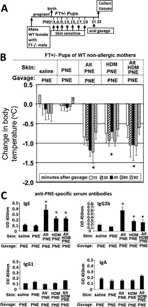
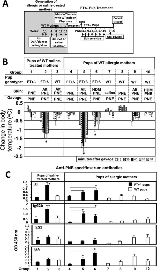
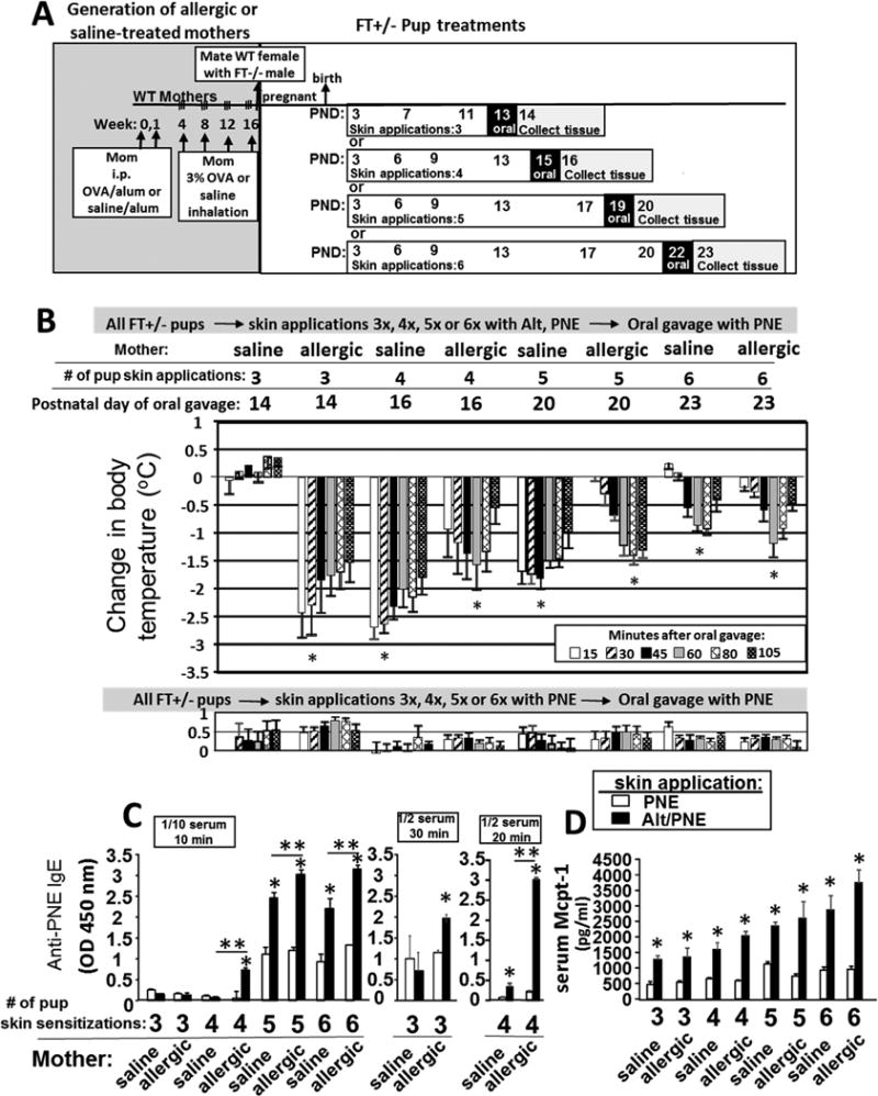
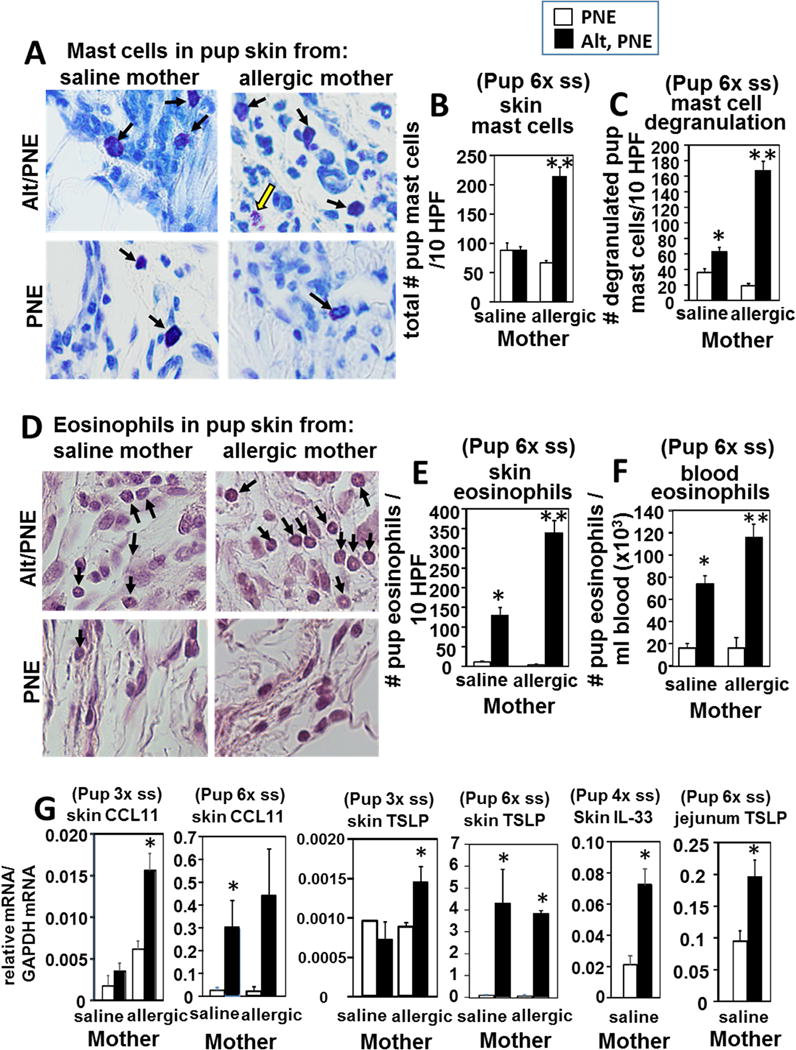
Background: Mechanisms for the development of food allergy in neonates are unknown but clearly linked in patient populations to a genetic predisposition to skin barrier defects. Whether skin barrier defects contribute functionally to development of food allergy is unknown.

Objective: The purpose of the study was to determine whether skin barrier mutations, which are primarily heterozygous in patient populations, contribute to the development of food allergy.

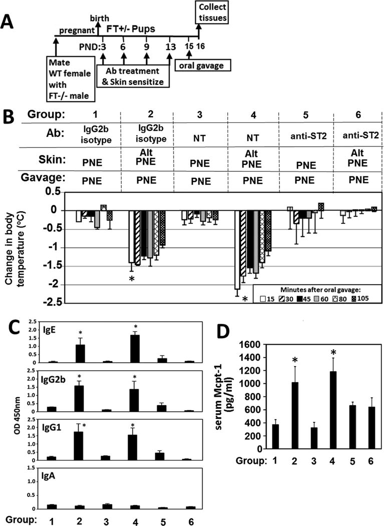
Methods: Mice heterozygous for the filaggrin (Flg)ft and Tmem79ma mutations were skin sensitized with environmental and food allergens. After sensitization, mice received oral challenge with food allergen, and then inflammation, inflammatory mediators, and anaphylaxis were measured.

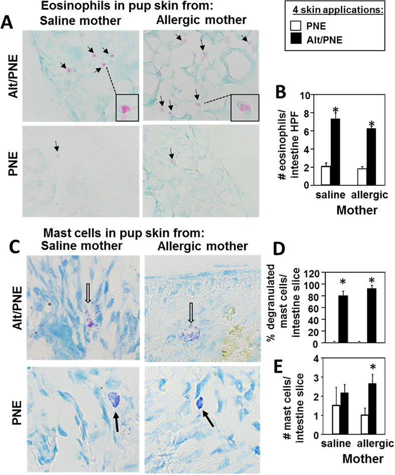
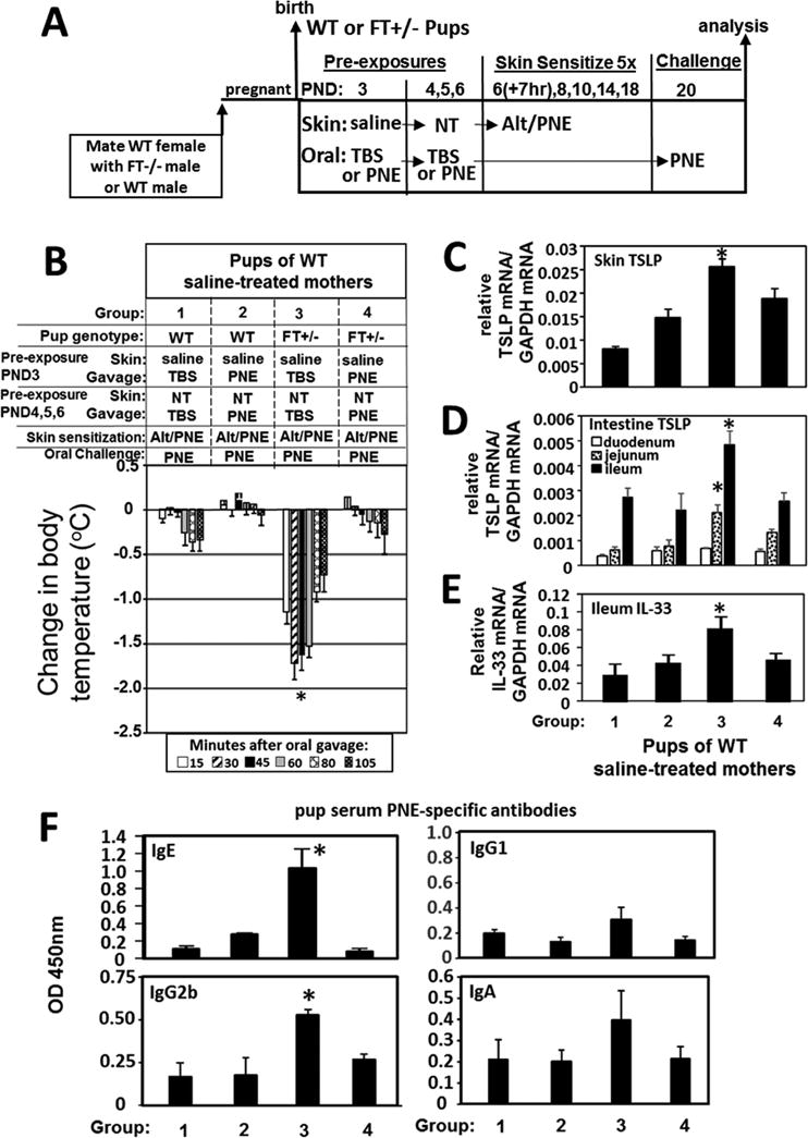
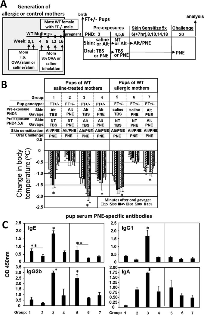
Results: We define development of inflammation, inflammatory mediators, and food allergen-induced anaphylaxis in neonatal mice with skin barrier mutations after brief concurrent cutaneous exposure to food and environmental allergens. Moreover, neonates of allergic mothers have increased responses to suboptimal sensitization with food allergens. Importantly, responses to food allergens by these neonatal mice were dependent on genetic defects in skin barrier function and on exposure to environmental allergens. ST2 blockade during skin sensitization inhibited the development of anaphylaxis, antigen-specific IgE, and inflammatory mediators. Neonatal anaphylactic responses and antigen-specific IgE were also inhibited by oral pre-exposure to food allergen, but interestingly, this was blunted by concurrent pre-exposure of the skin to environmental allergen.

Conclusion: These studies uncover mechanisms for food allergy sensitization and anaphylaxis in neonatal mice that are consistent with features of human early-life exposures and genetics in patients with clinical food allergy and demonstrate that changes in barrier function drive development of anaphylaxis to food allergen.

Keywords: Alternaria alternata; ST2; Sensitization; anaphylaxis; chicken egg ovalbumin; filaggrin; food allergy; house dust mite; mattrin; peanut; skin.


Figures

Figure 1
Figure 1
Skin sensitization of neonates with skin barrier gene mutations induced responsiveness to oral food allergen-induced anaphylaxis. (A) Timeline for mating and for FT+/− pup treatments with food and environmental allergens. (B) Food allergen-induced temperature (oC) changes on day 21. (C) Anti-peanut specific serum IgE, IgG1, IgG2b, and IgA as determined by ELISA. N=8-10/group. The data in all figures include both genders for pups because there were no differences in outcomes by gender (Supplement Data Figure 2). *, p<0.05 as compared to the saline skin-sensitized group and the PNE-only skin-sensitized group. Alt, Alternaria alternata extract; FT−/−, flaky tail mice homozygous for filaggrin and mattrin mutations; FT+/−, flaky tail mice heterozygous for filaggrin and for mattrin mutations; HDM, house dust mite extract; OVA, chicken egg ovalbumin; PND, postnatal day; PNE, peanut extract; WT, wild type.
Figure 2
Figure 2
Skin barrier gene mutations are required for oral food allergen-induced anaphylaxis and generation of anti-peanut specific IgE in FT+/− offspring of allergic and non-allergic mothers. (A) Timeline for mating of females with FT−/− males or WT males and for pup treatments with food and environmental allergens. (B) Food allergen-induced temperature changes on day 21. (C) Anti-peanut specific antibodies were measured by ELISA. Serum was from pups in Figure 2B. Anti-peanut specific serum IgE was measured using a 1/10 serum dilution and a 12 minute TMB color development. Anti-peanut specific serum IgG2b was measured using a 1/1000 serum dilution and a 12 minute TMB color development. Anti-peanut specific serum IgG1 was measured using a 1/20,000 serum dilution and a 6 minute TMB color development. Anti-peanut specific monomeric serum IgA was measured using a 1/10 serum dilution and a 30 minute TMB substrate color development. N=8-10/group. *, p<0.05 as compared to the corresponding PNE only skin-sensitized groups, ie as compared group 1 for FT+/− pups of saline treated mothers, as compared to group 4 of FT+/− pups of allergic mothers. For WT pups, there was no difference as compared to group 8. Alt, Alternaria alternata extract; FT−/−, flaky tail mice homozygous for filaggrin and mattrin mutations; FT+/−, flaky tail mice heterozygous for filaggrin and for mattrin mutations; HDM, house dust mite extract; PND, postnatal day; PNE, peanut extract; WT, wild type.
Figure 3
Figure 3
Allergic mothers transmitted to FT+/− pups an elevated responsiveness to oral food allergen anaphylaxis after suboptimal pup skin sensitization. (A) Timeline for induction of OVA-induced lung inflammation in females, mating of females with FT−/− (FLGft/ft/Tmem79ma/ma) males or WT males, and for pup treatments with food and environmental allergens. (B) Food allergen-induced temperature changes after oral gavage on days 13, 15, 19 or 22. (C) Serum was from pups in (B). In the panel on the left, anti-peanut specific serum IgE was measured using a 1/10 serum dilution and a 12 minute TMB substrate color development. In the two panels on the right, anti-peanut specific serum IgE was measured using a 1/2 serum dilution and a 20-30 minute TMB color development. (D) Serum Mcpt-1 was measured by ELISA. N=8-10/group. *, p<0.05 as compared to the corresponding PNE only skin-treated group. **, p<0.05 as compared to the indicated groups of Alt/PNE-treated pups from non-allergic saline-treated mothers.
Figure 4
Figure 4
Skin Inflammation and mediators of allergic responses are elevated in Alt/PNE skin sensitized FT+/− pups. Pup skin tissues from the site of skin sensitizations and pup blood from the pups in Figure 4B. (A) Shown are representative micrographs of toluidine-stained pup skin tissue sections from the site of 6 skin sensitizations and skin collected on PND22. Black arrows indicate mast cells. Yellow arrow indicates a degranulated mast cell. (B) Pups had 6 skin sensitizations (6×ss) and skin was collected on PND22. Presented is the number of skin mast cells in 10 high powered fields at 40× (HPF) per pup tissue section from 8-10 pups. (C) Pups had 6 skin sensitizations and skin was collected on PND22. Presented is the number of degranulated skin mast cells in 10 high powered fields at 40× (HPF) per pup tissue section from 8-10 pups. (D) Shown are micrographs of eosin-stained skin tissue section with a light hematoxylin counter stain from pups with 6 skin sensitizations and skin collected on PND22. Black arrows indicate eosinophils. (E) Number of eosinophils in 10 high powered fields at 40× (HPF) from pups with 6 six sensitizations and skin collected on PND22. (F) Number of blood eosinophils as determined by discomb staining and hemacytometer counting from pups with 6 skin sensitizations and blood collected on PND22. (G) RT-qPCR of pup skin TSLP and CCL11 for pup skin after 3 skin sensitization (3× ss) with skin collected PND12 or 6 skin sensitizations (6×ss) with skin collected on PND22. RT-qPCR of IL-33 after 4 skin sensitizations (4× ss) with skin collected PND16. RT-qPCR of TSLP for jejunum from pups with 6 skin sensitizations (6× ss) and intestine collection on PND22. N=8-10/group. *, p<0.05 as compared to the corresponding PNE only skin-treated group. **, p<0.05 as compared to the Alt/PNE-treated pups from non-allergic saline-treated mothers and compared to corresponding PNE skin-treated group from allergic mothers.
Figure 5
Figure 5
Intestinal Inflammation is elevated in Alt/PNE skin-sensitized FT+/− pups. Intestines were collected from pups from Figure 4B with 4 skin sensitizations. (A) Shown are micrographs of eosin-stained intestine tissue section with a light methyl-green counter stain. Black arrows indicate eosinophils. Box insert contains enlarged image of eosinophil indicated by dotted line. (B) Number of eosinophils per high powered 64× field (HPF) of pup intestine tissue section. (C) Shown are representative micrographs of toluidine-stained pup intestine tissue sections. Open arrows indicate degranulated purple mast cells. Closed black arrows indicate intact dark purple mast cells. (D) Percent degranulated mast cells per intestine tissue slice. (E) Number of cells per pup intestine tissue slice. N=8-10/group. *, p<0.05 as compared to the corresponding PNE only skin-treated group.
Figure 6
Figure 6
Anti-ST2 blocked development of oral food allergen-induced anaphylaxis. (A) Timeline for mating of females with FT−/− males or WT males, for antibody (Ab) treatment one hour before pup skin sensitization on PND3-13 with food and environmental allergens. FT+/− pups received an intraperitoneal injection of 8 8 μg anti-ST2 or IgG2b isotype control at 1 hr before skin sensitization #1, 12 μg antibody/g pup at 1 hr before skin sensitization #2 and #3, and then 15 μg antibody/g pup at 1 hr before skin sensitization #4. (B) Food allergen-induced temperature changes after oral gavage on postnatal day 15. (C) Anti-peanut specific IgE, IgG2b, IgG1 and monomeric IgA in serum from pups in panel B. (D) Serum Mcpt-1 was measured by ELISA. *, p<0.05 as compared to groups, 1, 3, 5 and 6.
Figure 7
Figure 7
Oral pre-exposure to PNE induced tolerance in FT+/− pups. (A) Timeline for induction of mating of females with FT−/− males or WT males, for pup pre-exposures on PND 3-6, and for pup skin treatments on PND6-18 with food and environmental allergens. Intestines were flushed with saline before RT-qPCR analysis (B) Food allergen-induced temperature changes after oral gavage on days 20. *, p<0.05 as compared to all other groups. For pups from panel B, shown is the RT-qPCR of (C) pup skin TSLP, (D) pup intestine TSLP, and (E) pup ileum IL-33. (F) Anti-peanut specific serum IgE, IgG2b, IgG1 and IgA from pups in panel 2 as determined by ELISA. TBS, Tris-buffered saline. *, p<0.05 as compared to groups 1, 2, and 4.
Figure 8
Figure 8
Oral pre-exposure to PNE to induce tolerance in FT+/− pups was less effective when their skin was pre-exposed to the ubiquitous environmental allergen Alt during oral pre-exposure to PNE. (A) Timeline for induction of OVA-induced lung inflammation in females, mating of females with FT−/− males, for pup pre-exposures on PND 3-6, for pup skin sensitization on PND6-18 with food and environmental allergens and for oral challenge on PND 20. (B) Food allergen-induced temperature changes after oral gavage on day 20. *, p<0.05 as compared to the group without the asterisk. (C) Anti-peanut specific serum IgE, IgG2b, IgG1 and monomeric IgA from pups in panel B as determined by ELISA. *, p<0.05 as compared to all groups without one asterisk. **, p<0.05 as compared to indicated group. N=8-10 pups/group.

References

    1. Wang J, Sampson HA. Food allergy. J Clin Invest. 2011;121:827–35. - PMC - PubMed
    1. DaVeiga SP. Epidemiology of atopic dermatitis: a review. Allergy Asthma Proc. 2012;33:227–34. - PubMed
    1. Shaker M. New insights into the allergic march. Curr Opin Pediatr. 2014;26:516–20. - PubMed
    1. Makhija MM, Robison RG, Caruso D, Cai M, Wang X, Pongracic JA. Patterns of allergen sensitization and self-reported allergic disease in parents of food allergic children. Ann Allergy Asthma Immunol. 2016;117:382–6. - PMC - PubMed
    1. Bousquet J, Anto J, Auffray C, Akdis M, Cambon-Thomsen A, Keil T, et al. MeDALL (Mechanisms of the Development of ALLergy): an integrated approach from phenotypes to systems medicine. Allergy. 2011;66:596–604. - PubMed

Publication types

MeSH terms