. 2018 Oct 15;180(Pt B):463-484.
doi: 10.1016/j.neuroimage.2018.01.075. Epub 2018 Feb 15.

Dynamic resting state fMRI analysis in mice reveals a set of Quasi-Periodic Patterns and illustrates their relationship with the global signal

Affiliations

Dynamic resting state fMRI analysis in mice reveals a set of Quasi-Periodic Patterns and illustrates their relationship with the global signal

Michaël E Belloy et al. Neuroimage. .

Abstract

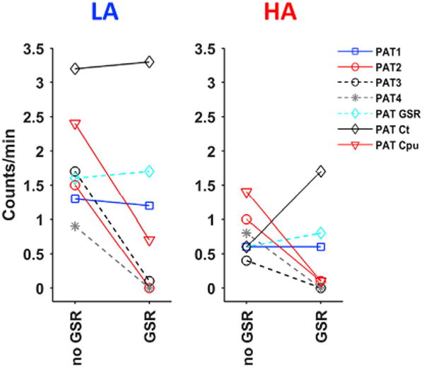
Time-resolved 'dynamic' over whole-period 'static' analysis of low frequency (LF) blood-oxygen level dependent (BOLD) fluctuations provides many additional insights into the macroscale organization and dynamics of neural activity. Although there has been considerable advancement in the development of mouse resting state fMRI (rsfMRI), very little remains known about its dynamic repertoire. Here, we report for the first time the detection of a set of recurring spatiotemporal Quasi-Periodic Patterns (QPPs) in mice, which show spatial similarity with known resting state networks. Furthermore, we establish a close relationship between several of these patterns and the global signal. We acquired high temporal rsfMRI scans under conditions of low (LA) and high (HA) medetomidine-isoflurane anesthesia. We then employed the algorithm developed by Majeed et al. (2011), previously applied in rats and humans, which detects and averages recurring spatiotemporal patterns in the LF BOLD signal. One type of observed patterns in mice was highly similar to those originally observed in rats, displaying propagation from lateral to medial cortical regions, which suggestively pertain to a mouse Task-Positive like network (TPN) and Default Mode like network (DMN). Other QPPs showed more widespread or striatal involvement and were no longer detected after global signal regression (GSR). This was further supported by diminished detection of subcortical dynamics after GSR, with cortical dynamics predominating. Observed QPPs were both qualitatively and quantitatively determined to be consistent across both anesthesia conditions, with GSR producing the same outcome. Under LA, QPPs were consistently detected at both group and single subject level. Under HA, consistency and pattern occurrence rate decreased, whilst cortical contribution to the patterns diminished. These findings confirm the robustness of QPPs across species and demonstrate a new approach to study mouse LF BOLD spatiotemporal dynamics and mechanisms underlying functional connectivity. The observed impact of GSR on QPPs might help better comprehend its controversial role in conventional resting state studies. Finally, consistent detection of QPPs at single subject level under LA promises a step forward towards more reliable mouse rsfMRI and further confirms the importance of selecting an optimal anesthesia regime.

Keywords: Default mode network; Dynamic rsfMRI; Global signal regression; Medetomidine/isoflurane anesthesia; Mouse; Quasi-periodic pattern (QPP).


Figures

Fig. 1
Fig. 1. Spectral range and resting state networks
All ICA and FC maps display thresholded T-values (one-sample T-test, p < 0.05 FDR-corrected). A-D) Single-slice high temporal resolution data. A) Group average multitaper power spectral density of the center brain slice for the low (LA, blue) and high (HA, red) anesthesia groups. Patches indicate standard deviations. Note the higher power under LA. The frequency range displays the highest spectral content, with the full range shown on top. Based on this observation, all data were filtered between 0.01 and 0.2 Hz. B) LA (top) and HA (bottom) RSNs determined with ICA. Top row text indicates similarity with resting state networks, lower row indicates overlap with anatomical parcellations (Paxinos and Franklin, 2007). C) ROI-based zFC matrix for LA (top right) and HA (bottom left). Significant differences are indicated with ‘S’ (two-sample T-test, FDR p < 0.05). ROIs are indicated on a representative EPI image. D) LA (top) and HA (bottom) seed-based FC maps, using left ROIs (C). Note for HA, the loss of FC, and for LA, the similarity with ICA-derived RSNs (B). E-F) Whole-brain low temporal resolution data. The matching slice, investigated in the high temporal resolution data, is indicated in blue. E) LA whole brain RSNs matching those shown in (B). Note the speculative mouse TPN and DMN, matching single slice lateral and cingulate ctx networks. Only two whole-brain striatal networks were observed, and two S1 networks instead of one. F) LA Seed-based FC maps illustrate similarity with RSNs (E). A third striatal network is now again observed. Abbreviations. LA, low anesthesia; HA, high anesthesia; ctx, cortex; Cg, Cingulate; S1, somatosensory area 1; FL, forelimb; HL, hindlimb; BF, barrel field; S2, somatosensory area 2; Cpu, caudate putamen; d, dorsal; vl, ventro-lateral; VP, ventral pallidum; Pir, piriform ctx; I, insular ctx; En; enthorhinal ctx; Tea, temporal association ctx; HC, hippocampus; TH, thalamus; DMN, default mode network; TPN, task positive network; RSN, resting state network.
Fig. 2
Fig. 2. Additional whole-brain resting state networks
This figure is complimentary to Fig. 1E and displays the remaining ICA-derived RSNs, obtained from the whole-brain low temporal resolution data. Note the observation of bilateral RSNs that display similarity with preceding mouse rsfMRI literature. Maps display thresholded T-values (one-sample T-test, p < 0.05 FDR-corrected).
Fig. 3
Fig. 3. Spatiotemporal patterns detected at the group level
A) Illustration of the Sliding Template Correlation (STC) time series associated with QPPs observed at different window sizes. Upper panel. Single STC excerpt at a window size of 12s. Red line indicates the threshold for pattern detection, with QPP occurrences indicated by black triangles. Lower Panel. Close-up of several STCs at different window sizes, illustrating phase offsets between detected patterns. Red indicates anti-phasic detections, versus similar phase detections in blue. B) Cross-correlation (cc) matrix of STCs at different window sizes. Lower triangle indicates max cc values, while upper triangle shows phase offsets (seconds) between detected patterns. Note the high cc from window size 12s upwards. C) Rows present QPPs determined for different window sizes of analysis (vertical axis), while their temporal unfolding is shown across the columns (horizontal axis; images interspersed by 1.5s). Images display normalized BOLD signals. QPPs are phased using the time delays of their STC cc (left panel). The resultant alignment can be visually appreciated. Note that the figure suggests that several types of QPPs could be observed (e.g. at 7.5s, 10.5s & 12s). At 12s we observed a full non-redundant pattern, displaying bilateral S2 towards medial Cg intensity propagation, followed by a low intensity wave (green square). Red square indicates redundancy or repeating parts of the cycle.
Fig. 4
Fig. 4. Detection of multiple Quasi-Periodic Patterns based on window size and visual inspection
A) Three different types of QPPs could be identified and are displayed at their respective ideal window sizes, after phase-alignment (1s intersperse). PAT1 is marked by contributions in cortical regions with opposing intensities. PAT2 and PAT3 display stronger involvement of Caudate Putamen, which are co-active with medial cortical regions. PAT2 does not display lateral cortical high intensities. Both PAT2 and PAT3 high intensities coincide with the S2-Cg intensity switch of PAT1”, instead of “coincide with PAT1’s S2-Cg intensity switch. B) Schematic illustration of the spatiotemporal flow of the three patterns. Circles indicate key regions that were used to visually classify patterns, while activity propagation is indicated by arrows. Red indicates high and blue low intensities. All involved brain regions are indicated in green on the middle illustration. C) STC cc matrices across all window sizes, for each pattern. PAT1 was more reliably detected at longer window sizes, PAT2 more at shorter ones. PAT3 appeared similarly correlated across most window sizes. D) Detection rate of each pattern, as determined by visual classification of a 100 patterns per window size. Note the bell-shape curve of PAT2 and PAT3 at shorter window sizes, and the U-curve for PAT1, which takes over after 12s (red circle). These curves illustrate skewed pattern detection dependent on window size. E) Fractional average correlation per window size. Red circles indicate the start of a plateau, representing the ideal window size. F) Occurrence rate across window sizes. Note the higher occurrence rates for PAT2 and PAT3 at shorter window sizes. G) Illustration of the overlap between non-phase-corrected STCs, determined for each pattern’s ideal window size. Although there is variation in peak timing and temporal correlation, individual patterns display coincident behavior with one another.
Fig. 5
Fig. 5. Hierarchical clustering confirms three Quasi-Periodic Patterns
All 500 individual QPPs, determined at each displayed window size, were hierarchically clustered using a maximal cross-correlation (cc) matrix based on: A) QPP spatial similarity. B) QPP temporal occurrence similarity, i.e. STC cc. Columns indicate the respective window size under investigation. Upper row panels show clustered cc matrices of the QPPs. Clusters were visually inspected and their content marked above the panels (MIX = mixture of all pattern subtypes). Note the clear presence of three clusters at shorter window sizes, especially via STC cc, confirming the prior visual classification. Lower row panels show the average sorted cc of each QPP with all other QPPs (black trace, STD indicated by grey patch). This serves as an indicator of overall QPP (dis)similarity, supporting the notion of different subtypes. Blue curves indicate the 10% fraction of QPPs that displayed the highest cc plateaus. Note the sharp transitions at shorter window sizes, indicating clear distinction between different pattern subtypes.
Fig. 6
Fig. 6. Global signal regression removes detection of PAT2 and PAT3, while preserving only PAT1. PAT2 and PAT3 display high similarity with the global signal
A) QPPs observed after GSR. The three displayed patterns are the same, but due to differences in phase detection, the starts and ends display higher intensities. P1 and P2 respectively refer to high and low intensities in the Cg. GSR P1 and P2 are shown phase-aligned to PAT GSR. A global CAP is shown below to illustrate its timing as falling between the S2-Cg switch. B) To illustrate the detection of only one pattern after GSR, hierarchical clustering was employed, but patterns were first sorted based on their temporal intensities in the Cg. Respective average Cg time series are displayed in red and blue, while black lines indicate unsorted patterns (center phase). A comparison is shown on the left under conditions of no GSR. Clusters were visually inspected and their content marked in red or blue to indicate relationship to Cg phase. C) Upper panel. Illustration of the overlap between non-phase-corrected STCs for PAT1-3 and PAT GSR. Note the high STC overlap and similarity between PAT1 and PAT GSR. Lower panel. All three apparent GSR patterns are displayed at the same timing as the above panel. Note their clear anti-phasic behavior, indicating they are the same. D) Left panel. STC cc between PAT1-3 and PAT GSR. Note the clear and low cc of PAT GSR with PAT2-3, suggesting that GSR removes their occurrences. Right panel. STC cc with the global signal. Note higher cc values for PAT2-3. Abbreviations. GSR, global signal regression; CAP, co-activation pattern.
Fig. 7
Fig. 7. Relationship with cortex, Caudate Putamen and global signal regression
A) QPPs observed without GSR, after GSR, with a cortical mask, and a Cpu mask. Patterns are shown phase-aligned with each other. Note the high similarity between GSR and cortical QPPs, lacking a clear Cpu contribution. With a Cpu-mask, a bilateral alternating high and low intensity could be observed in Cpu, with preserved coupling to the Cg area. Note the timing of the Cpu pattern between the GSR pattern’s S2-Cg switch. B) STCs of the patterns described in (A). Note the overlap between all STCs, except for that of the Cpu pattern, which synchronized and dephased through time. This illustrates how subcortical patterns could behave independently of cortical patterns, but still couple at specific time points, potentially contributing to the observation of patterns like PAT2 and PAT3. C) STC cc between patterns illustrated in (A - 3 lower panels) and whole brain patterns observed in Fig. 3C. Note the high cc with Cpu-masked QPPs at shorter window sizes and the high cc with cortical-masked QPPs at longer window sizes. GSR strongly lowered the cc at shorter window sizes, suggesting it diminished Cpu spatiotemporal dynamics. D) FA-values indicated the ideal window size for each QPP. Grey patch indicates the range of interest for the different patterns. E) Occurrence rates at all window sizes.
Fig. 8
Fig. 8. Pattern occurrence rate before and after global signal regression
All described QPPs were determined from the image series of 4 groups: LA – no GSR, LA – GSR, HA – no GSR, HA GSR. QPPs of one group were compared with the image series of others via sliding template correlation, to quantify occurrence rates across conditions. Panels display the occurrence rates of patterns before and after GSR, in their respective anesthesia groups. Both clearly indicate that PAT2-4 were no longer detected after GSR. Cpu QPP detections were lowered in the LA group and no longer seen in the HA group. PAT3 and PAT4, which were not visually identified in respectively the HA and LA group, were compared with the other anesthesia group in which they displayed the overall lowest occurrence rates. Abbreviations. LA, low anesthesia; HA, high anesthesia; GSR, global signal regression.
Fig. 9
Fig. 9. Single subject detection of Quasi-Periodic Patterns and the relationship with group analysis
Illustrations of QPPs detected for single subject three-slice images, with (left) and without (right) GSR: A) subject 11, high PAT1 contribution B) subject 8, high PAT1 & PAT2 contribution C) subject 4, high PAT2 contribution. Below each panel an excerpt of the subject’s STC and its STC, derived from the group-level analysis, are shown. The middle lowest panel shows the overlay of single subject STCs with and without GSR. A-B) Note the consistent high overlap for subject 11 and 8 across all panels. These subjects displayed strong cortical contributions in their QPPs. C) Subject 4’s QPP, without GSR, was dominated by Cpu intensities and showed less STC overlap. After GSR, a cortical component could be observed in the QPP and the STCs nicely overlapped. The subject’s STC after GSR overlapped with PAT1 at the group level, indicating removal of PAT2 and the Cpu contribution.

Similar articles

Cited by

References

    1. Abbas A, Majeed W, Thompson GJ, Keilholz SD. Phase of quasiperiodic pattern predicts performance on vigilance task in humans. Proc Int Soc Magn Reson Med. 2016:1192.
    1. Allen EA, Damaraju E, Plis SM, Erhardt EB, Eichele T, Calhoun VD. Tracking whole-brain connectivity dynamics in the resting state. Cerebr Cortex. 2014;24:663–676. doi: 10.1093/cercor/bhs352. - DOI - PMC - PubMed
    1. Anderson JS, Ferguson MA, Lopez-larson M, Yurgelun-todd D. Topographic maps of multisensory attention. Proc Nat Acad Sci. 2010;107 doi: 10.1073/pnas.1011616107/-/DCSupplemental. www.pnas.org/cgi/doi/10.1073/pnas.1011616107 https://doi.org/10.1073/pnas.1011616107/-/DCSupplemental . . www.pnas.org/cgi/doi/10.1073/pnas.1011616107. - DOI - DOI - PMC - PubMed
    1. Baudrie V, Laude D, Elghozi JL. Optimal frequency ranges for extracting information on cardiovascular autonomic control from the blood pressure and pulse interval spectrograms in mice. Am J Physiol Regul Integr Comp Physiol. 2007;292:R904–R912. doi: 10.1152/ajpregu.00488.2006. - DOI - PubMed
    1. Bianciardi M, Fukunaga M, van Gelderen P, Horovitz SG, de Zwart JA, Shmueli K, Duyn JH. Sources of functional magnetic resonance imaging signal fluctuations in the human brain at rest: a 7 T study. Magn Reson Imaging. 2009;27:1019–1029. doi: 10.1016/j.mri.2009.02.004. - DOI - PMC - PubMed

Publication types