Meta-Analysis
. 2018 Sep;22(7):2599-2614.
doi: 10.1007/s00784-018-2359-4. Epub 2018 Feb 17.

Diagnostic value of ultrasonography for the detection of disc displacements in the temporomandibular joint: a systematic review and meta-analysis

Affiliations
Meta-Analysis

Diagnostic value of ultrasonography for the detection of disc displacements in the temporomandibular joint: a systematic review and meta-analysis

Naichuan Su et al. Clin Oral Investig. 2018 Sep.

Abstract

Objectives: The aim was to assess the added diagnostic value of ultrasonography (US) for establishing the presence or absence of disc displacements (DDs) in temporomandibular joints (TMJs).

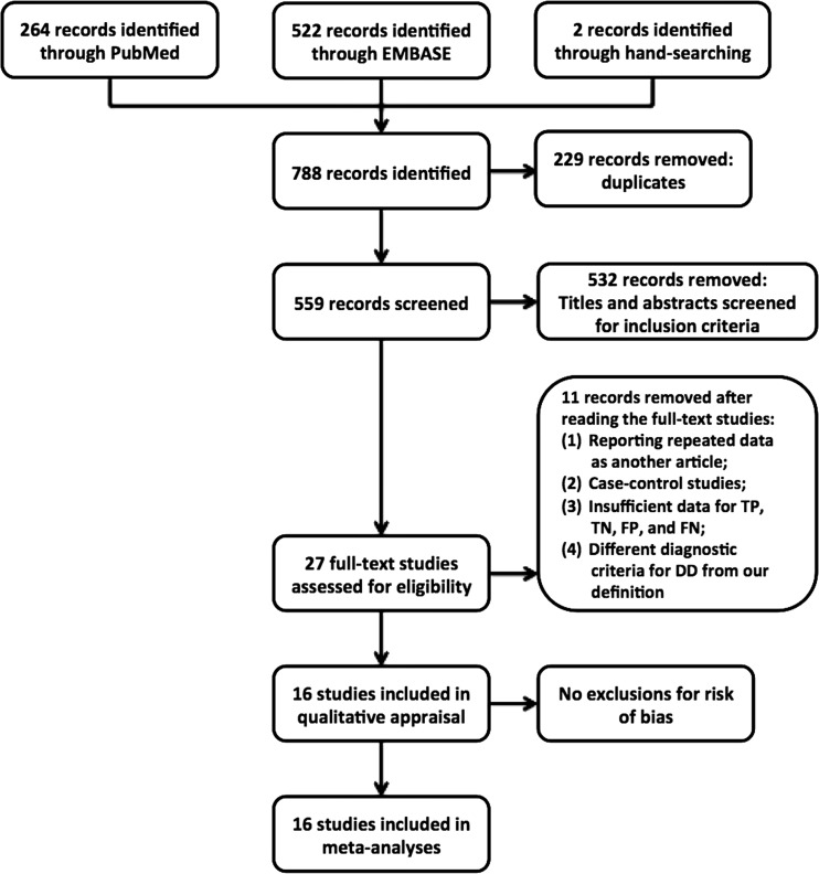
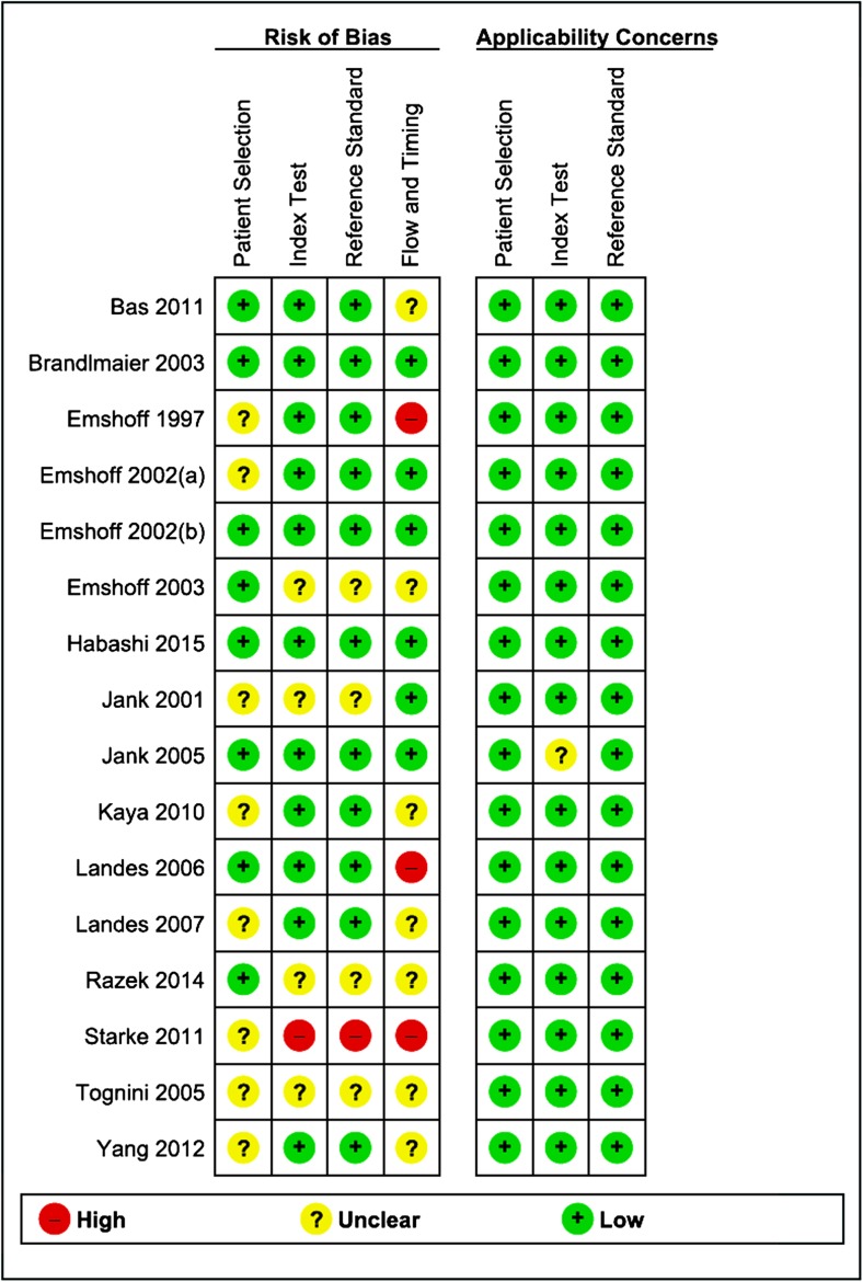
Materials and methods: Pubmed and EMBASE were searched electronically to identify diagnostic accuracy studies that assessed the diagnostic value of US for the diagnosis of DD, using Magnetic resonance imaging (MRI) as the reference standard. Meta-analyses were performed with Metadisc 1.4 and RevMan 5.3.

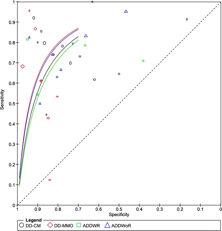
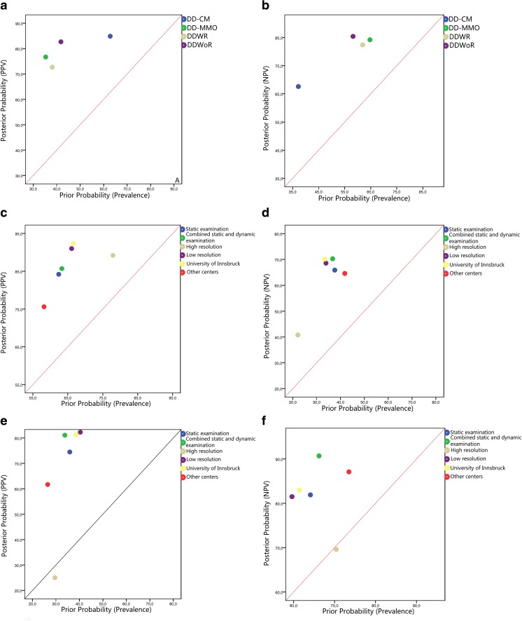
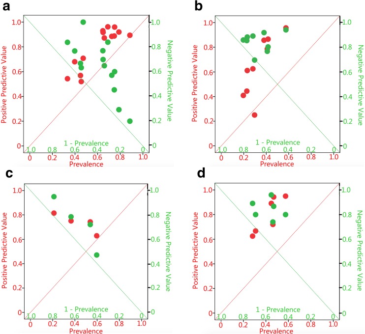
Results: A total of 16 studies qualified for meta-analyses. For the diagnosis of DD at closed mouth position (DD-CM) and DD at maximum mouth-opening position (DD-MMO), the added values of a positive result with US for ruling in DD-CM and DD-MMO were 22 and 41%, while those of a negative result with US for ruling out DD-CM and DD-MMO were 30 and 20%. For the diagnosis of DD with reduction (DDWR) and DD without reduction (DDWoR), the added values of a positive result in US for ruling in DDWR and DDWoR were 35 and 41%, while those of a negative result in US for ruling out DDWR and DDWoR were 21 and 27%.

Conclusions: Using MRI as reference standard, the added values of both positive predictive values and negative predictive values of US for ruling in and ruling out DDs are sufficient in the decision-making in dental practice.

Clinical relevance: US can be a good imaging tool to supplement clinical examination findings in patients with suspected DDs. Combined static and dynamic examinations using high-resolution US should be preferred.

Keywords: Magnetic resonance imaging; Meta-analysis; Temporomandibular joint disorders; Ultrasonography.


Conflict of interest statement

Conflict of interest

The authors declare that they have no conflict of interest.

Ethical approval

This article does not contain any studies with human participants or animals performed by any of the authors.

Informed consent

For this type of study, formal consent is not required.

Figures

Fig. 1
Fig. 1
Flow diagram of study inclusion
Fig. 2
Fig. 2
Summary of risk of bias assessment based on QUADAS 2
Fig. 3
Fig. 3
Summary reviewer operator characteristics (sROC) curves. Symbols represent included studies in meta-analyses (DD-CM disc displacement at closed mouth position, DD-MMO disc displacement at maximum mouth opening, DDWR disc displacement with reduction, DDWoR disc displacement without reduction)
Fig. 4
Fig. 4
Pooled posterior probabilities of ultrasonography for ruling in or ruling out disc displacement compared to prevalences of disc displacement (a positive predictive values of ultrasonography for ruling in the presence of disc displacement at closed mouth position (DD-CM), disc displacement at maximum mouth opening (DD-MMO), disc displacement with reduction (DDWR), and disc displacement without reduction (DDWoR); b negative predictive values of ultrasonography for ruling out the presence of DD-CM, DD-MMO, DDWR, and DDWoR; c positive predictive values of ultrasonography for ruling in the presence of DD-CM based on subgroups; d negative predictive values of ultrasonography for ruling out the presence of DD-CM based on subgroups; e positive predictive values of ultrasonography for ruling in the presence of DD-MMO based on subgroups; f negative predictive values of ultrasonography for ruling out the presence of DD-MMO based on subgroups). The diagonals represent the posterior probability (PPV or NPV) were the same as the prior probability (prevalence), and there was no added values for ruling in or ruling out the DDs at this diagonal
Fig. 5
Fig. 5
Positive and negative predictive values of ultrasonography compared to the prevalences of disc displacement at closed mouth position (DD-CM), disc displacement at maximum mouth opening (DD-MMO), disc displacement with reduction (DDWR), and disc displacement without reduction (DDWoR) for each included study. Red circles represent positive predictive values of US of the included studies and green circles represent negative predictive values of US of the included studies. (a DD-CM, b DD-MMO, c DDWR, d DDWoR). The red diagonals represent the positive predictive value were the same as prevalence, and there was no added value for ruling in the DDs at this diagonal. The green diagonals represent the negative predictive value were the same as the compliment of prevalence (1—prevalence), and there was no added value for ruling out the DDs at this diagonal

References

    1. Resende CM, Alves AC, Coelho LT, et al. Quality of life and general health in patients with temporomandibular disorders. Braz Oral Res. 2013;27(2):116–121. doi: 10.1590/S1806-83242013005000006. - DOI - PubMed
    1. Naeije M, Te Veldhuis AH, Te Veldhuis EC, et al. Disc displacement within the human temporomandibular joint: a systematic review of a “noisy annoyance”. J Oral Rehabil. 2013;40(2):139–158. doi: 10.1111/joor.12016. - DOI - PubMed
    1. Santos KC, Dutra ME, Warmling LV. Correlation among the changes observed in temporomandibular joint internal derangements assessed by magnetic resonance in symptomatic patients. J Oral Maxillofac Surg. 2013;71(9):1504–1512. doi: 10.1016/j.joms.2013.04.033. - DOI - PubMed
    1. Amaral Rde O, Damasceno NN, de Souza LA, et al. Magnetic resonance images of patients with temporomandibular disorders: prevalence and correlation between disk morphology and displacement. Eur J Radiol. 2013;82(6):990–994. doi: 10.1016/j.ejrad.2013.01.002. - DOI - PubMed
    1. Manfredini D, Arveda N, Guarda-Nardini L, et al. Distribution of diagnoses in a population of patients with temporomandibular disorders. Oral Surg Oral Med Oral Pathol Oral Radiol. 2012;114:e35–e41. doi: 10.1016/j.oooo.2012.03.023. - DOI - PubMed

MeSH terms

LinkOut - more resources