. 2018 Jun;17(3):e12733.
doi: 10.1111/acel.12733. Epub 2018 Feb 18.

Aging impacts CD103+ CD8+ T cell presence and induction by dendritic cells in the genital tract

Affiliations

Aging impacts CD103+ CD8+ T cell presence and induction by dendritic cells in the genital tract

Marta Rodriguez-Garcia et al. Aging Cell. 2018 Jun.

Abstract

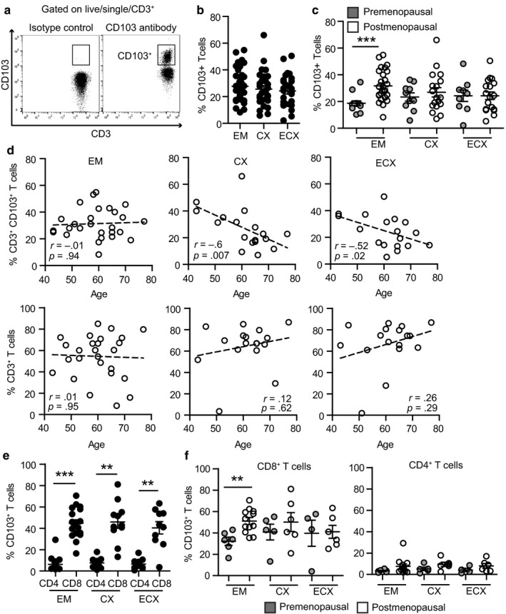
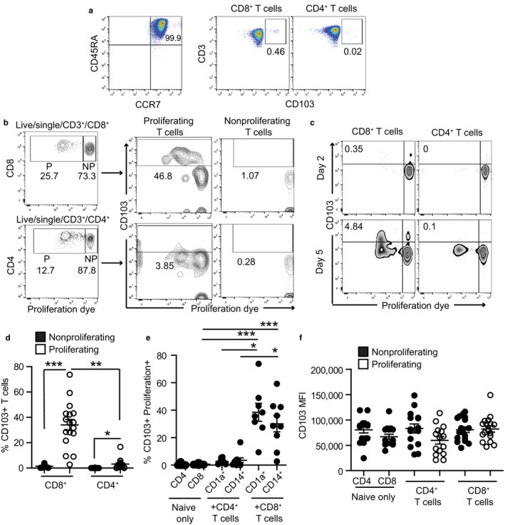
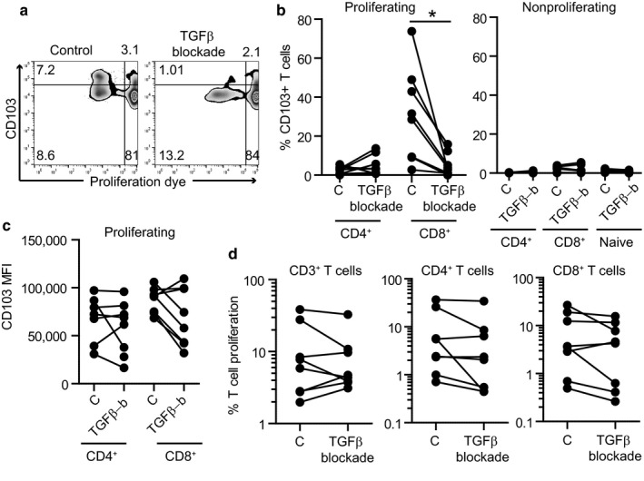
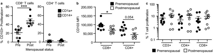
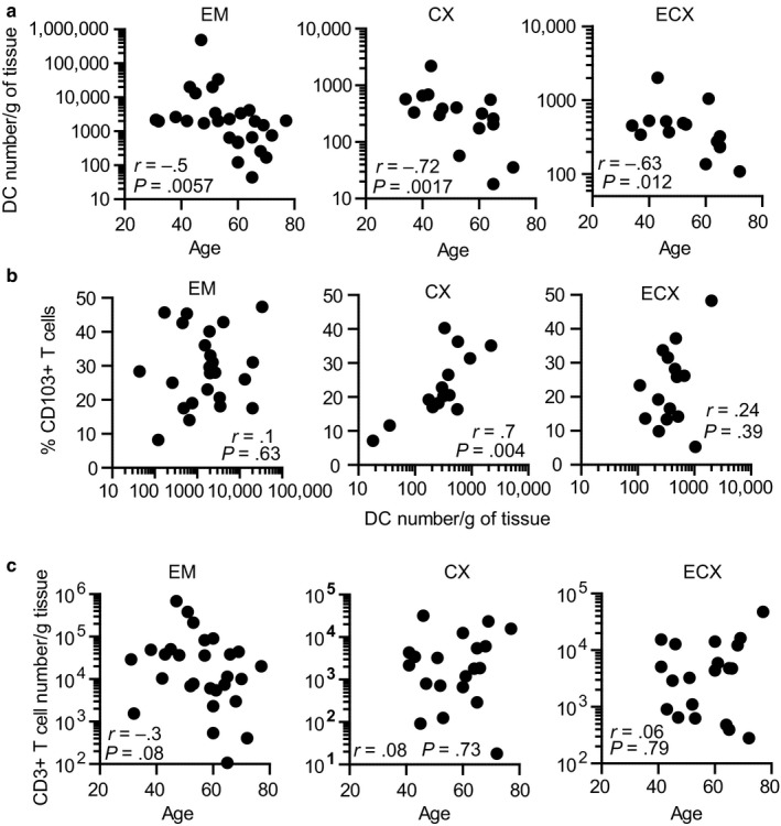
As women age, susceptibility to systemic and genital infections increases. Tissue-resident memory T cells (TRMs) are CD103+ CD8+ long-lived lymphocytes that provide critical mucosal immune protection. Mucosal dendritic cells (DCs) are known to induce CD103 expression on CD8+ T cells. While CD103+ CD8+ T cells are found throughout the female reproductive tract (FRT), the extent to which aging impacts their presence and induction by DCs remains unknown. Using hysterectomy tissues, we found that endometrial CD103+ CD8+ T cells were increased in postmenopausal compared to premenopausal women. Endometrial DCs from postmenopausal women were significantly more effective at inducing CD103 expression on allogeneic naïve CD8+ T cells than DCs from premenopausal women; CD103 upregulation was mediated through membrane-bound TGFβ signaling. In contrast, cervical CD103+ T cells and DC numbers declined in postmenopausal women with age. Decreases in DCs correlated with decreased CD103+ T cells in endocervix, but not ectocervix. Our findings demonstrate a previously unrecognized compartmentalization of TRMs in the FRT of postmenopausal women, with loss of TRMs and DCs in the cervix with aging, and increased TRMs and DC induction capacity in the endometrium. These findings are relevant to understanding immune protection in the FRT and to the design of vaccines for women of all ages.

Keywords: TGF-β; dendritic cells; female reproductive tract; menopause; resident memory T cells; sexually transmitted infections.


Figures

Figure 1
Figure 1
Menopausal status and aging differentially regulate CD103+ T cells in different sites of the female reproductive tract. (a) Representative plot of the gating strategy for CD103+ T cells within the CD3+ population. Left plot shows the isotype control and right plot the staining with anti‐CD103 antibody. (b) Percentage of CD103+ T cells within the CD3+ population in endometrium (EM = 40), endocervix (CX = 28), and ectocervix (ECX = 27) and (c) after separating pre‐ (gray; EM = 13, CX = 10, ECX = 9) and postmenopausal women (white; EM = 27, CX = 18, ECX = 18). Each dot represents a single patient. Mean ± SEM are shown; ***p < 0.001, nonparametric Kruskal–Wallis test with Dunn's post‐test correction for multiple comparisons. (d) Correlation between age and percentage of CD103+ T cells (top row) or percentage of CD3+ T cells (bottom row) in EM (n = 27), CX (n = 18), and ECX (n = 18) from postmenopausal women; Spearman correlation. (e) Percentage of CD103+ T cells within CD8+ and CD4+ T cells from EM (n = 18), CX (n = 11), and ECX (n = 10) and (f) after separating pre‐ and postmenopausal women. ***p < .001; **p < .01; Mann–Whitney U‐test
Figure 2
Figure 2
Endometrial DCs induce expression of CD103 on naïve T cells. (a) Representative plot of purity and CD103 expression on freshly isolated naïve T cells from blood before co‐culture (day 0). (b) Representative example of T‐cell proliferation rate and CD103 expression after allogeneic stimulation of naïve T cells with tissue DCs from EM. Upper row shows CD8+ T cells, and lower row shows CD4+ T cells. CD103 expression on proliferating (P, proliferation dye low) and nonproliferating (NP, proliferation dye high) cells is shown in middle and right panels, respectively. (c) Representative example of CD103 upregulation over time after allogeneic stimulation of naïve T cells with tissue DCs from EM. (d) Comparison of the percentage of CD103+ T cells within the proliferating or nonproliferating cell gates (n = 17). (e) Percentage of CD103+ T cells after allogeneic stimulation with EM DCs purified using CD1a+ (n = 8) or CD14+ (n = 9) magnetic bead selection. The graph shows proliferating CD103+ T cells, except in the naïve only control group, for which cells did not proliferate. (f) CD103 MFI (mean fluorescence intensity) on CD4+ CD103+ T cells (CD4+ T cells) or CD8+ CD103+T cells (CD8+ T cells) after allogeneic stimulation with EM DCs or naïve T cells cultured without DCs as a control (naïve only) (n = 17). *p < .05; **p < .01; ***p < .001; Kruskal–Wallis test with Dunn's post‐test correction for multiple comparisons
Figure 3
Figure 3
Endometrial DCs induce expression of CD103 on naïve CD8+ T cells through TGFβ signaling. (a) Representative plot of CD103 expression on proliferating and nonproliferating cells in the absence (control) or presence (TGFβ blockade) of the TGFβ signaling inhibitor SB431542. (b) Percentage of CD103 expression on proliferating (left) or nonproliferating (right) CD4+ and CD8+ T cells in the absence (C: control) or presence of the TGFβ signaling inhibitor SB431542 (TGFβ‐b) (n = 8). (c) CD103 mean fluorescence intensity on proliferating CD4+ and CD8+ T cells in the absence (C: control) or presence (TGFβ‐b) of the TGFβ signaling inhibitor SB431542 (n = 8). (d) Proliferation rate of total CD3+ T cells, CD4+ T cells, or CD8+ T cells after allogeneic stimulation with EM DCs in the presence of TGFβ signaling inhibitor (n = 8). Each dot represents a single patient. Mean ± SEM are shown. *p < .05; Wilcoxon‐matched pair test
Figure 4
Figure 4
Menopausal status regulates endometrial DC ability to induce CD103 expression on CD8+ T cells. (a) CD103+ T cell percentage, (b) CD103 mean fluorescence intensity, and (c) proliferation rate after allogeneic stimulation of naïve T cells with EM DCs from pre‐ (n = 9) or postmenopausal (n = 8) women. Results from CD1a+ and CD14+ DCs are shown combined. ***p < .001; *p < .05; Mann–Whitney U‐test
Figure 5
Figure 5
Dendritic cells (DC) numbers and CD103+ T cell percentage decline in cervix with aging. (a) Correlation between DC number and age (EM = 27; CX = 16; ECX = 15) and (b) DC number and CD103+ T cell percentage (EM = 25; CX = 15; ECX = 15). (c) Correlation between age and total CD3+ T cells per gram of tissue (EM = 28, CX = 20, ECX = 20). Each dot represents a single patient; Spearman correlation

References

    1. Casey, K. A. , Fraser, K. A. , Schenkel, J. M. , Moran, A. , Abt, M. C. , Beura, L. K. , … Masopust, D. (2012). Antigen‐independent differentiation and maintenance of effector‐like resident memory T cells in tissues. The Journal of Immunology, 188, 4866–4875. 10.4049/jimmunol.1200402 - DOI - PMC - PubMed
    1. CDC (2016). Diagnoses of HIV infection among adults aged 50 years and older in the United States and dependent areas, 2010–2014 In Surveillance Supplemental Report 2016;21(2).
    1. Cuburu, N. , Graham, B. S. , Buck, C. B. , Kines, R. C. , Pang, Y. Y. , Day, P. M. , … Schiller, J. T. (2012). Intravaginal immunization with HPV vectors induces tissue‐resident CD8+ T cell responses. The Journal of Clinical Investigation, 122, 4606–4620. 10.1172/JCI63287 - DOI - PMC - PubMed
    1. Duluc, D. , Gannevat, J. , Anguiano, E. , Zurawski, S. , Carley, M. , Boreham, M. , … Oh, S. (2013). Functional diversity of human vaginal APC subsets in directing T‐cell responses. Mucosal Immunology, 6, 626–638. 10.1038/mi.2012.104 - DOI - PMC - PubMed
    1. Erlebacher, A. (2013). Mechanisms of T cell tolerance towards the allogeneic fetus. Nature Reviews Immunology, 13, 23–33. 10.1038/nri3361 - DOI - PubMed

Publication types