. 2018 Mar 27;12(3):2560-2568.
doi: 10.1021/acsnano.7b08411. Epub 2018 Feb 22.

Mechanical Division of Cell-Sized Liposomes

Affiliations

Mechanical Division of Cell-Sized Liposomes

Siddharth Deshpande et al. ACS Nano. .

Abstract

Liposomes, self-assembled vesicles with a lipid-bilayer boundary similar to cell membranes, are extensively used in both fundamental and applied sciences. Manipulation of their physical properties, such as growth and division, may significantly expand their use as model systems in cellular and synthetic biology. Several approaches have been explored to controllably divide liposomes, such as shape transformation through temperature cycling, incorporation of additional lipids, and the encapsulation of protein division machinery. However, so far, these methods lacked control, exhibited low efficiency, and yielded asymmetric division in terms of volume or lipid composition. Here, we present a microfluidics-based strategy to realize mechanical division of cell-sized (∼6 μm) liposomes. We use octanol-assisted liposome assembly (OLA) to produce liposomes on chip, which are subsequently flowed against the sharp edge of a wedge-shaped splitter. Upon encountering such a Y-shaped bifurcation, the liposomes are deformed and, remarkably, are able to divide into two stable daughter liposomes in just a few milliseconds. The probability of successful division is found to critically depend on the surface area-to-volume ratio of the mother liposome, which can be tuned through osmotic pressure, and to strongly correlate to the mother liposome size for given microchannel dimensions. The division process is highly symmetric (∼3% size variation between the daughter liposomes) and is accompanied by a low leakage. This mechanical division of liposomes may constitute a valuable step to establish a growth-division cycle of synthetic cells.

Keywords: liposomes; membrane biophysics; microfluidics; octanol-assisted liposome assembly; synthetic biology; synthetic cell division.


Conflict of interest statement

The authors declare no competing financial interest.

Figures
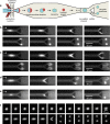
Figure 1
Figure 1
Mechanical division of liposomes. (a) Top-view schematic (not to scale) showing the experimental workflow leading to the mechanical division of liposomes. Double-emulsion droplets are formed at the production junction, which within minutes, mature into liposomes. By maintaining a hypertonic environment, the liposomes lose a specific volume, which sets up an intended high surface area-to-volume ratio. These “floppy” liposomes then pass through a narrow presplitter channel at a high velocity before they encounter the Y-shaped splitter, whereupon they can divide into two daughter liposomes. (b–e) Fluorescence time-lapse images (of the encapsulated dye, either Alexa Fluor 350 or Dextran-Alexa Fluor 647) showing different fates of liposomes upon encountering the splitter. (b) Division: A liposome gets deformed at the splitter and divides into two daughter liposomes. Note the similar size of the daughter liposomes indicating highly symmetric division. Also, there is no obvious increase in the background intensity after the splitting, indicating leakage-free division. (c) Bursting: Mother liposome dissociates due to the membrane rupture, spilling the inner contents into the environment. (d) Semi-division: Rarely, only one of the daughter cells survives the division process, while the other burst opens and dissociates. (e) Snaking: if small enough, the liposome passes through one of the Y-branches of the splitter, without either dividing or bursting. (f) Moving-frame region-of-interest showing an entire division event including the entry of the liposome into the narrow presplitter channel. The images underwent appropriate background subtraction, and further analyses were performed with similarly processed images. The lipid composition of liposomes is DOPC and Rh-PE (molar ratio of 99.9:0.1). Time difference Δt between successive frames is Δtdivision = 1.2 ms (panel b), Δtbursting = 1.2 ms (panel c), Δtsemi-division = 3 ms (panel d), Δtsnaking = 4 ms (panel e), Δtmoving-frame = 2 ms (panel f). Horizontal arrows indicate the flow direction. Mother liposomes appear deformed in the presplitter channel due to motion blurring.
Figure 2
Figure 2
Liposome fate upon encountering the splitter is determined by its size. (a) Mean diameter of daughter liposomes against that of corresponding mother liposomes. Red circles represent division events, while cyan squares represent semi-divisions. The line shows a linear fit with a slope m = 0.74 ± 0.01 (mean ± s.d., R2 = 0.89). For bursts (dark blue diamonds), data points are displayed at the horizontal axis, since no daughter liposomes were formed. Horizontal and vertical error bars indicate corresponding standard deviations. Ndivision = 151, Nburst = 602, Nsemi-division = 46. (b) Top-view schematics explaining how liposome size affects the splitting probability. A small enough liposome is moderately stretched at the splitter but still has enough excess surface area required for division. For a bigger liposome, the decrease in excess surface area is more drastic, thus increasing the probability of bursting. (c–f) The probability of different events, viz., division (c), bursting (d), semi-division (e), and snaking (f) as a function of liposome diameter, obtained from the experimental data (Ndivision = 151, Nburst = 602, Nsemi-division = 46, Nsnake = 159). The value of each bar denotes the fraction of the specific event occurring for that particular liposome diameter. Channel dimensions were kept constant, and the impact velocity varied within a similar range for all the events.
Figure 3
Figure 3
Mechanical division of liposomes is highly symmetric. (a) Plot showing the correlation between the diameters of the daughter liposomes, resulting from the same mother liposome. The linear fit, shown by the line, has a slope of 0.99 ± 0.01 (mean ± s.d., R2 = 0.88), emphasizing the highly symmetric nature of the division process. (b) Plot showing the correlation obtained between the total intensity of the two daughter liposomes obtained from the same mother liposome. The solid line, which is a linear fit to the data set, has a slope of m = 0.97 ± 0.01 (mean ± s.d., R2 = 0.86), indicating an equal distribution of the encapsulated material during the division process. Horizontal and vertical error bars indicate corresponding standard deviations. In total, 152 (panel a) and 151 (panel b) pairs of daughter liposomes were analyzed.
Figure 4
Figure 4
Liposome division is associated with a low leakage. (a) Plot of the total intensity of daughter liposomes against that of the corresponding mother liposomes (N = 78). The intensities are strongly correlated, with the solid line showing a linear fit with a slope of m = 0.87 ± 0.01 (mean ± s.d., R2 = 0.94), suggesting a leakage of 13% during the division process. Horizontal and vertical error bars indicate corresponding standard deviations. (b) Top-view schematic showing a dividing liposome at different time points. Summing up the fluorescence intensity along the x- (or y) axis for each region-of-interest generates a moving-frame xt (or yt) kymograph. (c) Average xt kymographs for division (upper, N = 131) and bursting (lower, N = 199) events. The xt kymograph of dividing liposomes gives a straight bright trace, while that of bursting liposomes results in a rapidly fading trace. (d) Average intensity profile stays constant in case of division (red solid line), while decays rapidly in case of bursting (blue dashed line). The plots are obtained from the corresponding average moving-frame xt kymographs. The shaded regions indicate the standard deviations. (e–f) Average moving-frame yt kymograph of division events (e) which clearly shows a constant dark background confirming a very low amount of leakage involved in the division process (N = 131). In comparison, a similar kymograph for bursting events (f) shows a considerable rise in the background as the encapsulated dye diffuses away into the environment (N = 199). The vertical dashed lines in b, c, e and f indicate the splitter position. Contrast has been enhanced for better visualization.

Similar articles

Cited by

References

    1. Angert E. R. Alternatives to Binary Fission in Bacteria. Nat. Rev. Microbiol. 2005, 3, 214–224. 10.1038/nrmicro1096. - DOI - PubMed
    1. Kysela D. T.; Brown P. J.; Casey Huang K.; Brun Y. V. Biological Consequences and Advantages of Asymmetric Bacterial Growth. Annu. Rev. Microbiol. 2013, 67, 417–435. 10.1146/annurev-micro-092412-155622. - DOI - PMC - PubMed
    1. Macara I. G.; Mili S. Polarity and Differential Inheritance-Universal Attributes of Life?. Cell 2008, 135, 801–812. 10.1016/j.cell.2008.11.006. - DOI - PMC - PubMed
    1. Caspi Y.; Dekker C. Divided We Stand: Splitting Synthetic Cells for Their Proliferation. Syst. Synth. Biol. 2014, 8, 249–269. 10.1007/s11693-014-9145-7. - DOI - PMC - PubMed
    1. Szostak J. W.; Bartel D. P.; Luisi L. Synthesizing Life. Nature 2001, 409, 387–390. 10.1038/35053176. - DOI - PubMed

Publication types

LinkOut - more resources