Review
. 2018 Feb 19;17(1):49.
doi: 10.1186/s12943-018-0780-6.

Chronic myeloid leukemia: the paradigm of targeting oncogenic tyrosine kinase signaling and counteracting resistance for successful cancer therapy

Affiliations
Review

Chronic myeloid leukemia: the paradigm of targeting oncogenic tyrosine kinase signaling and counteracting resistance for successful cancer therapy

Simona Soverini et al. Mol Cancer. .

Abstract

Deregulated activity of BCR-ABL1, a nonreceptor tyrosine kinase encoded by the fusion gene resulting from the t(9;22)(q34;q11) chromosomal translocation, is thought to be the driver event responsible for initiation and maintenance of chronic myeloid leukemia (CML). BCR-ABL1 was one of the first tyrosine kinases to be implicated in a human malignancy and the first to be successfully targeted. Imatinib mesylate, the first tyrosine kinase inhibitor (TKI) to be approved for therapeutic use, was hailed as a magic bullet against cancer and remains one of the safest and most effective anticancer agents ever developed. Second- and third-generation TKIs were later introduced to prevent or counteract the problem of drug resistance, that may arise in a small proportion of patients. They are more potent molecules, but have been associated to more serious side effects and complications. Patients achieving stable optimal responses to TKI therapy are predicted to have the same life expectancy of the general population. However, TKIs do not 'cure' CML. Only a small proportion of cases may attempt therapy discontinuation without experiencing subsequent relapse. The great majority of patients will have to assume TKIs indefinitely - which raises serious pharmacoeconomic concerns and is now shifting the focus from efficacy to compliance and quality of life issues. Here we retrace the steps that have led from the biological acquisitions regarding BCR-ABL1 structure and function to the development of inhibitory strategies and we discuss drug resistance mechanism and how they can be addressed.

Keywords: BCR-ABL1; Resistance; Tyrosine kinase; Tyrosine kinase inhibitors.


Conflict of interest statement

Ethics approval and consent to participate

Not applicable.

Consent for publication

Not applicable.

Competing interests

SS, GM: consultancy and honoraria from Incyte Biosciences, Novartis, Bristol-Myers Squibb.

LB, MM, MC: no competing interests.

Publisher’s Note

Springer Nature remains neutral with regard to jurisdictional claims in published maps and institutional affiliations.

Figures

Fig. 1
Fig. 1
Progression of CML from chronic phase (CP) to blastic phase (BP). Biologically, the transition is associated with the accumulation of additional hits in BCR-ABL1 itself (TKI-resistant kinase domain mutations) or in other genes/chromosomes. In the latter case, the degree of oncogenic addiction decreases, and inhibiting BCR-ABL1 alone may not be sufficient any more. This translates into an increase of drug resistance and in poor response to current therapies. ‘X’, ‘Y’ and ‘Z’ represent additional altered molecules other than BCR-ABL1
Fig. 2
Fig. 2
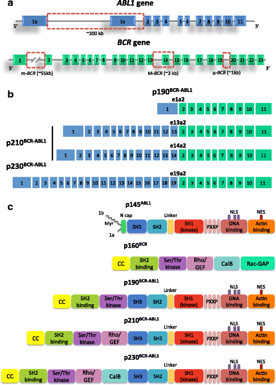
Genomic breakpoints in the BCR and ABL1 genes and resulting transcript types and proteins. a Translocation breakpoints in BCR most frequently fall in intron 13 or 14 (M-BCR) or in intron 1 (m-BCR), or in intron 19 (μ-BCR). In ABL1, the breakpoints are intronic as well, and most frequently fall in a large region comprised between exons 1b and 2. Exon 1a and 1b are mutually exclusive and are incorporated in the mature ABL1 mRNA as a result of alternative splicing. However, neither of the two is retained in BCR-ABL1 mRNA. b The most common fusion transcripts resulting from the translocation include e13a2 and e14a2, resulting from the M-BCR, both translated into the p210BCR-ABL1 isoform (typical of CML and of some cases of Ph + ALL); e1a2, resulting from the m-BCR and translated into the p190BCR-ABL1 isoform (typical of the majority of Ph + ALL); e19a2, resulting from the μ-BCR and translated into the p230BCR-ABL1 isoform (typical of a subset of CML once called chronic neutrophilic leukemias). c Domain organization of BCR, ABL1 and BCR-ABL1 proteins. BCR is a 160 kDa protein with a coiled-coil (CC) oligomerization domain, a domain thought to mediate binding to Src-homology 2 (SH2)-domain-containing proteins, a serine/threonine kinase domain, a region with homology to Rho guanine-nucleotide-exchange factor (Rho-GEF), a region thought to facilitate calcium-dependent lipid binding (CaLB) and a region showing homology to Rac GTPase activating protein (Rac-GAP). ABL1 is a 145 kDa protein that contains an N-cap (that in isoform 1b undergoes myristoylation, a post translation modification that attaches the fourteen-carbon saturated fatty acid myristate to the amino-terminal glycine of the protein), the tandem SH3, SH2 and SH1 (tyrosine-kinase) domains, four proline-rich SH3 binding sites (PXXP), three nuclear localization signals (NLSs), one nuclear exporting signal (NES), a DNA-binding domain, and an actin-binding domain. In all BCR-ABL1 protein isoforms, the CC domain of BCR is included, the myristoylated N cap is lost, and the ABL1 kinase domain is retained. National Center for Biotechnology Information (NCBI) accession numbers: ABL1 gene, NG_012034.1; BCR gene, NG_009244.1
Fig. 3
Fig. 3
Stategies for BCR-ABL1 inhibition. Displayed are the SH2 domain (green) and the SH1 (kinase) domain (blue). The inhibitor is in yellow. a ATP-competitive inhibitors like imatinib, nilotinib, dasatinib etc. bind in the cleft between the N-lobe and the C-lobe, at the bottom of which lies the ATP-binding site. b One mode of allosteric inhibition is to use small molecules mimicking myristate binding to the hydrophobic pocket located in the C-lobe. This is the mode of action of asciminib. c Another mode of allosteric inhibition is to use proteins (‘monobodies’) directed against the SH2-kinase interface
Fig. 4
Fig. 4
Regulation of the ABL1 tyrosine kinase. a All protein kinase domains have a highly conserved bilobed structure. The binding site for ATP and for the inhibitors is in a cleft between the 2 lobes. The phosphate-binding loop (P-loop) is highlighted in yellow. The phosphorylation state and conformation of the activation loop (A-loop; highlighted in red) determine whether the kinase is active or inactive. In all tyrosine kinases, the site of activating phosphorylation is generally a single Tyrosine residue located in the middle of the loop that once phosphorylated, can interact electrostatically with a neighboring Arginine residue, resulting in the stabilization of an extended and open conformation of the loop (right image). This conformation of the A-loop enables the access to the peptide substrate binding site. When the A-loop is unphosphorylated, it is folded inwards, blocking the peptide substrate binding site (left image). A second important regulatory feature of kinases is the conformation of a highly conserved aspartate-phenylalanine-glycine (DFG) motif (highlighted in orange) located at the N-terminal end of the A-loop. Images obtained with the Web-based 3D viewer NGL [113]. b Cartoon representation of ABL1 with the kinase domain (SH1), the SH2 and the SH3 domains. Alpha helices are in magenta, beta sheets in yellow. A myristic acid moiety in the myristate binding pocket is shown with a ball-and-stick representation. Binding of the myristoyl group to the myristate pocket induces a conformational change in the C-terminal helix of the kinase domain that is necessary for binding of the SH3-SH2 clamp, which keeps the kinase inactive. Image obtained with the web-based 3D viewer NGL [113] (Protein Data Bank [PDB] entry 1OPJ)
Fig. 5
Fig. 5
Overview of the mechanisms of resistance to BCR-ABL1 inhibition. According to the currently available data obtained in patients and/or cell lines, resistance may be due to (1) overexpression/increased activity of the efflux pump MDR1, and/or downmodulation/decreased activity of the influx pump hOCT1. This may result also from gene polymorphisms; (2) gene amplification and/or BCR-ABL1 mRNA and protein overexpression to levels that cannot be inhibited by achievable plasma concentrations of the TKI; (3) point mutations in the BCR-ABL1 kinase domain that interfere with TKI binding; (4) activation of alternative/downstream signaling pathways, e.g. of the SRC family kinases. Resistance mechanisms are not necessarily mutually exclusive

References

    1. Sawyers CL. Chronic myeloid leukemia. N Engl J Med. 1999;340:1330–1340. doi: 10.1056/NEJM199904293401706. - DOI - PubMed
    1. Shah NP, Advanced CML. Therapeutic options for patients in accelerated and blast phases. J Natl Compr Cancer Netw. 2008;6(Suppl 2):S31–SS6. - PubMed
    1. Soverini S, de Benedittis C, Mancini M, Martinelli G. Mutations in the BCR-ABL1 kinase domain and elsewhere in chronic myeloid leukemia. Clin Lymphoma Myeloma Leuk. 2015;15(Suppl):S120–S128. doi: 10.1016/j.clml.2015.02.035. - DOI - PubMed
    1. Chereda B, Melo JV. Natural course and biology of CML. Ann Hematol. 2015;94(Suppl 2):S107–S121. doi: 10.1007/s00277-015-2325-z. - DOI - PubMed
    1. Hehlmann R, Berger U, Pfirrmann M, Hochhaus A, Metzgeroth G, Maywald O, Hasford J, Reiter A, Hossfeld DK, Kolb HJ, et al. Randomized comparison of interferon alpha and hydroxyurea with hydroxyurea monotherapy in chronic myeloid leukemia (CML-study II): prolongation of survival by the combination of interferon alpha and hydroxyurea. Leukemia. 2003;17:1529–1537. doi: 10.1038/sj.leu.2403006. - DOI - PubMed

Publication types

MeSH terms

Substances