. 2018 Feb 19;17(1):44.
doi: 10.1186/s12943-018-0781-5.

miR-19b enhances proliferation and apoptosis resistance via the EGFR signaling pathway by targeting PP2A and BIM in non-small cell lung cancer

Affiliations

miR-19b enhances proliferation and apoptosis resistance via the EGFR signaling pathway by targeting PP2A and BIM in non-small cell lung cancer

Ulrich Baumgartner et al. Mol Cancer. .

Abstract

Background: Epidermal growth factor receptor (EGFR) mutations enable constitutive active downstream signaling of PI3K/AKT, KRAS/ERK and JAK/STAT pathways, and promote tumor progression by inducing uncontrolled proliferation, evasion of apoptosis and migration of non-small cell lung cancer (NSCLC). In addition, such EGFR mutations increase the susceptibility of patients with NSCLC to tyrosine kinase inhibitor (TKI) therapy, but treated patients will invariably relapse with resistant disease. A global understanding of underlying molecular mechanisms of EGFR signaling may improve the management of NSCLC patients.

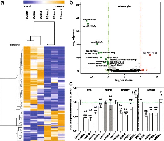
Methods: microarray analysis was performed to identify PI3K/AKT-regulated miRNAs. Phosphoproteomic analysis and cell based assays were performed using NSCLC cell lines lentivirally transduced with anti-miR or miR overexpressing constructs.

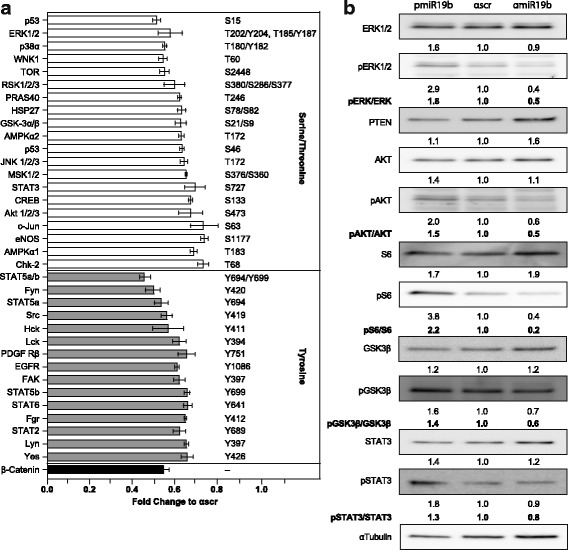
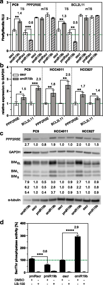
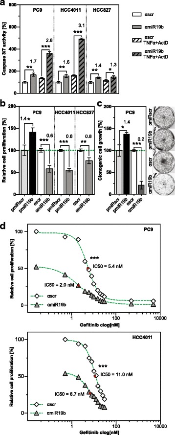
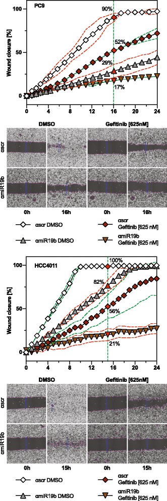
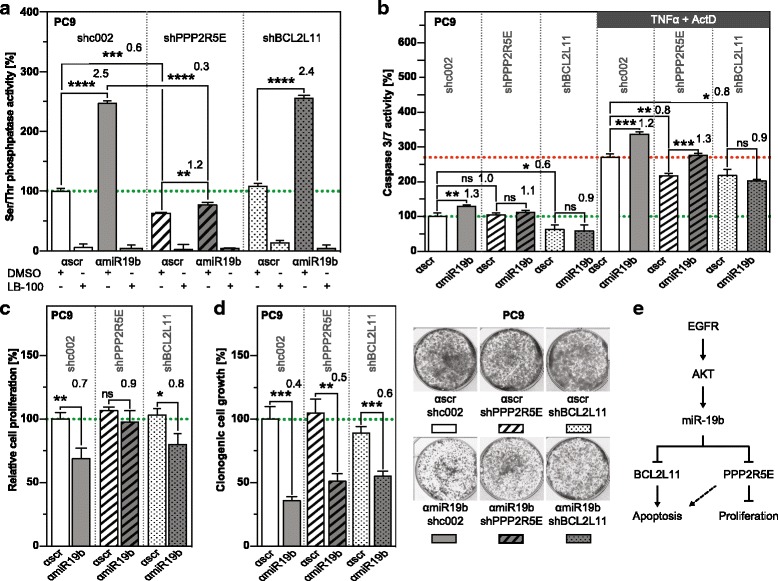
Results: Here, we show that 17 miRNAs including members of the miR-17~ 92 cluster are dysregulated following PI3K/AKT inhibition of EGFR mutant NSCLC cells. Bioinformatics analysis revealed that dysregulated miRNAs act in a concerted manner to enhance the activity of the EGFR signaling pathway. These findings were closely mirrored by attenuation of miR-17~ 92 family member miR-19b in NSCLC cell lines which resulted in reduced phosphorylation of ERK, AKT and STAT and effector proteins in EGFR mutant NSCLC cells. Consistent with this finding, cell cycle progression, clonogenic growth and migration were reduced and apoptosis was enhanced. Co-treatment of NSCLC cells with the tyrosine kinase inhibitor (TKI) gefitinib and anti-miR-19b construct reduced migration and clonogenic growth in a synergistic manner suggesting that EGFR and miR-19b act together to control oncogenic processes. Serine/threonine phosphatase PP2A subunit PPP2R5E and BCL2L11 encoding BIM were identified as major targets of miR-19b by target validation assays. Consistent with this finding, PP2A activity was strongly enhanced in NSCLC transduced with anti-miR-19b construct, but not in cells co-transduced with anti-miR-19b and shPPP2R5E, suggesting that PPP2R5E is a major constituent of the PP2A complex. Accordingly, enhanced proliferation by miR-19b was due to targeting PPP2R5E. In contrast, apoptosis resistance was mainly due to targeting BCL2L11.

Conclusion: Our results provide insight into the importance of targeting PPP2R5E and BCL2L11 by miR-19b in oncogenic processes of NSCLC. Attenuation of miR-19b expression could potentially be exploited in adjuvant therapy of EGFR mutant NSCLC.

Keywords: Apoptosis; Epidermal growth factor receptor; Non-small cell lung cancer; Proliferation; Serine-threonine phosphatase; microRNA.


Conflict of interest statement

Ethics approval and consent to participate

Not applicable.

Consent for publication

Not applicable.

Competing interests

The authors declare that they have no competing interests.

Publisher’s Note

Springer Nature remains neutral with regard to jurisdictional claims in published maps and institutional affiliations.

Figures

Fig. 1
Fig. 1
miR-19b is regulated by the PI3K/AKT branch of the EGFR signaling pathway. a Heat map clustering of miRNAs that are dysregulated by the PI3K inhibitor LY294002. PC9 cells were treated with 20 μM LY294002 or DMSO control for 72 h and expression of 800 miRNAs was analyzed by NanoString using the nCounter Human miRNA Expression Assay Kit (n = 3). b Volcano plot of dysregulated miRNAs described in (A) using a cutoff ±0.4 log2 fold change (FC) (n = 3). c miR-19b expression level by real-time qPCR relative to RNU48 in EGFR mutant NSCLC cells treated with 1.25 μM gefitinib, 1.25 uM afatinib, 20 μM LY294002 and 10 μM U0126 for 72 h. Results are presented as mean ± SD (n = 3). ns, not significant. *, p < 0.05; **, p < 0.01; ***, p < 0.001
Fig. 2
Fig. 2
Changes in phosphorylation elicited by miR-19b. a Phosphoprotein array of anti-miR19b–transduced PC9 cells relative to control (n = 4). Signal intensity of pairs of duplicate spots is indicated. Only proteins that give rise to a signal intensity at least two fold above the background and a fold change ≤ 0.8 relative to control are indicated. b Western blot analysis of pre-miR-19b and anti-miR-19b-transduced cells. Protein levels were normalized to α-tubulin and presented relative to the control. Numbers below the immunoblots indicate relative expression values. Signal ratio of phospho-specific antibody and pan-specific antibody for respective proteins are indicated in bold
Fig. 3
Fig. 3
PPP2R5E and BCL2L11 are direct targets of miR-19b. a Luciferase reporter assay of PC9 cells transduced with pre-miR19b (pmiR19b), pre-control (pmiRscr), anti-miR19b (αmiR19b) and anti-control (αscr) and transfected with constructs containing the wild-type (TS) or mutated (mTS) miR-19b target site from PPP2R5E or BCL2L11, respectively. Luciferase activity is presented as mean ± SD relative to Renilla activity (n = 3). b PPP2R5E and BCL2L11 mRNA levels by real-time qPCR in anti-miR19b–transduced NSCLC cells. Results are presented as mean fold change relative to the control ±SD, normalized to GAPDH (n = 3). c Western blot analysis of pre-miR-19b and anti-miR19b–transduced NSCLC cells using PPP2R5E and BCL2L11 specific antibodies. Protein levels were normalized to α-tubulin and GAPDH and presented relative to the control. d PP2A activity assay of pre-miR19b- and anti-miR19b–transduced cells relative to the control (n = 3). *, p < 0.05; **, p < 0.01; ***, p < 0.001
Fig. 4
Fig. 4
miR-19b regulates EGFR-induced cellular processes. a Spontaneous (solid columns) and TNFα/AktD-induced apoptosis (hatched columns) of EGFR mutant NSCLC transduced with anti-miR19b construct (n = 3). Apoptosis was analyzed by caspase3/7 cleavage assay 2 h post induction with TNFα/AktD. b BrdU incorporation and (c) clonogenic growth of pre-miR19b or anti-miR19b–transduced PC9 cells (n = 3). Cells were examined and imaged 8 days post-seeding. *, p < 0.05; **, p < 0.01; ***, p < 0.001, ****, p < 0.0001. d Clonogenic growth of anti-miR-19b and control-transduced PC9 (upper panel) and HCC4011 cells (bottom panel) at different concentrations of gefitinib. Cell numbers were determined 8 days after incubation with gefitinib. IC50 values were calculated from dose-response curves
Fig. 5
Fig. 5
EGFR and miR-19b act together to control cell migration. Wound closure assay of (a) PC9 and (b) HCC4011 cells. Wound closure was monitored over a period of 24 h in the presence or absence of 0.625 μM gefitinib using the imaging system Cell-IQ. Gefitinib was added immediately after injury of the monolayer. Results are presented as mean ± SD (n = 6)
Fig. 6
Fig. 6
Distinct roles of PPP2R5E and BCL2L11 in PP2A activity, proliferation and apoptosis of miR-19b-attenuated PC9 cells. a PP2A activity, (b) spontaneous (left panel) and TNFα/ActD-induced (right panel) apoptosis, (c) proliferation and (d) clonogenic growth of PC9 cells transduced with anti-miR-19b (grey columns) or anti-miR control (αscr, white columns) in combination with shcontrol (sh002, solid), shPPP2R5E (hatched) and shBCL2L11 (dotted) (n = 3). Analysis was performed as described in the legend to Fig. 4. e Model showing EGFR/AKT-induced miR-19b, its targets and cellular processes in which it is involved. ns, not significant. *, p < 0.05; **, p < 0.01; ***, p < 0.001, ****, p < 0.0001

References

    1. Ettinger DS, Akerley W, Borghaei H, Chang AC, Cheney RT, Chirieac LR, D'Amico TA, Demmy TL, Govindan R, Grannis FW, Jr, et al. Non-small cell lung cancer, version 2.2013. J Natl Compr Cancer Netw. 2013;11:645–653. doi: 10.6004/jnccn.2013.0084. - DOI - PubMed
    1. Felip E, Gridelli C, Baas P, Rosell R, Stahel R, Panel M. Metastatic non-small-cell lung cancer: consensus on pathology and molecular tests, first-line, second-line, and third-line therapy: 1st ESMO consensus conference in lung cancer; Lugano 2010. Ann Oncol. 2011;22:1507–1519. doi: 10.1093/annonc/mdr150. - DOI - PubMed
    1. Mok TS, Wu YL, Thongprasert S, Yang CH, Chu DT, Saijo N, Sunpaweravong P, Han B, Margono B, Ichinose Y, et al. Gefitinib or carboplatin-paclitaxel in pulmonary adenocarcinoma. N Engl J Med. 2009;361:947–957. doi: 10.1056/NEJMoa0810699. - DOI - PubMed
    1. Gazdar AF. Personalized medicine and inhibition of EGFR signaling in lung cancer. N Engl J Med. 2009;361:1018–1020. doi: 10.1056/NEJMe0905763. - DOI - PMC - PubMed
    1. Chong CR, Janne PA. The quest to overcome resistance to EGFR-targeted therapies in cancer. Nat Med. 2013;19:1389–1400. doi: 10.1038/nm.3388. - DOI - PMC - PubMed

Publication types

MeSH terms