Review
. 2018 Feb 19;17(1):45.
doi: 10.1186/s12943-018-0796-y.

Function of the c-Met receptor tyrosine kinase in carcinogenesis and associated therapeutic opportunities

Affiliations
Review

Function of the c-Met receptor tyrosine kinase in carcinogenesis and associated therapeutic opportunities

Yazhuo Zhang et al. Mol Cancer. .

Abstract

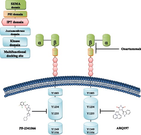
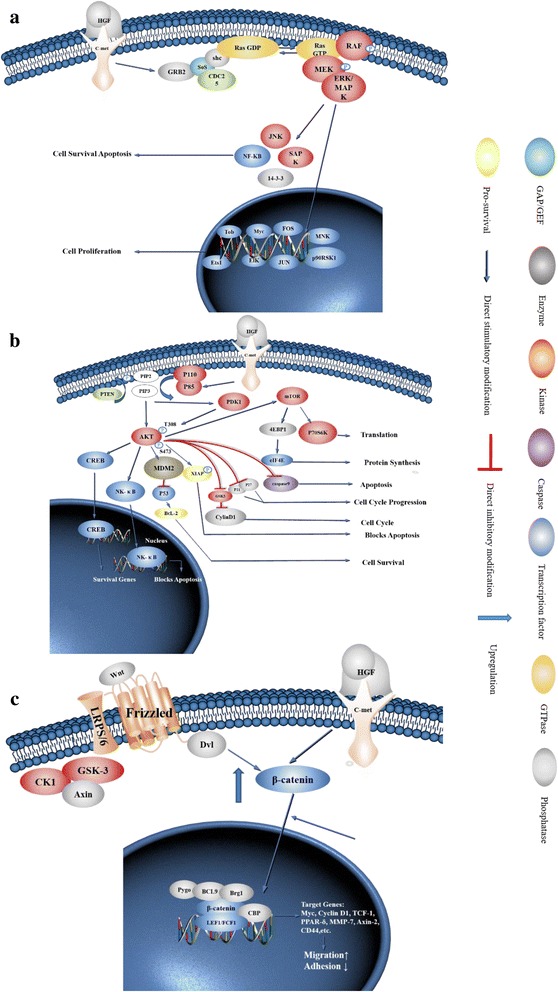
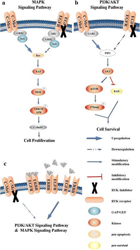
c-Met is a receptor tyrosine kinase belonging to the MET (MNNG HOS transforming gene) family, and is expressed on the surfaces of various cells. Hepatocyte growth factor (HGF) is the ligand for this receptor. The binding of HGF to c-Met initiates a series of intracellular signals that mediate embryogenesis and wound healing in normal cells. However, in cancer cells, aberrant HGF/c-Met axis activation, which is closely related to c-Met gene mutations, overexpression, and amplification, promotes tumor development and progression by stimulating the PI3K/AKT, Ras/MAPK, JAK/STAT, SRC, Wnt/β-catenin, and other signaling pathways. Thus, c-Met and its associated signaling pathways are clinically important therapeutic targets. In this review, we elaborate on the molecular structure of c-Met and HGF and the mechanism through which their interaction activates the PI3K/AKT, Ras/MAPK, and Wnt signaling pathways. We also summarize the connection between c-Met and RON and EGFR, which are also receptor tyrosine kinases. Finally, we introduce the current therapeutic drugs that target c-Met in primary tumors, and their use in clinical research.

Keywords: EGFR; HGF/c-Met; PI3K/AKT; RON; Ras/MAPK; Therapeutic strategy; Wnt.


Conflict of interest statement

Ethics approval and consent to participate

Not applicable.

Consent for publication

Not applicable.

Competing interests

The authors declare that they have no competing interests.

Publisher’s Note

Springer Nature remains neutral with regard to jurisdictional claims in published maps and institutional affiliations.

Figures

Fig. 1
Fig. 1
Structure of c-Met and binding sites for c-Met monoclonal antibody and small molecule inhibitors. c-Met is a heterodimer linked by an extracellular α chain and a transmembrane β chain. The β chain has a SEMA domain, a PSI domain, four IPT domains, a transmembrane domain, a juxtamembrane domain, a tyrosine kinase domain, and a C-terminal tail region. HGF is a heterodimer consisting of an α chain and a β chain linked via a disulfide bond, and forming six domains: the α chain contains a N-terminal hairpin domain and four Kringle domains and the β chain forms a serine protease analog domain lacking catalytic activity. The SEMA domain and the PSI domain in c-Met bind the β chain of HGF. The small molecule inhibitor PF-2341066 binds the TK domain of c-Met at Tyr312A, Lys345A, Pro317A, whereas the small molecule inhibitor ARQ197 forms a complex with the TK domain of c-Met at Pro1158A, Met1160A, Phe1123A, and onartuzumab forms a complex with the Sema-PSI domain of c-Met at Leu43B
Fig. 2
Fig. 2
Illustration of the molecular mechanism of c-Met downstream signaling pathways. a Binding of HGF and c-Met can induce conformational changes in c-Met, resulting in the activation of downstream Ras-Raf-MAPK and PI3K/AKT/mTOR signaling pathways. After autophosphorylation, PTK binds Gab2 and activates it. Gab2 activates SOS; SOS activates Ras and then Ras stimulates Raf, MEK, and MAPKs. Activated MAPKs can enter the nucleus to regulate the expression of transcription factors such as Elk1, Etsl, and c-Myc (among others) to modulate cell proliferation and apoptosis. b The PTK domain is the site of autophosphorylation and also provides a docking site for PI3K. With this interaction, PI3K converts PIP2 to PIP3, and then PIP3 binds to the signaling proteins AKT and PDK1; PDK activates AKT, and activated AKT not only translocates to the nucleus, but also activates GSK-3 and mTOR to regulate the expression of multiple transcription factors. c Wnt binds to the low-density lipoprotein receptor-related protein 5/6/Frizzled (LRP5/LRP6/Frizzled) co-receptor group and activates the Dishevelled protein (DSH/Dvl) resulting in inhibition of the degradation of β-catenin by the destruction complex (consisting of Axin, adenomatosis polyposis coli (APC), protein phosphatase 2A (PP2A), glycogen synthase kinase 3 (GSK3) and casein kinase 1α (CK1α)).Subsequently, β-catenin is transported to the nucleus via Rac1 and other factors and binds to the LEF/TCF transcription factors in the nucleus with BCL9/LGS and Pygo to promote expression of oncogenes such as Myc, Cyclin D1, and MMP-7. This process can promote the invasion and migration of cancer cells. Aberrant activation of HGF/c-Met in tumor cells can block the degradation of β-catenin by the destruction complex, resulting in a higher concentration of β-catenin in the cytoplasm, and can also promote the entry of β-catenin into the nucleus
Fig. 3
Fig. 3
Crosstalk between c-Met and EGFR. a, b The tyrosine kinase receptors EGFR and c-Met can initiate downstream PI3K/Akt signaling resulting in anti-apoptotic processes and Grb2/MAPK activation to promote the proliferation of tumor cells. Therefore, it is speculated that there might be an effect that allows c-Met to bypass the EGFR receptor to activate its downstream pathway, resulting in resistance to EGFR-TKI monotherapy. c c-Met-TKI monotherapy triggers upregulation of the EGFR ligand TGF-α, as well as upregulation of the EGFR protein family receptor ErbB3, which can contribute to one of the most potent dimers that can activate c-Met downstream pathways leading to acquired resistance in cancer cells

References

    1. Salgia R. MET in lung cancer: biomarker selection based on scientific rationale. Mol Cancer Ther. 2017;16:555–565. doi: 10.1158/1535-7163.MCT-16-0472. - DOI - PubMed
    1. Fu YT, Zheng HB, Zhou L, Zhang DQ, Liu XL, Sun H. Valproic acid, targets papillary thyroid cancer through inhibition of c-Met signalling pathway. Am J Transl Res. 2017;9:3138–3147. - PMC - PubMed
    1. Rucki AA, Xiao Q, Muth S, Chen J, Che X, Kleponis J, Sharma R, Anders RA, Jaffee EM, Zheng L. Dual Inhibition of Hedgehog and c-Met Pathways for Pancreatic Cancer Treatment. Mol Cancer Ther. 2017;16:2399-409. - PMC - PubMed
    1. Zhu L, Xiong X, Kim Y, Okada N, Lu F, Zhang H, Sun H. Acid sphingomyelinase is required for cell surface presentation of met receptor tyrosine kinase in cancer cells. J Cell Sci. 2016;129:4238–4251. doi: 10.1242/jcs.191684. - DOI - PMC - PubMed
    1. Liu WT, Jing YY, Yu GF, Chen H, Han ZP, Yu DD, Fan QM, Ye F, Li R, Gao L, et al. Hepatic stellate cell promoted hepatoma cell invasion via the HGF/c-Met signaling pathway regulated by p53. Cell Cycle. 2016;15:886–894. doi: 10.1080/15384101.2016.1152428. - DOI - PMC - PubMed

Publication types

MeSH terms