

Left ventricular structure and diastolic function by cardiac magnetic resonance imaging in hypertrophic cardiomyopathy
- PMID: 29455792
- PMCID: PMC5902823
- DOI: 10.1016/j.ihj.2016.12.021
Left ventricular structure and diastolic function by cardiac magnetic resonance imaging in hypertrophic cardiomyopathy
Abstract
Objective: Diastolic dysfunction is common in hypertrophic cardiomyopathy (HCM) and hypertensive heart disease (HHD), but its relationships with left ventricular (LV) parameters have not been well studied. Our objective was to assess the relationship of various measures of diastolic function, and maximum left ventricular wall thickness (MLVWT) and left ventricular mass index (LVMI) in HCM, HHD and normal controls using cardiac magnetic resonance imaging (CMR). We also assessed LV parameters and diastolic function in relation to late gadolinium enhancement (LGE) and right ventricular (RV) hypertrophy in HCM.
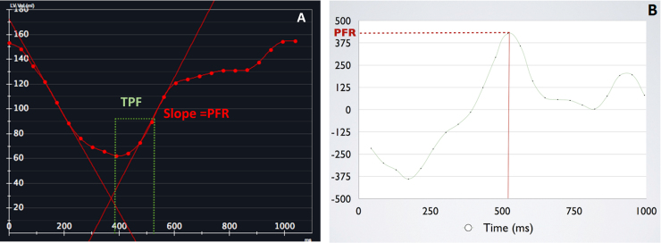
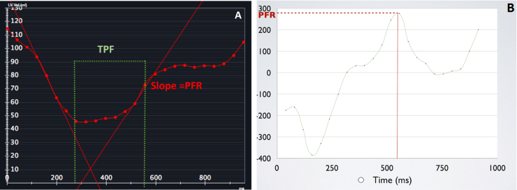
Methods: 41 patients with HCM, 21 patients with HHD and 20 controls were studied. Peak filling rate (PFR), time to peak filling (TPF), MLVWT and LVMI were measured using CMR. LGE and RV morphology were assessed in HCM patients.
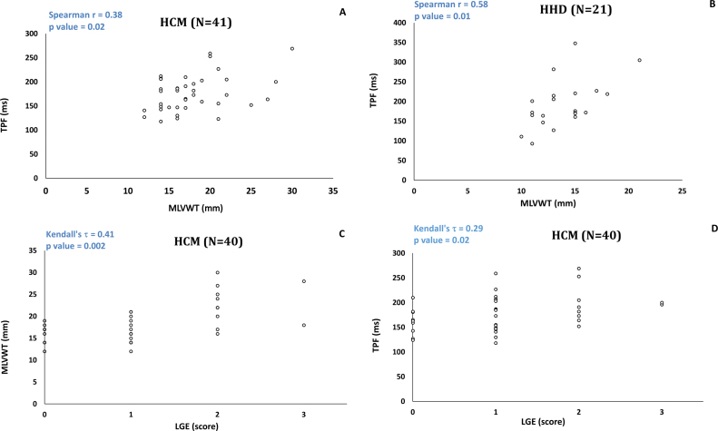
Results: MLVWT correlated with TPF in HCM (r=0.38; p=0.02), HHD (r=0.58; p=0.01) and controls (r=0.54; p=0.01); correlation between MLVWT and TPF was weaker in HCM than HHD. LVMI did not correlate with diastolic function. In HCM, LGE extent correlated with MLVWT (τ=0.41; p=0.002) and with TPF (τ=0.29; p=0.02). The HCM patients with RV hypertrophy had higher MLVWT (p<0.001) and TPF (p=0.03) than patients without RV hypertrophy.
Conclusion: MLVWT correlates with diastolic function (TPF) in HCM, HHD and controls. LVMI did not show significant correlation with TPF. The diastolic dysfunction in HCM is not entirely explained by wall thickening. LGE and RV involvement are associated with worse LV diastolic function, suggesting that these may be markers of more severe underlying myocardial disarray and fibrosis that contribute to diastolic dysfunction.
Keywords: Diastolic function; Hypertension; Hypertrophic cardiomyopathy; Left ventricle; Magnetic resonance imaging.
Copyright © 2016. Published by Elsevier B.V.
Figures
References
-
- Gaasch W.H., Zile M.R. Left ventricular diastolic dysfunction and diastolic heart failure. Annu Rev Med. 2004;55:373–394. - PubMed
-
- Yancy C.W., Jessup M., Bozkurt B. ACCF/AHA guideline for the management of heart failure. J Am Coll Cardiol. 2013;62(16):147–239. - PubMed
-
- Motoyasu M., Kurita T., Onishi K. Correlation between late gadolinium enhancement and diastolic function in hypertrophic cardiomyopathy assessed by magnetic resonance imaging. Circ J. 2008;72:378–383. - PubMed
-
- Małek Ł.A., Chojnowska L., Kłopotowski M. Left ventricular diastolic function assessed with cardiovascular magnetic resonance imaging and exercise capacity in patients with non-obstructive hypertrophic cardiomyopathy. Kardiol Pol. 2009;67:1–6. - PubMed
Publication types
MeSH terms
LinkOut - more resources
Full Text Sources
Other Literature Sources
