Review
. 2018 May;16(5):304-315.
doi: 10.1038/nrmicro.2017.173. Epub 2018 Feb 19.

Hopanoid lipids: from membranes to plant-bacteria interactions

Affiliations
Review

Hopanoid lipids: from membranes to plant-bacteria interactions

Brittany J Belin et al. Nat Rev Microbiol. 2018 May.

Abstract

Lipid research represents a frontier for microbiology, as showcased by hopanoid lipids. Hopanoids, which resemble sterols and are found in the membranes of diverse bacteria, have left an extensive molecular fossil record. They were first discovered by petroleum geologists. Today, hopanoid-producing bacteria remain abundant in various ecosystems, such as the rhizosphere. Recently, great progress has been made in our understanding of hopanoid biosynthesis, facilitated in part by technical advances in lipid identification and quantification. A variety of genetically tractable, hopanoid-producing bacteria have been cultured, and tools to manipulate hopanoid biosynthesis and detect hopanoids are improving. However, we still have much to learn regarding how hopanoid production is regulated, how hopanoids act biophysically and biochemically, and how their production affects bacterial interactions with other organisms, such as plants. The study of hopanoids thus offers rich opportunities for discovery.


Conflict of interest statement

Competing interests

The authors declare no competing interests.

Figures
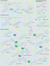
Figure 1 |
Figure 1 |. Pathways of sterol and hopanoid biosynthesis.
Sterols and hopanoids are synthesized from squalene through oxygen-dependent and oxygen-independent mechanisms, respectively. a | To produce the cholesterol precursor lanosterol, squalene is oxygenated by the squalene epoxidase enzyme to form 2,3-oxidosqualene, which is then cyclized by the oxidosqualene cyclase (OSC) family member lanosterol synthase. b | During hopanoid biosynthesis, squalene-hopene cyclase (SHC) enzymes directly cyclize squalene to form one of two C30 hopanoids — diploptene or diplopterol. c | Interestingly, many hopanoid-like compounds made by plants seem to originate from a hybrid cyclization, in which 2,3-oxidosqualene cyclization results in a molecule that contains a hopanoid core with a C3 oxygen signature. d | Bacterial C30 hopanoids, which are produced directly by SHC (as in part b), also can be modified to form C35 hopanoids. The first steps of C35 hopanoid formation are the addition of adenosine by hopanoid biosynthesis-associated radical SAM protein HpnH (step 1) and the removal of adenine by putative hopanoid-associated phosphorylase HpnG (step 2), which generates a ribosylhopanoid with a ribose side chain. This ribose moiety can be readily interconverted between the cyclic and acyclic forms (step 3), from which the signature-extended hopanoid bacteriohopanetetrol (BHT) can be produced by an unknown enzyme (step 4). The side chains of the extended hopanoids can be further altered by the hpn gene products hopanoid biosynthesis-associated glycosyltransferase protein HpnI, hopanoid biosynthesis-associated protein HpnK, hopanoid biosynthesis-associated radical SAM protein HpnJ and aminotransferase HpnO to generate N-acetylglucosaminyl BHT, glucosaminyl BHT, BHT cyclitol ether and aminobacteriohopanetriol (steps 5,6,7 and 8). Hopanoids can also be methylated at the C2 or C3 positions by hopanoid 2-methyltransferase HpnP and hopanoid C3 methylase HpnR, respectively (steps 9 and 10); although only BHT methylation is shown, it can occur for any C35 or C30 hopanoid. For details of triterpenoid stereochemistry, we refer the reader to REF. . Part d is adapted from REF. , CC BY.
Figure 2 |
Figure 2 |. Regulation of lipid raft formation by hopanoids.
a | The main constituents of bacterial membranes are amphiphilic lipids, which contain polar headgroups and hydrophobic tails that preferentially self-associate to form lipid bilayers. All bacteria have a cytoplasmic membrane (CM) that delimits the cytoplasm and is enclosed by a protective and rigid layer of peptidoglycan. In Gram-positive bacteria, a thick layer of peptidoglycan encloses the CM and forms the basis of the cell surface. In Gram-negative bacteria, the CM is also referred to as the inner membrane (IM) and the peptidoglycan is surrounded further by an additional asymmetric bilayer, the outer membrane (OM). The inner leaflet of the OM comprises glycerophospholipids, and the external leaflet mainly comprises lipopolysaccharides (LPSs), which can cover up to 75% of the cell exterior,. LPS structures are highly strain-specific, but they can be roughly divided into three domains: a core oligosaccharide and a O-specific polysaccharide (or O-chain) that together form a hydrophilic heteropolysaccharide, and a lipophilic, membrane-anchoring domain, termed lipid A. The hopanoids shown in the figure are present in some, but not all, Gram-negative and Gram-positive bacteria. b | The distributions of specific lipid classes between, and within, the leaflets of the IM and OM determine their physical and biochemical properties. In addition to containing distinct functional groups, lipids differ in the lengths and degrees of saturation of their hydrophobic tails. These properties tune the effective molecular geometries of lipids and in turn determine their packing within the bilayer. A high proportion of unsaturated and variable-length lipids, which pack poorly in the membrane, is associated with a more fluid, liquid-disordered (Ld) phase. When more planar saturated lipids interact with cholesterol (or some hopanoids), they can self-associate within membranes to form a less fluid, more tightly and regularly packed liquid-ordered (Lo) phase. The interactions leading to the formation of an Lo phase in model membranes are thought to provide the basis for the assembly of raft domains in eukaryotic membranes. The Lo phase is also associated with longer lipid tails, as the increased surface of hydrophobic interactions increases their stability. In Gram-negative bacteria, hopanoids are enriched in the OM where they preferentially interact with lipid A,, and in some organisms, such as Bradyrhizobium sp. BTAi1, they greatly enhance the mechanical integrity of the membrane, as evidenced in transmission electron micrographs. Because hopanoids have a rigid, planar ring structure, it is presumed that they generally are found in Lo microdomains, perhaps regulating the relative position of LPSs, thereby facilitating LPS recognition during host–bacteria interactions,. These membrane microdomains could also affect the localization and dynamics of proteins, restricting their roles in membrane trafficking, signalling and metabolism to functional sub-compartments. The ability of hopanoids to promote lipid rafts has been demonstrated in model membrane vesicles containing eukaryotic phosphatidylcholine (DOPC) and sphingomyelin (SM), in which diploptene addition results in the formation of low-fluidity microdomains. Part b is reproduced from REF. , Macmillan Publishers Limited, and with permission from REF. , Proceedings of the National Academy of Sciences.
Figure 3 |
Figure 3 |. Hopanoid-rich vesicles in Frankia spp.
a | Root nodules induced by Frankia spp. on Alnus incana subsp. rugosa. Sample is ~1.5 cm in diameter. b | Filamentous structure of Frankia symbionts containing spherical nitrogen-fixing vesicles. c | Detail of cultured Frankia vesicles containing nitrogenase (scale bar = 500 nm). d | Multilamellate, hopanoid-rich lipid envelope (arrowheads) of the Frankia nitrogenase vesicles (scale bar = 50 nm) (arrows show the bacterial cell wall). Parts a and b are courtesy of David R. Benson, University of Connecticut, USA, and part c is amended from Journal of Bacteriology, 173, 2061–2067 doi: 10.1128/jb.173.6.2061–2067.1991 (1991) with permission from American Society for Microbiology (REF. 138). Part d is reproduced with permission from REF. , Proceedings of the National Academy of Sciences.
Figure 4 |
Figure 4 |. Chemical structure of hopanoid-lipid A from Bradyrhizobium spp.
a | Hexa-acylated lipid A from Escherichia coli contains a bis-phosphorylated (light blue) glucosamine disaccharide backbone (green), which is asymmetrically substituted by six acyl chains. b | Bradyrhizobium spp. hopanoid-lipid A (HoLA) molecules are a mixture of penta-to-hepta-acylated species, which differ in the number, length and nature of acyl substitutes on the saccharide backbone and are devoid of phosphate groups. Bradyrhizobium spp. lipid A has a 2,3-diaminoglucose (DAG) disaccharide backbone (violet), a galacturonic acid (GalA) residue (yellow) on the vicinal DAG and an α-(1, 6)-mannose disaccharide (orange) on the distal DAG. The acyl chains are asymmetrically distributed on the sugar skeleton; the two secondary very long chain fatty acids (VLCFAs; red), one of which is not stoichiometrically hydroxylated at C2, are subsititued at the ω−1 position by a hopanepolyol acid and by an acetyl (Ac) group (blue), respectively. The primary acyl chains on the E. coli and Bradyrhizobium spp. lipid A backbone have the R configuration at the chiral centre, whereas the VLCFAs have the S configuration at the ω−1 position. The non-stoichiometric 2-hydroxylation of the fatty acids occurs with the S configuration. c | A conceptual scheme illustrating the outer membrane (OM) of Bradyrhizobium spp. is shown. Lipid A (red) insertion decreases the fluidity and increases the mechanical strength of the membrane; VLCFAs entirely span the lipid bilayer so that the covalently linked hopanoid (red) is positioned in the inner leaflet; collectively, this would be expected to order both leaflets, with VLCFAs contributing to tightly link the two. Regular phospholipids are shown in green and free hopanoid molecules are shown in blue.

References

    1. Fahy E, Sud M, Cotter D & Subramaniam S LIPID MAPS online tools for lipid research. Nucleic Acids Res 35, W606–W612 (2007). - PMC - PubMed
    1. Rohmer M, Bouvier P & Ourisson G Molecular evolution of biomembranes: structural equivalents and phylogenetic precursors of sterols. Proc. Natl Acad. Sci. USA 76, 847–851 (1979). - PMC - PubMed
    1. Burlingame AL, Haug P & Belsky T Calvin M Occurrence of biogenic steranes and pentacyclic triterpanes in an Eocene shale (52 millions years) and in an early Pre-Cambrian shale (2.7 billion years): a preliminary report. Proc. Natl Acad. Sci. USA 54, 1406–1412 (1965). - PMC - PubMed
    1. Brocks JJ & Banfield J Biomarker evidence for green and purple sulphur bacteria in a stratified Paleoproterozoic sea. Nature 437, 866–870 (2005). - PubMed
    1. Ourisson G & Albrecht P Geohopanoids: the most abundant natural products on Earth? Acc. Chem. Res 25, 398–402 (1992).

Publication types

LinkOut - more resources