. 2018 Mar;8(3):445-448.
doi: 10.3892/mco.2018.1547. Epub 2018 Jan 10.

Xanthogranulomatous pituitary adenoma: A case report and literature review

Affiliations

Xanthogranulomatous pituitary adenoma: A case report and literature review

Guihong Li et al. Mol Clin Oncol. 2018 Mar.

Abstract

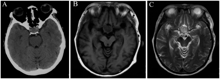
Xanthogranuloma, also referred to as cholesterol granuloma or xanthogranulomatous reaction, is a granulomatous lesion that is infrequently found in the sellar and parasellar regions. Xanthogranulomatous pituitary adenoma is relatively rare and, thus, the etiology, diagnosis, management and prognosis of this condition remain incompletely understood. We herein report the case of a 56-year-old female patient who presented to our institution with intermittent headache, vomiting and distending pain in the bilateral orbital regions. Brain magnetic resonance imaging revealed a sellar mass with a heterogeneous signal. The mass was subtotally resected, and histopathological examination confirmed the diagnosis of xanthogranulomatous pituitary adenoma. Although the patient's symptoms were relieved following surgical treatment, intractable hyponatremia and diabetes insipidus developed and she received hormone replacement therapy. At the last follow-up (November 2016), the patient remained recurrence-free. A total of 14 cases of pituitary adenoma with concomitant xanthogranuloma were identified in the literature, and the clinical and radiological manifestations are discussed. Sellar xanthogranuloma is usually associated with craniopharyngioma or Rathke's cleft cyst; however, it may also occur in isolation. Xanthogranulomatous pituitary adenomas are infrequent, making their diagnosis challenging. Surgical resection is the preferred treatment, and attention should be paid to postoperative hypopituitarism and development of diabetes insipidus.

Keywords: diabetes insipidus; pituitary adenoma; sellar region; xanthogranuloma.


Figures

Figure 1.
Figure 1.
Representative computed tomography (CT) and magnetic resonance imaging (MRI) scans performed preoperatively. (A) Brain CT scan showing a sellar mass with heterogeneous hyperintensity. Brain MRI showing an intra- and suprasellar oval mass involving the hypothalamus and the foramen of Monro. The structures of the pituitary gland and pituitary stalk could not be clearly identified. The mass was (B) heterogeneously isointense on a representative T1WI and (C) heterogeneously hyperintense on a representative T2WI.
Figure 2.
Figure 2.
Representative images of histopathological and immunohistochemical staining of resected tumor tissue. (A) Histopathological examination confirmed the diagnosis of plurihormonal xanthogranulomatous pituitary adenoma, showing pituitary adenoma cells, cholesterol clefts, hemosiderin deposits, macrophages and foam cells (hematoxylin and eosin stain; original magnification, ×100). Immunohistochemical staining showed the tumor was positive for (B) chromogranin A (magnification, ×100) and (C) synaptophysin (magnification, ×100).
Figure 3.
Figure 3.
Postoperative magnetic resonance imaging confirmed partial tumor resection. The residual tumor above the optic chiasma exhibited (A) heterogeneous isointensity on a representative T1-weighted image and (B) heterogeneous hyperintensity on a representative T2-weighted image, with (C) significant contrast enhancement.

Similar articles

Cited by

References

    1. Nishioka H, Shibuya M, Ohtsuka K, Ikeda Y, Haraoka J. Endocrinological and MRI features of pituitary adenomas with marked xanthogranulomatous reaction. Neuroradiology. 2010;52:997–1002. doi: 10.1007/s00234-010-0675-8. - DOI - PubMed
    1. Paulus W, Honegger J, Keyvani K, Fahlbusch R. Xanthogranuloma of the sellar region: A clinicopathological entity different from adamantinomatous craniopharyngioma. Acta Neuropathol. 1999;97:377–382. doi: 10.1007/s004010051001. - DOI - PubMed
    1. Shirataki K, Okada S, Matsumoto S. Histopathological study of the ‘cholesterol granuloma reaction’ in the sellar and juxta-sellar tumors. No To Shinkei. 1988;40:133–139. (In Japanese) - PubMed
    1. Yokoyama S, Goto M, Hirano H, Hirakawa W, Noguchi S, Hirahara K, Kadota K, Asakura T. Pituitary adenoma with cholesterol clefts. Endocr Pathol. 1998;9:91–95. doi: 10.1007/BF02739956. - DOI - PubMed
    1. Miyajima Y, Oka H, Utsuki S, Fujii K. Rathke's cleft cyst with xanthogranulomatous change-case report. Neurol Med Chir. 2011;51:740–742. doi: 10.2176/nmc.51.740. - DOI - PubMed