. 2017:1:977-982.
doi: 10.1038/s41551-017-0168-8. Epub 2017 Dec 12.

A dextran-based probe for the targeted magnetic resonance imaging of tumours expressing prostate-specific membrane antigen

Affiliations

A dextran-based probe for the targeted magnetic resonance imaging of tumours expressing prostate-specific membrane antigen

Guanshu Liu et al. Nat Biomed Eng. 2017.

Abstract

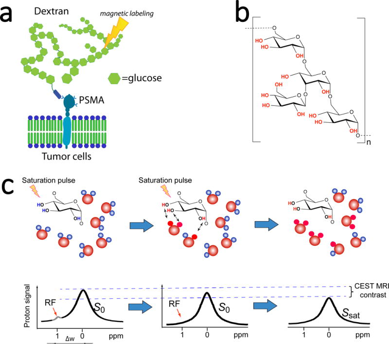
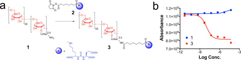
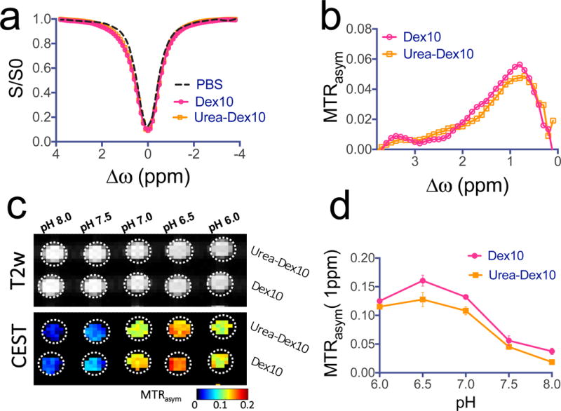
Safe imaging agents able to render the expression and distribution of cancer receptors, enzymes or other biomarkers would facilitate clinical screening of the disease. Here, we show that diamagnetic dextran particles coordinated to a urea-based targeting ligand for prostate-specific membrane antigen (PSMA) enable targeted magnetic resonance imaging (MRI) of the PSMA receptor. In a xenograft model of prostate cancer, micromolar concentrations of the dextran -ligand probe provided sufficient signal to specifically detect PSMA-expressing tumours via chemical exchange saturation transfer MRI. The dextran-based probe could be detected via the contrast originating from dextran hydroxyl protons, thereby avoiding the need of chemical substitution for radioactive or metallic labelling. Because dextrans are currently used clinically, dextran-based contrast agents may help extend receptor-targeted imaging to clinical MRI.


Conflict of interest statement

Competing interests The authors declare no competing financial interests.

Figures

Figure 1
Figure 1. Development of dextran-based contrast agents for PSMA-receptor MR imaging
a) Illustration of PSMA targeting and MRI detection of dextran-based agents that uses only RF-based magnetic labelling (signal saturation) without the need for metallic or radioactive labels. b) The chemical composition of dextran. c) Illustration of CEST MRI detection of natural dextran, which is achieved by the continuous transfer of saturated protons (red) from hydroxyl groups to surrounding water molecules, generating a reduction in the water signal (MRI contrast) proportional to the dextran concentration and rate of exchange.
Figure 2
Figure 2. Synthesis and characterization of PSMA-targeting dextran
a) Synthetic scheme of PSMA targeting Dex10 urea-Dex10 (compound 3); b) PSMA receptor binding abilities of Dex10 (compound 1) and urea-Dex10 (compound 3) from a fluorescence-based assay for the inhibition on the hydrolysis of N-acetylaspartylglutamate (NAAG) to glutamate by the lysates of PSMA-positive human prostate adenocarcinoma (LNCaP) cells for 2 hours. Ki = 2 nM for urea-Dex 10.
Figure 3
Figure 3. CEST MRI detection of dextrans
a) Z-spectra and b) MTRasym plots showing the CEST effects of PSMA-targeted urea-Dex10 and non-targeted Dex10 at 1 mg/mL, pH 7.4 and 37°C. c) T2-weighted and CEST (MTRasym) images of 1 mg/mL urea-Dex10 and 1 mg/mL Dex10 at different pH values. d) Comparison of the pH dependence of the CEST signal at 1 ppm of urea-Dex10 and Dex10 at 1 mg/mL, pH 7.4 and 37°C, indicating the functionalization of Dex10 does not alter CEST effects. All measurements were acquired using a rectangular RF pulse with strength B1 = 4.7 μT and length tsat = 3 seconds. Molar concentrations of 1 mg/mL correspond to approximately 90 and 100 μM of urea-Dex10 and Dex10, respectively. Error bars are the standard deviations of three separate measures.
Figure 4
Figure 4. Changes in the dynamic CEST signal in PSMA(+) and PSMA(−) tumours
a) T2-weighted image and dynamic CEST maps at 1 ppm after the injection of 375 mg/kg urea-10KD-dextran (injection volume =100 μL). b) Mean changes in the CEST signal in PSMA(+) and PSMA(−) tumours in one of the mice for which time dependence was measured. CEST signal enhancement was quantified by ∆MTRasym = MTRasym(t)- MTRasym(t=0), where the error bars are the standard errors of the CEST signal of all the pixels in each tumour. All CEST images were acquired using a 1.8 μT and 3-second-long CW pulse. c) Average CEST signal in the tumour for five mice before (blue) and one hour after (red) the injection of urea-Dex10. The signal difference is shown in black. Error bars are standard deviations of the CEST signal of all five tumours. d) Bar plots showing the mean changes in CEST signal as quantified by ∆MTRasym (1 h) in each type tumour (n = 5 and 3 for urea-Dex10 and non-targeted Dex10 respectively). * : P<0.05 (Student’s t test, two-tailed and unpaired). e) In vivo fluorescence image of a representative mouse showing a distinctive tumour uptake of urea-Dex10 at 60 minutes after injection. f) Sections of PSMA(+) PC3-PIP (top) and PSMA(−) PC3-flu (bottom) tumours stained with anti-PSMA. Images were acquired at 40× magnification. g) Fluorescence microscopy of nuclei (blue, stained with DAPI) dextran (red, NIR-600-labeled). Scale bar= 500 μm for the left three panels and 100 μm for the most right panels, which are the zoomed view of area enclosed in dashed green box in the image on the left. On the right, a scatter plot shows the comparison of the normalized mean fluorescence intensity of three different field of views in the tumours.

Similar articles

Cited by

References

    1. Mahajan A, et al. Bench to bedside molecular functional imaging in translational cancer medicine: to image or to imagine? Clin Radiol. 2015;70:1060–1082. - PubMed
    1. Hajdu I, et al. Cancer cell targeting and imaging with biopolymer-based nanodevices. Int J Pharm. 2013;441:234–241. - PubMed
    1. Cohen B, Dafni H, Meir G, Harmelin A, Neeman M. Ferritin as an endogenous MRI reporter for noninvasive imaging of gene expression in C6 glioma tumors. Neoplasia. 2005;7:109–117. - PMC - PubMed
    1. Artemov D, Mori N, Ravi R, Bhujwalla ZM. Magnetic resonance molecular imaging of the HER-2/neu receptor. Cancer Res. 2003;63:2723–2727. - PubMed
    1. Tse BW, et al. PSMA-targeting iron oxide magnetic nanoparticles enhance MRI of preclinical prostate cancer. Nanomedicine (Lond) 2015;10:375–386. - PubMed