. 2017 Dec:4:78-86.
doi: 10.1016/j.cobme.2017.09.011. Epub 2017 Oct 6.

Functional Near Infrared Spectroscopy: Enabling Routine Functional Brain Imaging

Affiliations

Functional Near Infrared Spectroscopy: Enabling Routine Functional Brain Imaging

Meryem A Yücel et al. Curr Opin Biomed Eng. 2017 Dec.

Abstract

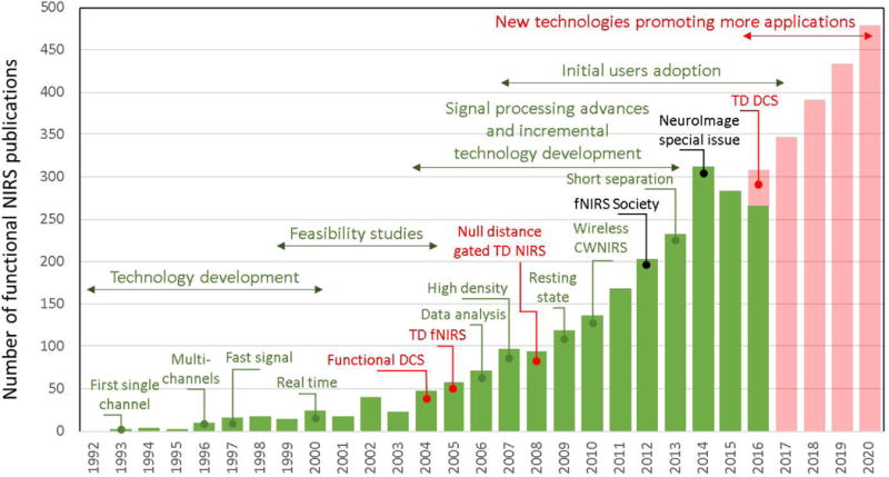
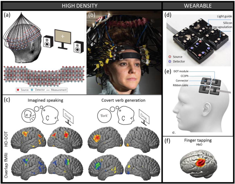
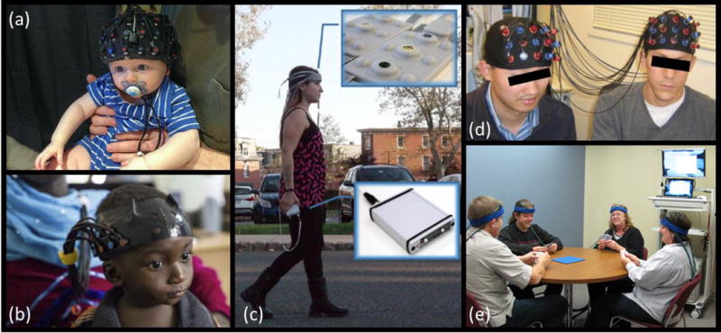
Functional Near-Infrared Spectroscopy (fNIRS) maps human brain function by measuring and imaging local changes in hemoglobin concentrations in the brain that arise from the modulation of cerebral blood flow and oxygen metabolism by neural activity. Since its advent over 20 years ago, researchers have exploited and continuously advanced the ability of near infrared light to penetrate through the scalp and skull in order to non-invasively monitor changes in cerebral hemoglobin concentrations that reflect brain activity. We review recent advances in signal processing and hardware that significantly improve the capabilities of fNIRS by reducing the impact of confounding signals to improve statistical robustness of the brain signals and by enhancing the density, spatial coverage, and wearability of measuring devices respectively. We then summarize the application areas that are experiencing rapid growth as fNIRS begins to enable routine functional brain imaging.


Figures

Figure 1
Figure 1. Volume of fNIRS publications per year
The graph illustrates the exponential growth of the technology applications since its first implementation in 1993 [1][2][3], and highlights what we believe are the major contributions to the field over the past 25 years. The establishment of the fNIRS society and the corresponding NeuroImage special issue [4] mark an increase in publications by users (in black). While the vast majority of current fNIRS applications rely on the continuous-wave (CW) modality, we marked in red some important technological innovations that go beyond this traditional CWNIRS approach: Time-Domain NIRS (TD-NIRS) [10] and Diffuse Correlation spectroscopy (DCS) [11][12]. We believe these new technologies will in turn promote the development of new applications in the future, as we illustrate in our predictions for the growth of the field in red bars.
Figure 2
Figure 2. Probe advances. a-b-c) High-density probe
(a) Schematic and (b) photograph of a high-density probe with 96 sources and 92 detectors yielding over 1200 overlapping measurement channels. (c) The HD-DOT measurements, combined with anatomical light propagation modeling and reconstruction, enable mapping with high resolution on the cortical surface of the hemodynamic response to different cognitive brain tasks. The HD-DOT spatial response (blue) and the independently measured fMRI BOLD response (yellow) show very good spatial overlap (green). Figure panels a, b and c were modified with permission from [13] (d-e-f) Wearable probe. (d) Photograph of a modular wearable device consisting of 4 independent DOT modules each constructed from 30 × 30 mm printed circuit board, 4 photodiodes and 2 dual-wavelength sources. (e) Schematic representation of the wearable device positioned on the scalp over the primary somatomotor cortices (f) Group-average mapping of the hemodynamic response to a finger tapping task using the wearable probe presented above and image reconstruction based on a subject-registered atlas. Figure panels d, e and f were modified with permission from [19].
Figure 3
Figure 3. Applications of fNIRS to various neuroscience studies
(a) Example of fNIRS cap on the head of a 7-month old infant sitting on his parent’s lap. Photograph courtesy of Dr. Katherine Perdue, Boston Children’s Hospital. (b) fNIRS headgear on a 13-month-old infant. Modified with permission from [42], photo credit to the Bill and Melinda Gates Foundation. (c) Battery operated and wireless unit allows untethered outdoor measurement during mobility studies. Modified with permission from [49]. (d) Hyperscanning fNIRS experiment simultaneously measuring brain activity in two people while they play a computer-based cooperation game side by side. Modified with permission from [59]. (e) An example of experimental setup for fNIRS hyperscanning of 4 volunteers playing a card game. Photograph courtesy of Arthur DiMartino, TechEn, Inc.

References

    1. Villringer A, Planck J, Hock C, Schleinkofer L, Dirnagl U. Near infrared spectroscopy (NIRS): A new tool to study hemodynamic changes during activation of brain function in human adults. Neurosci Lett. 1993;154:101–104. - PubMed
    1. Hoshi Y, Tamura M. Detection of dynamic changes in cerebral oxygenation coupled to neuronal function during mental work in man. Neurosci Lett. 1993;150:5–8. - PubMed
    1. Kato T, Kamei A, Takashima S, Ozaki T. Human visual cortical function during photic stimulation monitoring by means of near-infrared spectroscopy. J Cereb Blood Flow Metab. 1993;13:516–520. - PubMed
    1. Boas DA, Elwell CE, Ferrari M, Taga G. Twenty years of functional near-infrared spectroscopy: Introduction for the special issue. Neuroimage. 2014;85:1–5. This paper gives an introduction to a special issue on fNIRS containing 58 papers, providing a broad snapshot of the status of the field. - PubMed
    1. Buxton RB. Interpreting oxygenation-based neuroimaging signals: the importance and the challenge of understanding brain oxygen metabolism. Front Neuroenergetics. 2010;2:8. - PMC - PubMed