. 2020 Jul;11(4):240-247.
doi: 10.1080/21541248.2018.1438024. Epub 2018 Mar 21.

Shedding new light on RhoA signalling as a drug target in vivo using a novel RhoA-FRET biosensor mouse

Affiliations

Shedding new light on RhoA signalling as a drug target in vivo using a novel RhoA-FRET biosensor mouse

Max Nobis et al. Small GTPases. 2020 Jul.

Abstract

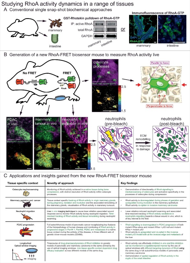
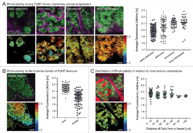
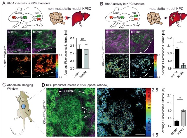
The small GTPase RhoA is a master regulator of signalling in cell-extracellular matrix interactions. RhoA signalling is critical to many cellular processes including migration, mechanotransduction, and is often disrupted in carcinogenesis. Investigating RhoA activity in a native tissue environment is challenging using conventional biochemical methods; we therefore developed a RhoA-FRET biosensor mouse, employing the adaptable nature of intravital imaging to a variety of settings. Mechanotransduction was explored in the context of osteocyte processes embedded in the calvaria responding in a directional manner to compression stress. Further, the migration of neutrophils was examined during in vivo "chemotaxis" in wound response. RhoA activity was tightly regulated during tissue remodelling in mammary gestation, as well as during mammary and pancreatic carcinogenesis. Finally, pharmacological inhibition of RhoA was temporally resolved by the use of optical imaging windows in fully developed pancreatic and mammary tumours in vivo. The RhoA-FRET mouse therefore constitutes a powerful tool to facilitate development of new inhibitors targeting the RhoA signalling axis.

Keywords: FLIM; FRET; Intravital imaging; Rho-GTPases; RhoA; biosensors; breast cancer; extracellular matrix; pancreatic cancer.


Figures

Figure 1.
Figure 1.
Studying RhoA activity dynamics in a range of tissues. (A) Conventional single snap-shot based biochemical approaches to analysing RhoA activity in two examples tissues of the mammary gland and intestine. These included bead-based pulldown of RhoA-GTP in tissue lysate and a recently developed immunofluorescence of fixed tissue samples using a RhoA-GTP specific antibody [15,19]. scale bars, 25 μm (B) With the generation of the new RhoA-FRET biosensor mouse RhoA activity could be monitored live in osteocytes of the calvaria, in vivo in pancreatic ductal adenocarcinomas, mammary tumours and during neutrophil migration (RhoA-FRET biosensor, green; collagen-derived second harmonic generation (SHG) signal, magenta) with corresponding fluorescence lifetime imaging microscopy (FLIM) images of RhoA activity (high RhoA activity: blue to green; low RhoA activity: yellow to red). scale bars, 50 μm (C) A summary of the new insights gained by the use of the new RhoA-FRET biosensor mouse in a variety of tissues and applications. Adapted from Nobis et al. 2017, Cell Reports and adapted from Servier Medial Art, licensed under the Creative Commons Attribution 3.0 Unported license (https://creativecommons.org/licenses/by/3.0/).
Figure 2.
Figure 2.
Spatially defined RhoA acitivty during the progression of PyMT-driven mammary carcinomas. (A) RhoA-FRET mice crossed to MMTV-polyoma-middle-T antigen (PyMT) mice allow for the tracking of RhoA activity during the progression of invasive mammary carcinoma (n = 1 mouse, 280 cells). (B) RhoA activity is increased at the invasive borders of primary PyMT tumours (white dashed line) compared to tumour core regions (n = 1 mouse, 180 cells). (C) Intravenous injeciton of a contrast dye (Qtracker655) allows for monitoring of RhoA activity in cancer cells in relation to their proximity to local vasculature (n = 2 mice, 130 cells). Dots, single cells; line, mean; error bars, SD; scale bars, 50 µm.
Figure 3.
Figure 3.
In vivo imaging of RhoA activity in the pancreas and KPC tumours reveals spatial activation at the invasive border of mutant p53 driven KPC tumours. (A) RhoA is inactive in non-invasive p53-null PDACs both at the tumour center and borders (white dashed line) (n = 2 mice, 163 cells). (B) RhoA acitvity is increased at the invasive border of p53 mutant (p53R172H/+) tumours compared to tumour center regions (n = 2 mice, 77 cells). (C) Schematic of an abdominal imaging window (AIW) to examine RhoA activity in the pancreas and in primary pancreatic tumours. (D) RhoA activity during tumour progression of primary mutant p53 driven PDACs imaged intravitally through optical windows (n = 3 mice, 293 cells). Columns, mean; error bars, SEM; *p < 0.05; **p < 0.01; scale bars, 50 µm.

Similar articles

Cited by

References

    1. Karlsson R, Pedersen ED, Wang Z, et al. Rho GTPase function in tumorigenesis. Biochim Biophys Acta. 2009;1796:91–8. doi:10.1016/j.bbcan.2009.03.003. PMID:19327386. - DOI - PubMed
    1. Hodge RG, Ridley AJ.. Regulating Rho GTPases and their regulators. Nat Rev Mol Cell Biol. 2016;17:496–510. doi:10.1038/nrm.2016.67. PMID:27301673. - DOI - PubMed
    1. Conway JRW, Warren SC, Timpson P. Context-dependent intravital imaging of therapeutic response using intramolecular FRET biosensors. Methods. 2017;128:78–94. doi:10.1016/j.ymeth.2017.04.014. PMID:28435000. - DOI - PubMed
    1. Conway JRW, Carragher NO, Timpson P. Developments in preclinical cancer imaging: innovating the discovery of therapeutics. Nat Rev Cancer. 2014;14:314–28. doi:10.1038/nrc3724. PMID:24739578. - DOI - PubMed
    1. Nobis M, Warren SC, Lucas MC, et al. Moving molecular mobility and activity to an intravital imaging setting: implications for cancer modelling and targeting. J Cell Sci. 2018. doi:10.1242/jcs.206995. - DOI - PubMed

Publication types

Substances

LinkOut - more resources