. 2018 Mar 13;57(10):1640-1651.
doi: 10.1021/acs.biochem.7b01100. Epub 2018 Feb 21.

Selective Recognition of RNA Substrates by ADAR Deaminase Domains

Affiliations

Selective Recognition of RNA Substrates by ADAR Deaminase Domains

Yuru Wang et al. Biochemistry. .

Abstract

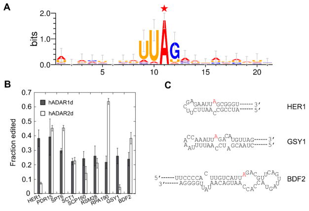
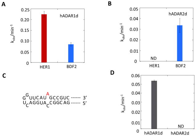
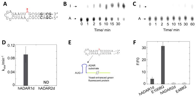
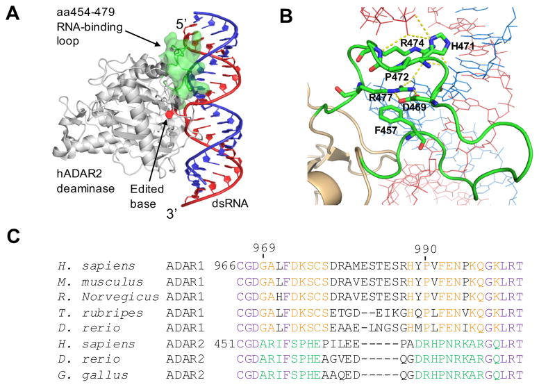
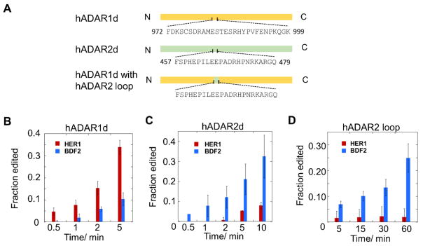
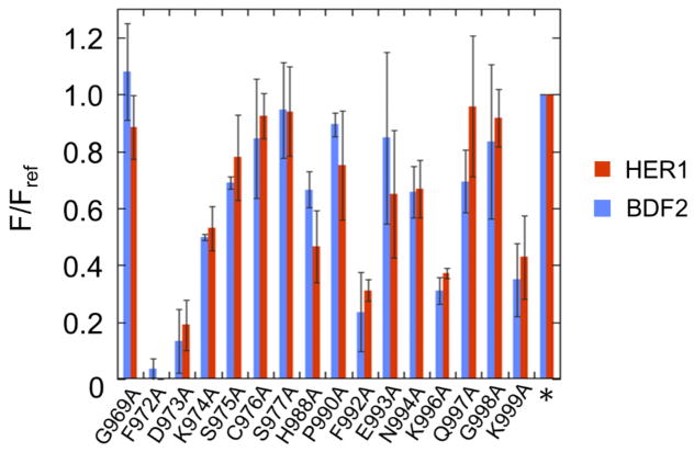
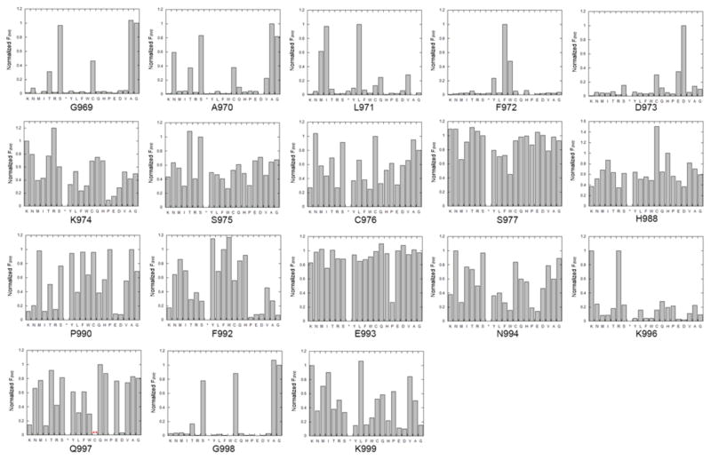
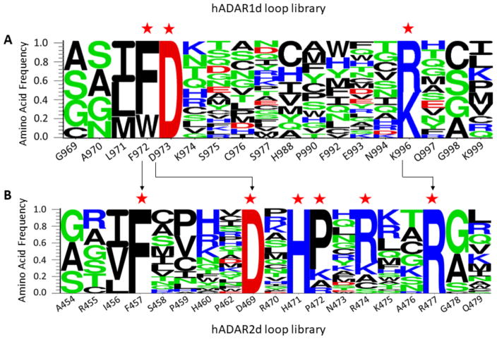
Adenosine deamination is one of the most prevalent post-transcriptional modifications in mRNA and is catalyzed by ADAR1 and ADAR2 in humans. ADAR1 and ADAR2 have different substrate selectivity, which is believed to mainly originate from the proteins' deaminase domains (hADAR1d and hADAR2d, respectively). RNA-seq of the Saccharomyces cerevisiae transcriptome subjected to ADAR-catalyzed RNA editing identified substrates with common secondary structure features preferentially edited by hADAR1d over hADAR2d. The relatively small size and efficient reaction of one of these substrates suggested it could be useful for further study of the hADAR1d reaction. Indeed, a short hairpin stem from the S. cerevisiae HER1 mRNA was efficiently deaminated by hADAR1d and used to generate an hADAR1d-specific fluorescent reporter of editing activity. Using substrates preferred by either hADAR1d or hADAR2d in vitro, we found that a chimeric protein bearing an RNA-binding loop from hADAR2d grafted onto hADAR1d showed ADAR2-like selectivity. Finally, a high-throughput mutagenesis analysis (Sat-FACS-Seq) of conserved residues in an RNA-binding loop of hADAR1d revealed essential amino acids for function, advancing our understanding of RNA recognition by this domain.


Conflict of interest statement

The authors declare no competing financial interest.

Figures

Figure 1
Figure 1
Sequence motif surrounding candidate editing sites identified from RNA-seq and confirmation of editing by Sanger sequencing. A) Nearest neighbor and next nearest neighbor preference for hADAR1d revealed by yeast candidate substrates. The edited A is labeled with a red star. B) Sanger sequencing confirms in vivo editing on yeast candidate sites by hADAR1d and hADAR2d. The experiment was performed in triplicate. Error bar refers to SD, n≥3. C) Secondary structures of HER1 mRNA (top), GSY1 mRNA (middle), and BDF2 mRNA (bottom) surrounding the edited A (colored in red).
Figure 2
Figure 2
In vitro deamination reveals that HER1 RNA and Gabra3 RNA are hADAR1d preferred substrates. A) Deamination on HER1 RNA and BDF2 RNA by hADAR1d, with 35 nM protein and 10 nM RNA. Error bar indicates SD, n≥3. B) Deamination on HER1 RNA and BDF2 RNA by hADAR2d, with 35 nM protein and 10 nM RNA. Error bar indicates SD, n≥3. There are two off-target sites in the BDF2 RNA other than the one under study; however, the editing at the two sites are very low compared to the main site (Figure S2). ND: non-detectable. C) Local secondary structure of Gabra3 substrate predicted by Mfold. D) In vitro deamination on Gabra3 RNA by hADAR1d and hADAR2d with 35 nM protein and 10 nM RNA. Error bar indicates SD, n≥3. ND: non-detectable.
Figure 3
Figure 3
Deamination of hADAR1d and hADAR2d on a 33 nt HER1 substrate and construction of a HER1-based reporter. A) Structure of the 33 nt HER1 substrate predicted by Mfold. Positions where are modified from the native HER1 RNA are bolded. B) and C) Editing of the 33 nt HER1 substrate (10 nM) by 150 nM hADAR1d (B) and 150 nM hADAR2d (C) at different time points. D) Plot of kobs for hADAR1d and hADAR2d editing the 33 nt HER1 substrate. Error bar indicates SD, n≥3. ND: non-detectable. E) Components of the HER1 fluorescent reporter. F) Characterization of the HER1 reporter with different ADAR proteins. F/F0 is the ratio of sample fluorescence divided by negative control (inactive mutant) fluorescence. Error bar indicates SD, n≥3.
Figure 4
Figure 4
The ADAR 5’ binding loop (22). A) Crystal structure of hADAR2 deaminase domain bound to dsRNA substrate (22). The editing site nucleotide is flipped out of the RNA duplex. The 5’ RNA binding loop is highlighted in green. B) Close-up view of interactions between the ADAR2 5’ binding loop and the RNA (21,22). Residues F457 (corresponding to F972 in ADAR1), D469, H471, P472, R474, and R477 are highlighted. C) Sequence alignment around the 5’ binding loops of ADAR1s (A1) and ADAR2s (A2) from different organisms. The conservation patterns are labeled in different colors. Purple: conserved in both ADAR1 and ADAR2; red: conserved in ADAR1; green: conserved in ADAR2; black: not conserved.
Figure 5
Figure 5
A loop swapping experiment shows that the 5’ RNA binding loops contribute to ADAR selectivity. A) Construction of the loop chimera protein. Residues in the 5' binding loop regions are shown. B) hADAR1d E1008Q (2 nM) acting on the HER1 and BDF2 substrates (10 nM). C) hADAR2d E488Q (2 nM) acting on the HER1 and BDF2 substrates (10 nM). D) Loop chimera protein (10 nM) acting on the HER1 and BDF2 substrates (10 nM). Error bar indicates SD, n≥3.
Figure 6
Figure 6
Alanine scan of the ADAR1 5’ binding loop using both HER1 reporter (Figure 3E) and BDF2 reporter (21). The star indicates hADAR1d E1008Q. F/Fref is the ratio of sample fluorescence divided by the parent protein (hADAR1d E1008Q) fluorescence. Error bar indicates SD, n≥3.
Figure 7
Figure 7
Fluorescence activated cell sorting for hADAR1d 5’ binding loop library. A) Top: cells expressing the yeGFP reporter and hADAR1d E912A were used to define background fluorescence and above-background fluorescence categories. Bottom: parameters corresponding to each sorting gate. B) Top: cells expressing the yeGFP reporter and hADAR1d loop library were sorted into different categories based on different levels of fluorescence. Bottom: parameters corresponding to each sorting gate.
Figure 8
Figure 8
Normalized average fluorescence corresponding to each of the twenty common amino acids at 18 positions in the hADAR1 5’ binding loop. The star refers to stop codon. For the Q997 position, no codon sequence for cysteine was identified in either the input library or the active clones.
Figure 9
Figure 9
Logo plots summarizing results of Sat-FACS-Seq analyses of residues present in A) the 5’ binding loop of the ADAR1 deaminase domain and B) the 5’ binding loop of the ADAR2 deaminase domain (21). Positions where the wild type residue is highly preferred are labeled with red stars. Arrows indicate residues that are likely to be functionally equivalent.

Similar articles

Cited by

References

    1. Carlile TM, Rojas-Duran MF, Zinshteyn B, Shin H, Bartoli KM, Gilbert WV. Pseudouridine profiling reveals regulated mRNA pseudouridylation in yeast and human cells. Nature. 2014;515:143–146. - PMC - PubMed
    1. Gilbert WV, Bell TA, Schaening C. Messenger RNA modifications: form, distribution, and function. Science. 2016;352:1048–1412. - PMC - PubMed
    1. Li X, Xiong X, Wang K, Wang L, Shu X, Ma S, Yi C. Transcriptome-wide mapping reveals reversible and dynamic N1-methyladenosine methylome. Nat Chem Biol. 2016;12:311–316. - PubMed
    1. Mauer J, Luo X, Blanjoie A, Jiao X, Grozhik AV, Patil DP, Linder B, Pickering BF, Vasseur JJ, Chen Q, et al. Reversible methylation of m6Am in the 5' cap controls mRNA stability. Nature. 2017;541:371–375. - PMC - PubMed
    1. Zhao BS, Roundtree IA, He C. Post-transcriptional gene regulation by mRNA modifications. Nat Rev Mol Cell Biol. 2017;18:31–42. - PMC - PubMed

Publication types