. 2018 May:303:120-133.
doi: 10.1016/j.expneurol.2018.02.012. Epub 2018 Feb 16.

Modest enhancement of sensory axon regeneration in the sciatic nerve with conditional co-deletion of PTEN and SOCS3 in the dorsal root ganglia of adult mice

Affiliations

Modest enhancement of sensory axon regeneration in the sciatic nerve with conditional co-deletion of PTEN and SOCS3 in the dorsal root ganglia of adult mice

Zachary R Gallaher et al. Exp Neurol. 2018 May.

Abstract

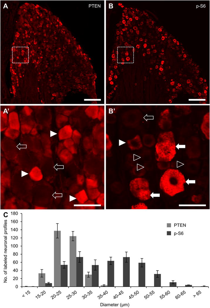
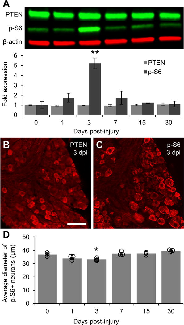
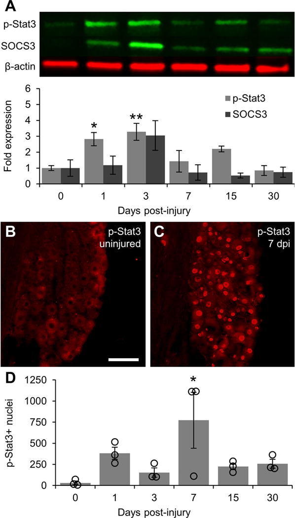
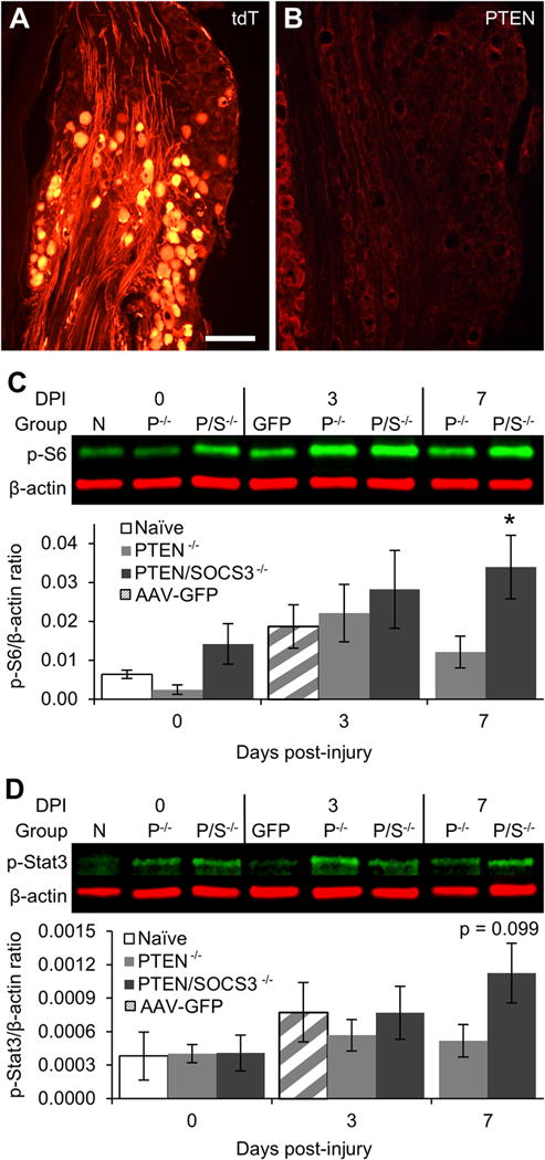
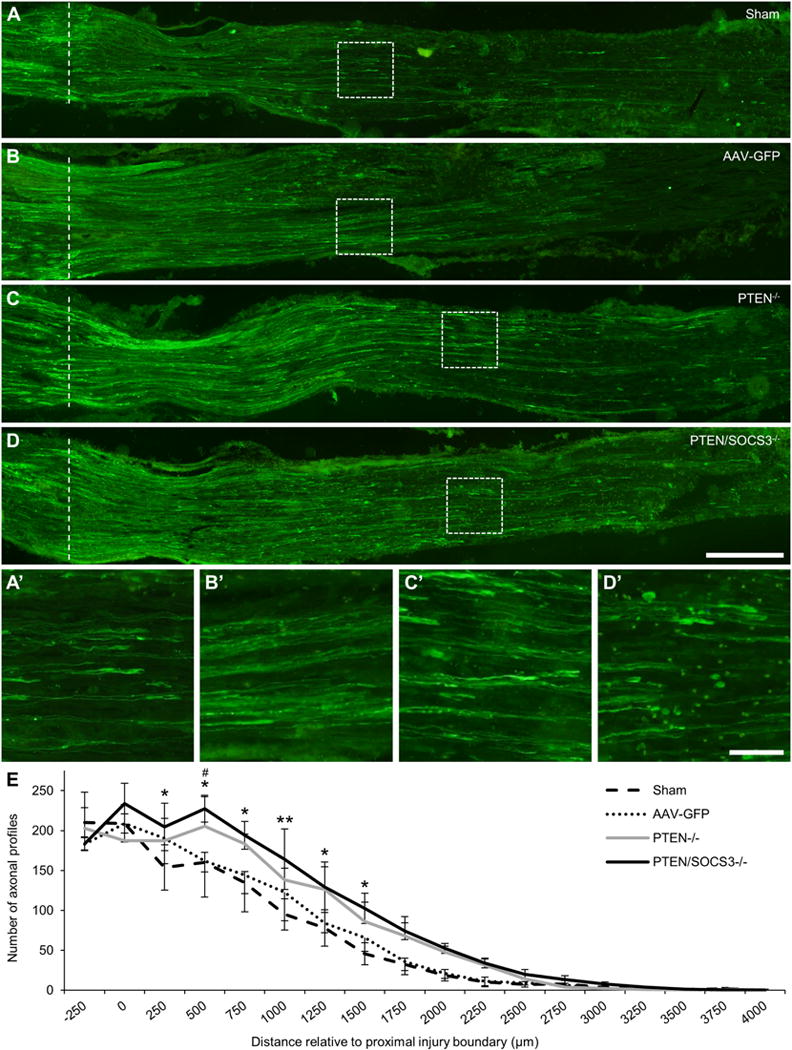
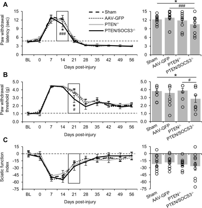
Axons within the peripheral nervous system are capable of regeneration, but full functional recovery is rare. Recent work has shown that conditional deletion of two key signaling inhibitors of the PI3K and Jak/Stat pathways-phosphatase and tensin homolog (PTEN) and suppressor of cytokine signaling-3 (SOCS3), respectively-promotes regeneration of normally non-regenerative central nervous system axons. Moreover, in studies of optic nerve regeneration, co-deletion of both PTEN and SOCS3 has an even greater effect. Here, we test the hypotheses (1) that PTEN deletion enhances axon regeneration following sciatic nerve crush and (2) that PTEN/SOCS3 co-deletion further promotes regeneration. PTENfl/fl and PTEN/SOCS3fl/fl mice received direct injections of AAV-Cre into the fourth and fifth lumbar dorsal root ganglia (DRG) two weeks prior to sciatic nerve crush. Western blot analysis of whole cell lysates from DRG using phospho-specific antibodies revealed that PTEN deletion did not enhance or prolong PI3K signaling following sciatic nerve crush. However, PTEN/SOCS3 co-deletion activated PI3K for at least 7 days post-injury in contrast to controls, where activation peaked at 3 days. Quantification of SCG10-expressing regenerating sensory axons in the sciatic nerve after crush injury revealed longer distance regeneration at 3 days post-injury with both PTEN and PTEN/SOCS3 co-deletion. Additionally, analysis of noxious thermosensation and mechanosensation with PTEN/SOCS3 co-deletion revealed enhanced sensation at 14 and 21 days after crush, respectively, after which all treatment groups reached the same functional plateau. These findings indicate that co-deletion of PTEN and SOCS3 results in modest but measureable enhancement of early regeneration of DRG axons following crush injury.

Keywords: Dorsal root ganglia; Nerve crush; PTEN; Regeneration; Ribosomal protein S6; SOCS3; Sciatic nerve; Stat3.


Figures

Figure 1
Figure 1. Immunohistochemical analysis of PTEN and p-S6 in uninjured DRG
A-B: Representative 20x stitched images of phosphatase and tensin homolog (PTEN, A) and phosphorylated ribosomal protein S6 (p-S6, B) expression in the L4 dorsal root ganglia. Area in dashed box within A is shown in A’ to further demonstrate the localization of PTEN in small diameter neuronal profiles (arrowheads) and a lack of expression in large neuronal shadows (hollow arrows). Area in dashed box within B is shown in B’ to demonstrate localization of p-S6 primarily in large-diameter neuronal profiles (arrows) and not in small diameter profiles (hollow arrowheads). Examples of the less frequent small diameter profiles containing p-S6 (arrowhead) and large diameter profiles with low p-S6 expression (hollow arrow) are also shown. C: Quantification of PTEN and p-S6 neuronal profiles in every 6th section throughout the whole DRG (n = 3). Only profiles containing a nuclear shadow were quantified and ordered in 5 μm bins according to the diameter of the cell body along its long axis. The distribution patterns of these two proteins indicate that they are expressed in two separate neuronal sub-populations, PTEN in small diameter C-type neurons and p-S6 in large diameter A-type neurons. The mutual exclusion of these two proteins is consistent with the role of PTEN as an inhibitor of PI3K signaling upstream of mTOR activation and subsequent S6 phosphorylation. Scale bar = 200 μm in A and B. Scale bar = 50 μm in A’ and B’.
Figure 2
Figure 2. Activation of PI3K signaling following sciatic nerve crush
A: Representative Western blot images of PTEN and p-S6 staining from L4/5 DRG lysates with graphical representation of data normalized to 0 day uninjured controls (n = 3 DRG pools per time point). β-actin was used as a loading control. PI3K signaling is greatly elevated at 3 days post-injury as shown by a five-fold increase in the level of p-S6 as compared to controls. p-S6 levels in DRG lysates returned to baseline within 7 days of injury. B: Representative image of PTEN expression in DRG 3 days post-injury showing that injury does not alter the localization of PTEN in small diameter neurons. C: Representative image of p-S6 in DRG 3 days post-injury showing sustained phosphorylation of S6 in large diameter neurons and increased S6 phosphorylation in smaller diameter neurons. D: Graphical representation of the average diameter of neurons positively stained for p-S6 (n = 3 per time point). Individual data points are indicated by hollow circles. ** p < 0.01 vs 0 day uninjured controls in A. * p < 0.05 vs 0 day uninjured controls and 7, 15, and 30 days post-injury in B. Scale bar = 100 μm.
Figure 3
Figure 3. Activation of Jak/Stat signaling following sciatic nerve crush
A: Representative Western blot images of p-Stat3 and SOCS3 from L4/5 DRG lysates with graphical representation of data normalized to 0 day uninjured controls (n = 3 DRG pools per time point). β-actin was used as a loading control. Jak/Stat signaling is activated at early time points after injury as shown by elevated p-Stat3 levels at 1 and 3 days post-injury. As with PI3K signaling, Jak/Stat signaling returns to baseline within 7 days of injury following an upward trend in SOCS3 expression that does not reach significance. B: Representative image of p-Stat3 expression in 0 day uninjured control DRG showing a lack of p-Stat3-positive nuclei. C: Representative image of p-Stat3 expression 7 days post-injury showing a large number of p-Stat3-positive nuclei in both small and large diameter neurons. D: Graphical representation of the number of p-Stat3-positive nuclei quantified in every 6th section throughout the whole DRG (n = 3 per time point). Individual data points are indicated by hollow circles. * p < 0.05, ** p < 0.01 vs 0 day uninjured controls. Scale bar = 100 μm.
Figure 4
Figure 4. Effect of PTEN and PTEN/SOCS3 co-deletion on activation of PI3K and Jak/Stat signaling
A: Representative image of tdT expression in DRG two weeks after injection of AAV-Cre directly into the L4 DRG of a Cre-reporter mouse. The image shows tdT-positive neurons within the injection center (bottom 2/3 of image) and tdT-negative neurons that were not successfully transfected (top 1/3). Overall, injections resulted in the successful transfection of approximately 35% of neurons within injected DRG. B: Representative image of PTEN expression following direct injection of AAV-Cre into the L4 DRG of a PTENfl/fl mouse showing a lack of PTEN immunoreactivity in neurons within the injection center with sustained PTEN expression in small diameter neurons outside the injection center (far left of image). C: Representative Western blot images of p-S6 staining from L4/5 DRG lysates at 0, 3, or 7 days post-injury (DPI) with graphical representation of data shown as ratio of p-S6 over β-actin loading control. Naïve DRG (N) were both uninjected and uninjured. For PTEN−/− (P−/−) and PTEN/SOCS3−/− (P/S−/−) groups, mice received direct injections of AAV-Cre into the L4/5 DRG two weeks prior to sacrifice (0 day) or sciatic nerve crush (3 and 7 days) to allow for run-down of protein present at the time of injury. Vector control mice (GFP) received AAV-GFP injections two weeks prior to sciatic nerve crush and sacrifice 3 DPI. D: Representative Western blot images of p-Stat3 from L4/5 DRG lysates presented as in C. Intraganglionic injections of both AAV-Cre and AAV-GFP appear to dampen the level of p-S6 and p-Stat3 at 3 DPI, the previously observed peak activation time point. However, PTEN/SOCS3 co-deletion leads to activate signaling in the PI3K pathway as shown by significantly elevated levels of p-S6 to at least 7 days post-injury (C). PTEN/SOCS3 co-deletion also displayed a trend towards increased p-Stat3 levels at this time point (D). * p < 0.05 vs 0 day naïve controls. Scale bar = 200 μm.
Figure 5
Figure 5. Effect of PTEN and PTEN/SOCS3 co-deletion on regeneration of sensory axons in the sciatic nerve following nerve crush
A-D: Representative 20x stitched images of SCG10-expressing regenerating sensory axons in sciatic nerves 3 days after sciatic nerve crush from sham (A), AAV-GFP (B), PTEN−/− (C), and PTEN/SOCS3−/− mice (D). The proximal boundary of the injury site is marked with a dotted line. Areas within dashed boxes are shown in A’-D’ to show regenerating axons and growth cone-like swellings near the primary front of axonal regeneration in each case. E: Graph of axon counts performed in every 6th section at 250 μm intervals relative to the proximal injury boundary (labeled as 0; n = 3 per group). For clarity, SEM is shown in only the up direction for PTEN−/− and the down direction for AAV-GFP. While both PTEN deletion alone and PTEN/SOCS3 co-deletion significantly enhanced total axonal regeneration (p < 0.001 PTEN/SOCS3−/− vs sham and AAV-GFP; p < 0.01 PTEN−/− vs sham; p < 0.05 vs AAV-GFP), PTEN/SOCS3 co-deletion increased the number of regenerating axons at the distances indicated. * p < 0.05, ** p < 0.01 PTEN/SOCS3−/− vs sham, # p < 0.05 PTEN/SOCS3−/− vs AAV-GFP. Scale bar = 250 μm in A-D; scale bar = 50 μm in A’-D’.
Figure 6
Figure 6. Effect of PTEN and PTEN/SOCS3 co-deletion on functional recovery following nerve crush
A-C: Graphical representation of functional recovery of noxious thermosensation (A), mechanosensation (B), and motor function (C). Measurements were taken two weeks prior to DRG injections or sham surgery (baseline, BL), 4 hours prior to crush (0), and at eight weekly time points after crush injury. To highlight the significant differences between the responses of PTEN/SOCS−/− mice and other groups, the lower standard error bars for PTEN−/− in A and for AAV-GFP in B have been omitted. The bar graphs to the right show the data for the boxed time point with individual data points represented by hollow circles. PTEN/SOCS3 co-deletion in DRG neurons accelerated functional recovery as shown by separation of this group from other groups at 14 days for noxious thermosensation and 21 days post-injury for mechanosensation. * p < 0.05 PTEN/SOCS3−/− vs sham, ### p < 0.001 PTEN/SOCS3−/− vs AAV-GFP in A. * p < 0.05 PTEN/SOCS3−/− vs sham, # p < 0.001 PTEN/SOCS3−/− vs PTEN −/− in B.

Similar articles

Cited by

References

    1. Abe N, Borson SH, Gambello MJ, Wang F, Cavalli V. Mammalian Target of Rapamycin (mTOR) Activation Increases Axonal Growth Capacity of Injured Peripheral Nerves. J Biol Chem. 2010;285:28034–28043. doi: 10.1074/jbc.M110.125336. - DOI - PMC - PubMed
    1. Bauer S, Kerr BJ, Patterson PH. The neuropoietic cytokine family in development, plasticity, disease and injury. Nat Rev Neurosci. 2007;8:221–232. doi: 10.1038/nrn2054. - DOI - PubMed
    1. Boyd JG, Gordon T. Neurotrophic factors and their receptors in axonal regeneration and functional recovery after peripheral nerve injury. Mol Neurobiol. 2003;27:277–323. doi: 10.1385/MN:27:3:277. - DOI - PubMed
    1. Burfoot MS, Rogers NC, Watling D, Smith JM, Pons S, Paonessaw G, Pellegrini S, White MF, Kerr IM. Janus Kinase-dependent Activation of Insulin Receptor Substrate 1 in Response to Interleukin-4, Oncostatin M, and the Interferons. J Biol Chem. 1997;272:24183–24190. doi: 10.1074/jbc.272.39.24183. - DOI - PubMed
    1. Byrne AB, Walradt T, Gardner KE, Hubbert A, Reinke V, Hammarlund M. Insulin/IGF1 Signaling Inhibits Age-Dependent Axon Regeneration. Neuron. 2014;81:561–573. doi: 10.1016/j.neuron.2013.11.019. - DOI - PMC - PubMed

Publication types

MeSH terms

Substances