

Isolated unilateral temporalis muscle hypertrophy in a child: a case report with literature review
- PMID: 29458353
- PMCID: PMC5817789
- DOI: 10.1186/s12887-018-1061-7
Isolated unilateral temporalis muscle hypertrophy in a child: a case report with literature review
Abstract
Background: Temporalis muscle hypertrophy is a rare entity of masticatory muscle hypertrophy. All types of masticatory muscle hypertrophies have been documented of which temporalis muscle hypertrophy is one. Temporalis muscle hypertrophy is most commonly bilateral and usually associated with other types of masticatory muscles hypertrophy such as masseter or pterygoid hypertrophy. However, isolated unilateral temporalis muscle hypertrophy is extremely rare and only 9 cases have been reported to date in English literature since 1990 with only two patients less than 18 years. There is no exact etiology identified and the diagnosis is made by muscle biopsy combined with imaging study to exclude other possibilities. Age at presentation is ranges from 15 to 65 years with involvement of both sexes. We report the youngest child who is a seven year old girl with right side isolated unilateral temporalis muscle hypertrophy.
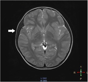
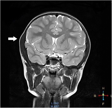
Case presentation: In this patient, we discuss the youngest child with isolated unilateral temporalis muscle hypertrophy and literature review to date. The patient is a seven year old female presenting with painless swelling of the right temporalis muscle. There had no features of inflammation, trauma, neoplasm or history of parafunctions such as bruxism. The child was not complaining significantly headache or visual disturbances as well. She had undergone radiological assessment with ultrasound scan and contrast MRI. The diagnosis was confirmed by muscle biopsy which shows normal muscle architecture. She was managed conservatively with regular follow up.
Conclusion: Isolated unilateral temporalis muscle hypertrophy is extremely rare in children. However this case raises the importance of considering alternative diagnoses despite the condition being rare in the pediatric population.
Keywords: IUTMH in pediatrics; Isolated unilateral temporalis muscle hypertrophy; Sri Lanka.
Conflict of interest statement
Authors’ information
JCR (MBBS, DCH) is a trainee MD pediatrics affiliated with the Teaching Hospital Kandy Sri Lanka. CW (MBBS,DCH,MD,FCCP) is a consultant pediatrician at the Teaching Hospital Kandy Sri Lanka. GR (MBBS, MD) is a consultant radiologist at the Teaching Hospital Kandy Sri Lanka.
Ethics approval and consent to participate
Not applicable
Consent for publication
Written informed consent was obtained from the parents of the patient for publication of this Case report and any accompanying images. A copy of the written consent is available for review by the Editor-In-Chief of this journal.
Competing interests
The authors declare that they have no competing interests.
Publisher’s Note
Springer Nature remains neutral with regard to jurisdictional claims in published maps and institutional affiliations.
Figures
References
-
- Legg JW. Enlargement of the temporal and masseter muscles on both sides. Trans Pathol Soc L. 1880;31:361–366.
-
- Kessel LJ. Benign bilateral masseteric hypertrophy with temporal muscle involvement. Oral Surgery, Oral Med. Oral Pathol. [Internet]. 1970 [cited 2016 Feb 5];30:450–3. Available from: https://www.sciencedirect.com/science/article/pii/0030422070901568 - PubMed
-
- Kalish GH. Hypertrophy of the Masseter or Temporalis Muscles or Both. Arch. Pediatr. Adolesc. Med. [Internet]. American Medical Association; 1971 [cited 2016 Feb 4];121:346. Available from: https://jamanetwork.com/journals/jamapediatrics/article-abstract/503983?... - PubMed
-
- Da Silva K, Mandel L. Bilateral temporalis muscle hypertrophy: a case report. Oral Surg. Oral Med. Oral Pathol. Oral Radiol. Endod. [Internet]. 2006 [cited 2015 Sep 16];102:e1–3. Available from: http://www.oooojournal.net/article/S1079-2104(06)00077-1/fulltext - PubMed
-
- Kim HJ, Yum KW, Lee SS, Heo MS, Seo K. Effects of botulinum toxin type a on bilateral masseteric hypertrophy evaluated with computed tomographic measurement. Dermatologic Surg. 2003;29:484–489. - PubMed
Publication types
MeSH terms
LinkOut - more resources
Full Text Sources
Other Literature Sources
Medical
