Review
. 2018 Feb 20;16(1):4.
doi: 10.1186/s12947-018-0122-2.

Cardiac fluid dynamics meets deformation imaging

Affiliations
Review

Cardiac fluid dynamics meets deformation imaging

Matteo Dal Ferro et al. Cardiovasc Ultrasound. .

Abstract

Cardiac function is about creating and sustaining blood in motion. This is achieved through a proper sequence of myocardial deformation whose final goal is that of creating flow. Deformation imaging provided valuable contributions to understanding cardiac mechanics; more recently, several studies evidenced the existence of an intimate relationship between cardiac function and intra-ventricular fluid dynamics. This paper summarizes the recent advances in cardiac flow evaluations, highlighting its relationship with heart wall mechanics assessed through the newest techniques of deformation imaging and finally providing an opinion of the most promising clinical perspectives of this emerging field. It will be shown how fluid dynamics can integrate volumetric and deformation assessments to provide a further level of knowledge of cardiac mechanics.

Keywords: Cardiac fluid dynamics; Deformation imaging; Hemodynamic forces; Intraventricular pressure gradient; Speckle tracking.


Conflict of interest statement

Ethics approval and consent to participate

The entire study was performed in accordance with the Helsinki declaration; all subjects provided written informed consent (N.O 43/2009, prot 2161). Ethics committee approval was not required.

Consent for publication

Not applicable.

Competing interests

GP and GT receive indirect support from TomTec Gmbh. All other authors declare no competing interest, financial or otherwise.

Publisher’s Note

Springer Nature remains neutral with regard to jurisdictional claims in published maps and institutional affiliations.

Figures

Fig. 1
Fig. 1
The knowledge of endocardial borders in regular B-mode (left side, is an generic exemplary image) along the entire heartbeat can be used for a comprehensive assessment of cardiac mechanics based on volumes, deformation, and flow (right side). Volume curve and EF provide a primary measure of cardiac function; strain represents a second level of information whose clinical value was demonstrated in literature; a flow force provides a further level of knowledge
Fig. 2
Fig. 2
Strain properties in the first population. Curved bands in the GLS-GCS plane represent the regions with constant value of EF. DCM patients present a reduction of EF and of both strain parameters with respect to the Controls. OHCM patients present a conserved or slightly increased EF with respect to controls, thus they tend to displace on the right along the curves of constant EF, which correspond to a reduction of GLS and slight increase of GCS. The OHCM patient indicated with a white dot corresponds to that with highest obstruction
Fig. 3
Fig. 3
Longitudinal flow forces profiles in the first population. DCM patients present a reduction of flow forces (already normalized with volume). OHCM patients present flow forces with comparable entity than Controls, although with a larger variability (notice the scale difference in the third graph)
Fig. 4
Fig. 4
Systolic ejection force (or normalized force impulse) in the first population. DCM patients present a reduction of flow force. Most OHCM patients present OHCM patients present flow forces with comparable entity than Controls with the exceptions of few cases. The OHCM patient indicated with a white dot corresponds to that with highest obstruction
Fig. 5
Fig. 5
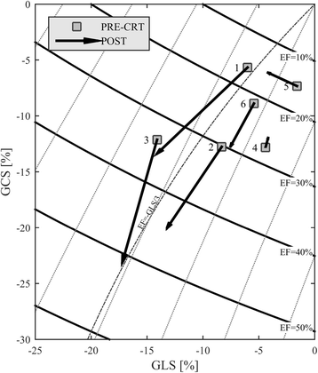
Strain properties in the CRT patients. Curved bands in the GLS-GCS plane represent the regions with constant value of EF. Responder patients (1–3) present an evident improvement in EF and strain values increases accordingly. Non-responder patients (4–6) display minor improvement in EF and strain
Fig. 6
Fig. 6
Polar histogram of the distribution of flow forces during systole. Results are reported PRE- and POST-CRT for a responder patient (#1) and a non-responder (#4), the transversal scale is magnified (2×) to improve visual readability. This representation displays that the therapeutic improvement in alignment of flow force is found in the responder patient only
Fig. 7
Fig. 7
Changes from PRE- and POST-CRT or the ratio between systolic flow force components during systole, measure by root mean square (RMS). Responder patients (1–3) present an evident improvement of force alignment with therapy as indicated by the increase longitudinal-to-transversal ratio. Non-responder patients (4–6) display no or minor improvement in force alignment

References

    1. Kilner PJ, Yang GZ, Wilkes AJ, Mohiaddin RH, Firmin DN, Yacoub MH. Asymmetric redirection of flow through the heart. Nature. 2000;404:759–761. doi: 10.1038/35008075. - DOI - PubMed
    1. Pedrizzetti G, Domenichini F. Nature optimizes the swirling flow in the human left ventricle. Phys Rev Lett. 2005;95:1–4. doi: 10.1103/PhysRevLett.95.108101. - DOI - PubMed
    1. Mangual JO, Kraigher-Krainer E, De Luca A, Toncelli L, Shah A, Solomon S, et al. Comparative numerical study on left ventricular fluid dynamics after dilated cardiomyopathy. J Biomech. 2013;46:1611–1617. doi: 10.1016/j.jbiomech.2013.04.012. - DOI - PubMed
    1. Carlhäll CJ, Bolger A. Passing strange flow in the failing ventricle. Circ Hear Fail. 2010;3:326–331. doi: 10.1161/CIRCHEARTFAILURE.109.911867. - DOI - PubMed
    1. Son J-W, Park W-J, Choi J-H, Houle H, Vannan MA, Hong G-R, et al. Abnormal left ventricular vortex flow patterns in association with left ventricular apical thrombus formation in patients with anterior myocardial infarction. Circ J. 2012;76:2640–2646. doi: 10.1253/circj.CJ-12-0360. - DOI - PubMed