. 2018 Feb 20;37(1):32.
doi: 10.1186/s13046-018-0696-4.

FAK-ERK activation in cell/matrix adhesion induced by the loss of apolipoprotein E stimulates the malignant progression of ovarian cancer

Affiliations

FAK-ERK activation in cell/matrix adhesion induced by the loss of apolipoprotein E stimulates the malignant progression of ovarian cancer

Huiling Lai et al. J Exp Clin Cancer Res. .

Erratum in

Abstract

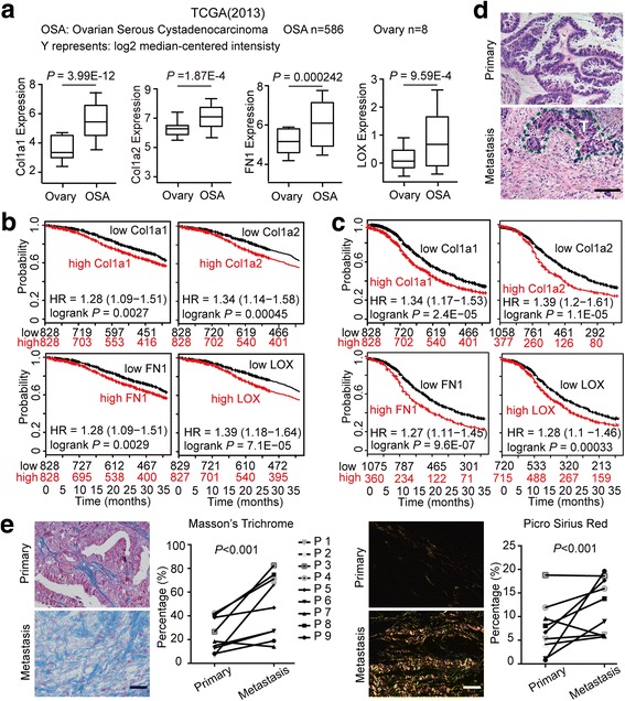
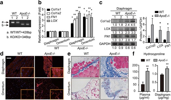
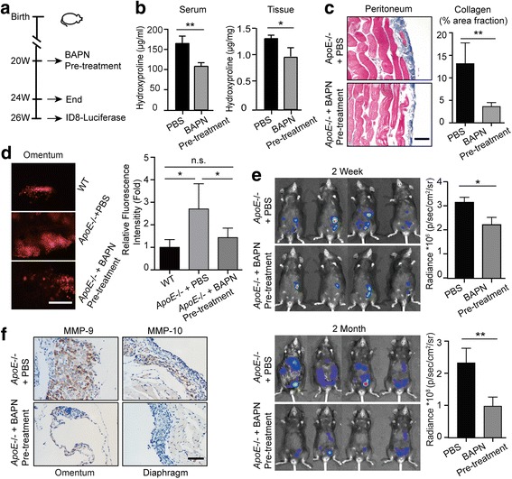
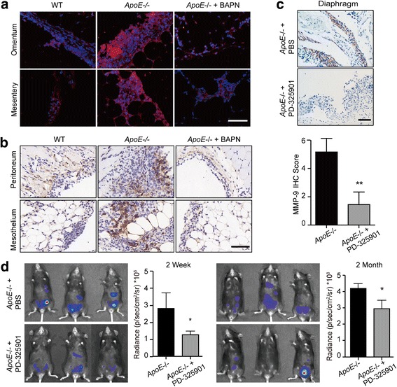
Background: Extracellular matrix (ECM) is a mediator of tumor progression. However, whether the alterations of the intraperitoneal ECM prior to tumor establishment affects the malignant progression of ovarian cancer remains elusive.

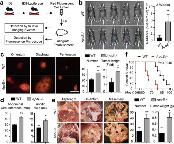
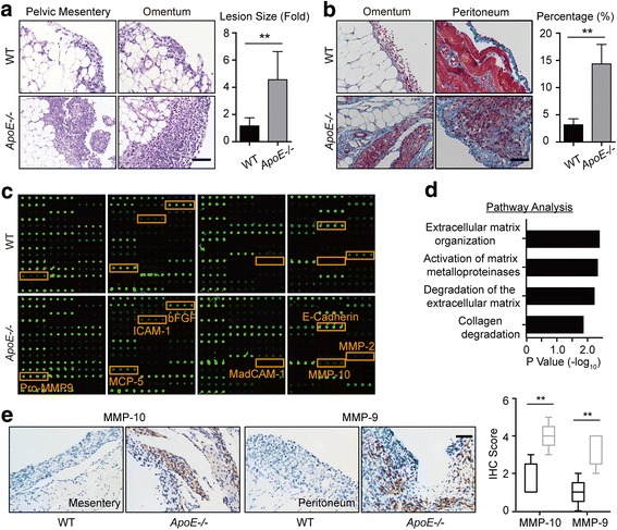
Methods: Apolipoprotein (ApoE) knock-out mice was used to analyze the intraperitoneal ECM alterations by quantification of the major components of ECM. ID8 cells were implanted in vivo to generate allografts and human ovarian cancer cell lines were characterized in vitro to assess the effects of ECM alterations on the malignant progression of ovarian cancer. Adhesion assay, immunochemistry, cytokines profile, proliferation assay, transwell invasion assay and western blot were used to determine the malignant phenotype of ovarian cancer cells.

Results: ApoE loss induced increased ECM deposition, which stimulated the adhesions of ovarian cancer cells. The adhesion-mediated focal adhesion kinase (FAK) signaling enhanced the invasive behaviors of ovarian cancer cells through activation of a ERK-MMP linkage. This ECM-induced signaling cascade was further confirmed in human ovarian cancer cell lines in vitro. Furthermore, reversal of the ECM accumulation with BAPN or abrogation of adhesion-induced ERK activation in ovarian cancer cells with MEK inhibitors (MEKi) was found to effectively delay ovarian cancer progression.

Conclusions: These findings identify the FAK-ERK activation in cell/matrix adhesion in the malignant progression of ovarian cancer and the efficiency of BAPN or MEKi for tumor suppression, providing an impetus for further studies to explore the possibility of new anticancer therapeutic combinations.

Keywords: Adhesion; Apolipoprotein E; Extracellular matrix; Ovarian cancer; Tumor progression.


Conflict of interest statement

Ethics approval and consent to participate

All mouse experiments were carried out in accordance with a protocol approved by the Ethics Committee of Tongji Hospital, Tongji Medical College. Human tissues were donated for research purposes by patients undergoing oophorectomy surgery at Tongji Hospital (Wuhan, China). Ethical approval was granted by the Ethics Committee of Tongji Hospital.

Consent for publication

Not applicable.

Competing interests

The authors have no competing interest to disclose.

Publisher’s Note

Springer Nature remains neutral with regard to jurisdictional claims in published maps and institutional affiliations.

Figures

Fig. 1
Fig. 1
ECM alteration is associated with the tumorigenesis and progression of human ovarian cancer. (a) Col1a1, Col1a2, FN1 and LOX transcript expression levels in normal ovary and ovarian cancer tissues in published TCGA data (Oncomine). Center line represents median values, box limits are the 25th and 75th percentiles, and whiskers represent the minimum and maximum values. (b, c) Kaplan-Meier analyses of the 3-year OS (b) and PFS (c) in ovarian cancer patients according to Col1a1, Col1a2, FN1 and LOX expression levels. These data were dichotomized at the median value into high and low expressing groups. (d) Representative images of H&E staining exhibit the histology of paired primary and metastatic lesions from ovarian cancer patients. The dashed line (green) delimits the tumor tissues. T represents tumor. (e) Representative images and positive-stained percentage of Masson’s Trichrome (left) and Picrosirius Red (right) staining of primary lesions and metastases from ovarian cancer patients. Bar represents 50 μm
Fig. 2
Fig. 2
ApoE loss leads to altered peritoneal ECM composition. (a) Agarose gel electrophoresis of ApoE gene. (b) The mRNA expression of Collagen1, FN1 and LOX in the excised diaphragm and omentum of WT and ApoE−/− mice. (c) Western blot analysis of Col1a2, LOX and FN1 protein levels in the diaphragm. 1 and 2 represent different samples. (d) Frozen sections were immunoassayed for LOX (red) and nuclei were stained with Hoechst (blue). (e) The representative images of Masson’s Trichrome staining in the diaphragm and omentum from WT and ApoE−/− mice. (f) The plasma and diaphragm were analyzed for hydroxyproline content. Error bars represent the SD of triplicate experiments. Bar represents 50 μm. *P < 0.05; **P < 0.005
Fig. 3
Fig. 3
The remodeled peritoneal microenvironment accelerates ovarian cancer progression. (a) Flowchart of the in vivo experiments. (b) Luciferase bioluminescence was determined in WT and ApoE−/− mice two weeks after ID8 allografts engraftment. Graph represents the mean radiance for each group (n = 5). (c) The omentum, peritoneum and diaphragm of WT and ApoE−/− mice were excised and analyzed under a fluorescence microscope (left). The number and weight of the tumor lesions were measured (right). Bar represents 200 μm. (d) The tumor burden was assayed by the abdominal circumference and ascetic fluid content. (e) Representative images (left) and quantification (right) of tumor lesions dispersed in the diaphragm, omentum and mesentery. Dashed circles delimit the tumor tissues. (f) Survival rate of WT and ApoE−/− mice with ID8 allografts. Error bars represent the SD of the experimental data from five mice. *P < 0.05; **P < 0.005
Fig. 4
Fig. 4
Remodeled ECM enhances the invasive behaviors of ovarian cancer cells. (a) H&E stain of the tumor lesions from WT and ApoE−/− mice at two weeks post ID8 engraftment (left). Graph represents the mean size of the lesions calculated from ten random fields (right). (b) Masson’s Trichrome stain of tumor lesions (left). The mean percentage of positive regions in ten random fields was calculated (right). (c) The cytokines/chemokines profile in the supernatants of ascites from WT and ApoE−/− mice. Four groups of mouse cytokine dot-blot arrays are shown. Dot-blots with significant changes are shown in boxed areas (red). (d) The top four pathways enriched among the molecules with significant changes using KEGG pathway analysis. (e) MMP-10 and MMP-9 protein expression in tumor lesions, determined by blinded IHC analysis (left). Box plot of the IHC score of MMP-10 and MMP-9 (right). Box represents the 25th–75th percentile while whiskers indicate the 5th–95th percentile. The black box represents tumor lesions from WT mice and the grey box represents tumor lesions from ApoE−/− mice. Each experiment included data from 5 mice. Bar represents 50 μm. *P < 0.05; **P < 0.005
Fig. 5
Fig. 5
BAPN treatment delays ovarian cancer progression by reducing adhesions. (a) Experimental design: PBS or BAPN was intraperitoneally administrated to 20-weeks-old female ApoE−/− mice each day and continued for four weeks. A cohort of mice was sacrificed for further experiments. For the remaining mice, the drug treatment was stopped for two weeks before the establishment of ID8 allografts. (b) Hydroxyproline was measured in the plasma and diaphragm. (c) Masson’s Trichrome stain after BAPN treatment (left). The positive-staining percentage of 10 random fields was calculated (right). Bar represents 50 μm. (d) Cells adhesive to the omentum were analyzed four hours after ID8 intraperitoneal injection by fluorescence microscopy (left). The adhesive cells were determined from the total fluorescent intensity after digestion (right). Bar represents 200 μm. (e) In vivo luciferase measured at two weeks (top) and two months (bottom) post establishment in ApoE−/− mice with PBS or BAPN pre-treatment. Quantification of luminescence is represented as the radiance. (f) MMP-9 expression measured by IHC in tumor lesions of ApoE−/− mice with PBS or BAPN treatment. Each experiment includes data from 4 mice. Bar represents 50 μm. *P < 0.05; **P < 0.005
Fig. 6
Fig. 6
Remodeled ECM promotes malignancy of ovarian cancer via FAK-ERK-MMP activation. (a) Specimens from Fig. 5D were immunoassayed for p-FAKY397 (red), and nuclei were stained with Hoechst (blue). (b) IHC of active ERK (p-p44/42 MAPKThr202/Tyr204) in tumor lesions from WT and ApoE−/− mice with PBS or BAPN treatment two weeks after ID8 engraftment. (c) PBS or PD-325901 was administrated to ApoE−/− mice once ID8-Luciferase cells were intraperitoneally injected and treatment continued for one month. The representative images (top) and quantification data (bottom) of MMP-9 protein expression in tumor lesions two months after ID8 engraftment. (d) In vivo luciferase expression was determined two weeks or two months post treatment. Luminescence (right panel) is represented as the radiance (p/s/cm2/sr). Each experiment included data from 4 mice. Bar represents 50 μm. *P < 0.05; **P < 0.005

References

    1. Siegel RL, Miller KD, Jemal A. Cancer statistics, 2017. CA Cancer J Clin. 2017;67(1):7–30. doi: 10.3322/caac.21387. - DOI - PubMed
    1. Lengyel E. Ovarian cancer development and metastasis. Am J Pathol. 2010;177(3):1053–1064. doi: 10.2353/ajpath.2010.100105. - DOI - PMC - PubMed
    1. Lu P, Takai K, Weaver VM, Werb Z. Extracellular matrix degradation and remodeling in development and disease. Cold Spring Harb Perspect Biol. 2011;3(12) - PMC - PubMed
    1. Pickup MW, Mouw JK, Weaver VM. The extracellular matrix modulates the hallmarks of cancer. EMBO Rep. 2014;15(12):1243–1253. doi: 10.15252/embr.201439246. - DOI - PMC - PubMed
    1. Cox TR, Erler JT. Remodeling and homeostasis of the extracellular matrix: implications for fibrotic diseases and cancer. Dis Model Mech. 2011;4(2):165–178. doi: 10.1242/dmm.004077. - DOI - PMC - PubMed

MeSH terms

Substances