. 2018 Feb 20;11(1):6.
doi: 10.1186/s13041-018-0349-8.

Down-regulation of ghrelin receptors on dopaminergic neurons in the substantia nigra contributes to Parkinson's disease-like motor dysfunction

Affiliations

Down-regulation of ghrelin receptors on dopaminergic neurons in the substantia nigra contributes to Parkinson's disease-like motor dysfunction

Yukari Suda et al. Mol Brain. .

Abstract

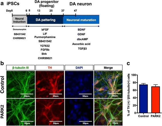
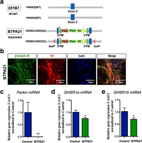
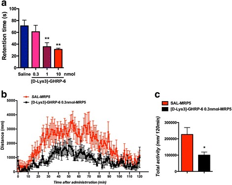
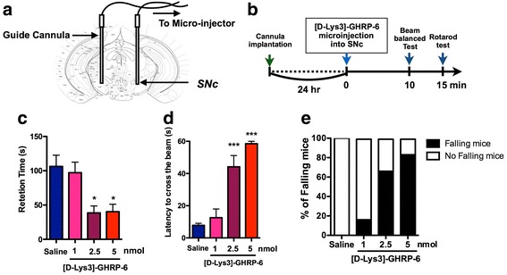
Ghrelin exerts a wide range of physiological actions throughout the body and appears to be a promising target for disease therapy. Endogenous ghrelin receptors (GHSRs) are present in extrahypothalamic sites including the substantia nigra pars compacta (SNc), which is related to phenotypic dysregulation or frank degeneration in Parkinson's disease (PD). Here we found a dramatic decrease in the expression of GHSR in PD-specific induced pluripotent stem cell (iPSC)-derived dopaminergic (DAnergic) neurons generated from patients carrying parkin gene (PARK2) mutations compared to those from healthy controls. Consistently, a significant decrease in the expression of GHSR was found in DAnergic neurons of isogenic PARK2-iPSC lines that mimicked loss of function of the PARK2 gene through CRISPR Cas9 technology. Furthermore, either intracerebroventricular injection or microinjection into the SNc of the selective GHSR1a antagonist [D-Lys3]-GHRP6 in normal mice produced cataleptic behaviors related to dysfunction of motor coordination. These findings suggest that the down-regulation of GHSRs in SNc-DA neurons induced the initial dysfunction of DA neurons, leading to extrapyramidal disorder under PD.

Keywords: Dopamine neuron; GHSR; Ghrelin; Parkinson’s disease; iPS.


Conflict of interest statement

Ethics approval and consent to participate

All of the experimental procedures for cell differentiation and analysis were approved by the respective Ethics Committees of Keio University School of Medicine (Approval Number: 20–16-28) and Hoshi University School of Medicine (Approval Number: 28–008).

Consent for publication

Written informed consent to publish was obtained from the patient and the healthy donor.

Competing interests

The authors declare that they have no competing interests.

Publisher’s Note

Springer Nature remains neutral with regard to jurisdictional claims in published maps and institutional affiliations.

Figures

Fig. 1
Fig. 1
Ghrelin receptor (GHSR) expression in dopaminergic neurons derived from control and PARK2-specific iPSCs. a Schematic of the induction of a DA-enriched culture protocol. b Double-labeling for the dopaminergic neuron marker tyrosine hydroxylase (TH, red) and neurons (βIII-tubulin, green) of control and PARK2-specific iPSC-derived dopaminergic neurons. Scale bar = 50 μm. c Quantitative data of the percentage of TH positive cells per βIII-tubulin positive cells
Fig. 2
Fig. 2
Ghrelin receptor (GHSR) expression in dopaminergic neurons derived from control and PARK2-specific iPSCs. a-b mRNA expression of GHSR1a (a) and GHSR1b (b) between control and Parkinson’s disease-specific iPS cells derived-dopaminergic neurons. **p < 0.01, ***p < 0.001 vs. control iPS cell-derived dopaminergic neurons. c Immunocytochemical analysis for TH (green) and ghrelin receptor (red) in control and PARK2 iPS cell-derived dopaminergic neurons
Fig. 3
Fig. 3
Decreased expression of GHSR in isogenic PARK2-KIKO iPSC-derived DA neurons. a Generation of isogenic PARK2-KIKO iPSCs. Schematic illustration of the gene-editing strategy for knock-in of the stop codon and the puromycin resistance gene into control iPSCs (201B7). b Double-labeling for the DA neuron marker tyrosine hydroxylase (TH, red) and the neuronal marker β-tubulin III (TUJ1, green) of control and PARK2-KIKO iPSC-derived dopaminergic neurons. c Expression level of Parkin mRNA in differentiated DA neurons derived from the control and PARK2KIKO iPSC groups. **p < 0.01 vs. control. d-e The expression levels of GHSR1a (d) and GHSR1b (e) in differentiated dopaminergic neurons derived from the control and PARK2-KIKO iPSC groups. *p < 0.05 vs. control
Fig. 4
Fig. 4
Effects of intracerebroventricular injection of the selective GHSR1a antagonist [D-Lys3]-GHRP-6 on motor coordination. a Average latency to fall in the rotarod test. **p < 0.01 vs. SAL. b Time-course change in the locomotor-enhancing effect of morphine (5 mg/kg, s.c.) after treatment with [D-Lys3]-GHRP-6 at 0.3 nmol (n = 7) or saline (n = 8). Each point represents the mean activity distance for 1 min with SEM. c Total locomotor activity induced by morphine (5 mg/kg, s.c.) after treatment with [D-Lys3]-GHRP-6 at 0.3 nmol (n = 7) or saline (n = 8). Each column represents the mean total activity distance for 120 min with SEM. *p < 0.05 vs. SAL-MRP5
Fig. 5
Fig. 5
Effects of intra-SNc injection of [D-Lys3]-GHRP-6 on motor coordination. a Microinjection sites of [D-Lys3]-GHRP-6 in the SNc. Plates show coronal sections of the mouse brain. b Schedule for the experiment. c Accelerated rotarod test (4-20 rpm). The line graph shows the average latency to fall in the rotarod test for 15 min after bilateral microinjection of [D-Lys3]-GHRP-6 (1, 2.5 or 5 nmol / each site) or saline (n = 6/group) into the SNc. *p < 0.05 vs. Saline. d-e The balance beam test was performed 10 min after the microinjection of [D-Lys3]-GHRP-6 (1, 2.5 or 5 nmol / each site) or saline (n = 6/group) into the SNc. ***p < 0.001 vs. Saline

References

    1. Abbas N, Lücking CB, Ricard S, Dürr A, Bonifati V, De Michele G, Bouley S, Vaughan JR, Gasser T, Marconi R, et al. A wide variety of mutations in the parkin gene are responsible for autosomal recessive parkinsonism in Europe. French Parkinson's Disease Genetics Study Group and the European Consortium on Genetic Susceptibility in Parkinson's Disease. Hum Mol Genet. 1999;8:567–574. doi: 10.1093/hmg/8.4.567. - DOI - PubMed
    1. Kitada T, Asakawa S, Hattori N, Matsumine H, Yamamura Y, Minoshima S, Yokochi M, Mizuno Y, Shimizu N. Mutations in the parkin gene cause autosomal recessive juvenile parkinsonism. Nature. 1998;392:605–608. doi: 10.1038/33416. - DOI - PubMed
    1. Dawson TM. Parkin and defective ubiquitination in Parkinson's disease. J Neural Transm Suppl. 2006:209–13. - PubMed
    1. Sulzer D. Multiple hit hypotheses for dopamine neuron loss in Parkinson's disease. Trends Neurosci. 2007;30:244–250. doi: 10.1016/j.tins.2007.03.009. - DOI - PubMed
    1. Tanaka K, Suzuki T, Hattori N, Mizuno Y. Ubiquitin, proteasome and parkin. Biochim Biophys Acta. 2004;1695:235–247. doi: 10.1016/j.bbamcr.2004.09.026. - DOI - PubMed

Publication types

MeSH terms