. 2018 Mar;67(3):364-375.
doi: 10.1099/jmm.0.000677. Epub 2018 Jan 11.

Denture-associated biofilm infection in three-dimensional oral mucosal tissue models

Affiliations

Denture-associated biofilm infection in three-dimensional oral mucosal tissue models

Daniel J Morse et al. J Med Microbiol. 2018 Mar.

Abstract

Purpose: In vitro analyses of virulence, pathogenicity and associated host cell responses are important components in the study of biofilm infections. The Candida-related infection, denture-associated oral candidosis, affects up to 60 % of denture wearers and manifests as inflammation of palatal tissues contacting the denture-fitting surface. Commercially available three-dimensional tissue models can be used to study infection, but their use is limited for many academic research institutions, primarily because of the substantial purchase costs. The aim of this study was to develop and evaluate the use of in vitro tissue models to assess infections by biofilms on acrylic surfaces through tissue damage and Candida albicans virulence gene expression.

Methodology: In vitro models were compared against commercially available tissue equivalents (keratinocyte-only, SkinEthic; full-thickness, MatTek Corporation). An in vitro keratinocyte-only tissue was produced using a cancer-derived cell line, TR146, and a full-thickness model incorporating primary fibroblasts and immortalised normal oral keratinocytes was also generated. The in vitro full-thickness tissues incorporated keratinocytes and fibroblasts, and have potential for future further development and analysis.

Results: Following polymicrobial infection with biofilms on acrylic surfaces, both in-house developed models were shown to provide equivalent results to the SkinEthic and MatTek models in terms of tissue damage: a significant (P<0.05) increase in LDH activity for mixed species biofilms compared to uninfected control, and no significant difference (P>0.05) in the expression of most C. albicans virulence genes when comparing tissue models of the same type.

Conclusion: Our results confirm the feasibility and suitability of using these alternative in vitro tissue models for such analyses.

Keywords: biofilm; candidosis; denture stomatitis; infection; oral mucosa; tissue model.


Conflict of interest statement

The authors declare that there are no conflicts of interest.

Figures
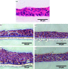
Fig. 1.
Fig. 1.
Representative light microscopy images of the in vitro keratinocyte-only tissue model, sectioned at 5 µm thickness. (a) SkinEthic reconstituted human oral epithelia (RHOE) tissue; (b) in vitro keratinocyte-only tissue seeded with 5×10⁵ cells cultured for 5 days; (c) in vitro keratinocyte-only tissue seeded with 5×10⁵ cells cultured for 10 days; (d) in vitro keratinocyte-only tissue seeded with 1×10⁶ cells cultured for 5 days; (e) in vitro keratinocyte-only tissue seeded with 1×10⁶ cells cultured for 10 days. Stained with haematoxylin and eosin. The scale bar represents 50 µm.
Fig. 2.
Fig. 2.
Light microscopy image showing a very thin epithelium when culturing is performed with medium covering the cells (e.g. not at the air–liquid interface). Stained with haematoxylin and eosin. The scale bar represents 50 µm.
Fig. 3.
Fig. 3.
(a) In vitro full-thickness mucosal tissue using primary oral cells. Clear distinct layers of fibroblast-populated lamina propria (LP) in collagen, with good stratification and differentiation of the epithelia (SE). (b) In vitro full-thickness mucosal tissue using primary fibroblasts, with TR146 cancer-derived keratinocytes. Note the substantial lack of cellular maturation and organisation, and the reduced epithelial stratification compared with tissues developed using immortalised normal oral keratinocytes. Stained with haematoxylin and eosin. The scale bar represents 50 µm.
Fig. 4.
Fig. 4.
Biofilm-induced tissue damage as measured by the lactate dehydrogenase activity assay of infections. The results are expressed as fold change, normalised against an acrylic-only (no biofilm) control. (a) SkinEthic RHOE, (b) in vitro keratinocyte-only tissue, (c) MatTek EpiOral full-thickness tissue and (d) in vitro full-thickness tissue. All of the tissues show a similar pattern of damage; the largest increase in tissue damage is induced by mixed-species biofilms, compared to single-species and uninfected control acrylic coupons.
Fig. 5.
Fig. 5.
Expression of putative Candida albicans virulence genes of mixed-species biofilms post-tissue infection. The results are expressed as the fold change of samples relative to the housekeeping gene ACT1 against a normalised C. albicans-only biofilm control. No significant differences in C albicans gene expression were observed between tissue models of the same type (e.g. SkinEthic RHOE versus in vitro keratinocyte only, or MatTek EpiOral full-thickness versus in vitro full-thickness tissues) with the exception of full-thickness tissues for the ALS1 gene.
Fig. 6.
Fig. 6.
Typical light microscopy images of (a) full-thickness tissue after infection with acrylic coupons only (no biofilm), showing no damage to epithelium; (b) full-thickness tissue after infection with bacteria-only biofilm, showing slight tissue damage and clustering of biofilms on the epithelial surface (indicated by arrows); (c) full-thickness tissue after infection with Candida albicans-only biofilm, showing substantial epithelial damage and biofilms clustered on the surface of the epithelium (indicated by arrows); (d) full-thickness tissue after infection with mixed-species biofilm, showing extensive epithelial damage and microbial invasion through the epithelium (indicated by arrows). Stained with haematoxylin and eosin. The scale bar represents 50 µm.
Fig. 7.
Fig. 7.
Typical light microscopy images of (a) MatTek full-thickness tissue after infection with acrylic coupons only (no biofilm), showing no damage to epithelium; (b) MatTek full-thickness tissue after infection with bacteria-only biofilm, showing very slight tissue damage and clustering of biofilms on the epithelial surface (indicated by arrows); (c) MatTek full-thickness tissue after infection with Candida albicans-only biofilm, showing slight epithelial damage and biofilms clustered on the surface of the epithelium (indicated by arrows); (d) MatTek full-thickness tissue after infection with mixed-species biofilm, showing slight epithelial damage and biofilms clustered on the surface of the epithelium (indicated by arrows). Stained with haematoxylin and eosin. The scale bar represents 50 µm.
Fig. 8.
Fig. 8.
Representative light microscope image of the mixed-species biofilm infection of MatTek full-thickness tissues, demonstrating tissue damage caused by invading Candida albicans and bacteria as a result of biofilm infection. Stained with haematoxylin and eosin. The scale bar represents 50 µm.

References

    1. Flemming HC, Wingender J. The biofilm matrix. Nat Rev Microbiol. 2010;8:623–633. doi: 10.1038/nrmicro2415. - DOI - PubMed
    1. Costerton JW, Stewart PS, Greenberg EP. Bacterial biofilms: a common cause of persistent infections. Science. 1999;284:1318–1322. doi: 10.1126/science.284.5418.1318. - DOI - PubMed
    1. Sutherland IW. The biofilm matrix—an immobilized but dynamic microbial environment. Trends Microbiol. 2001;9:222–227. doi: 10.1016/S0966-842X(01)02012-1. - DOI - PubMed
    1. Costerton JW, Cheng KJ, Geesey GG, Ladd TI, Nickel JC, et al. Bacterial biofilms in nature and disease. Annu Rev Microbiol. 1987;41:435–464. doi: 10.1146/annurev.mi.41.100187.002251. - DOI - PubMed
    1. Hall-Stoodley L, Stoodley P. Developmental regulation of microbial biofilms. Curr Opin Biotechnol. 2002;13:228–233. doi: 10.1016/S0958-1669(02)00318-X. - DOI - PubMed

Substances

LinkOut - more resources