Review
. 2018:111:1-59.
doi: 10.1016/bs.apcsb.2017.08.003. Epub 2017 Oct 6.

Homo- and Heterodimerization of Proteins in Cell Signaling: Inhibition and Drug Design

Affiliations
Review

Homo- and Heterodimerization of Proteins in Cell Signaling: Inhibition and Drug Design

Sitanshu S Singh et al. Adv Protein Chem Struct Biol. 2018.

Abstract

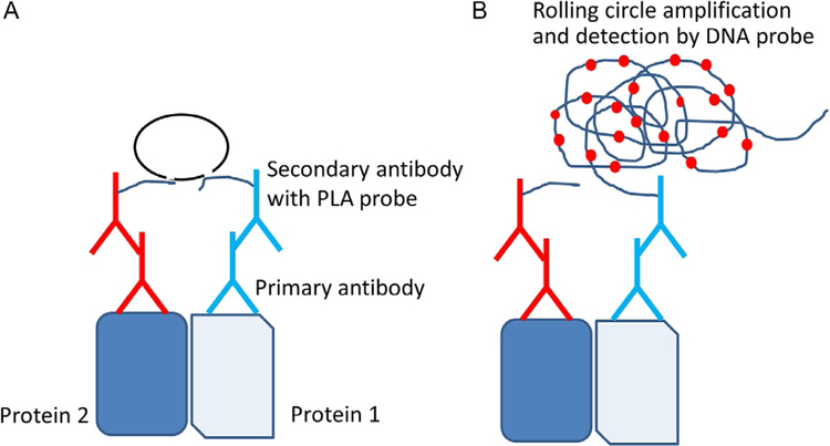
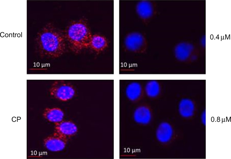
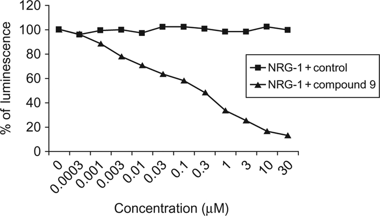
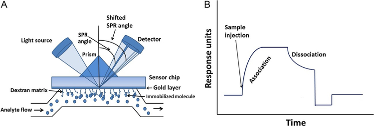
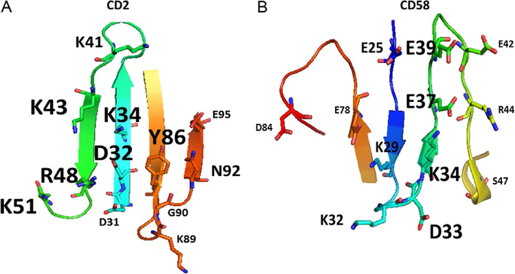
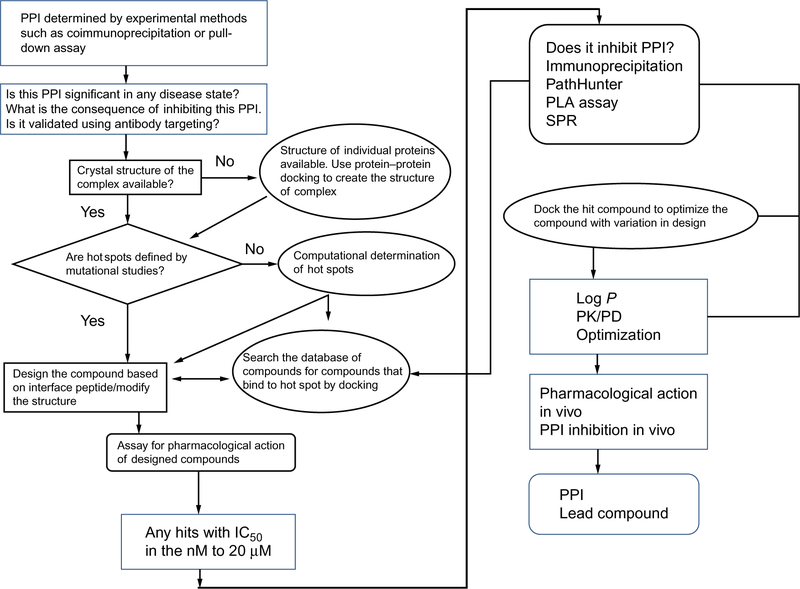
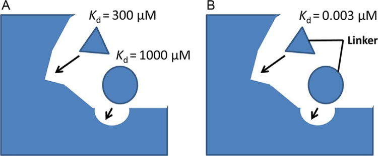
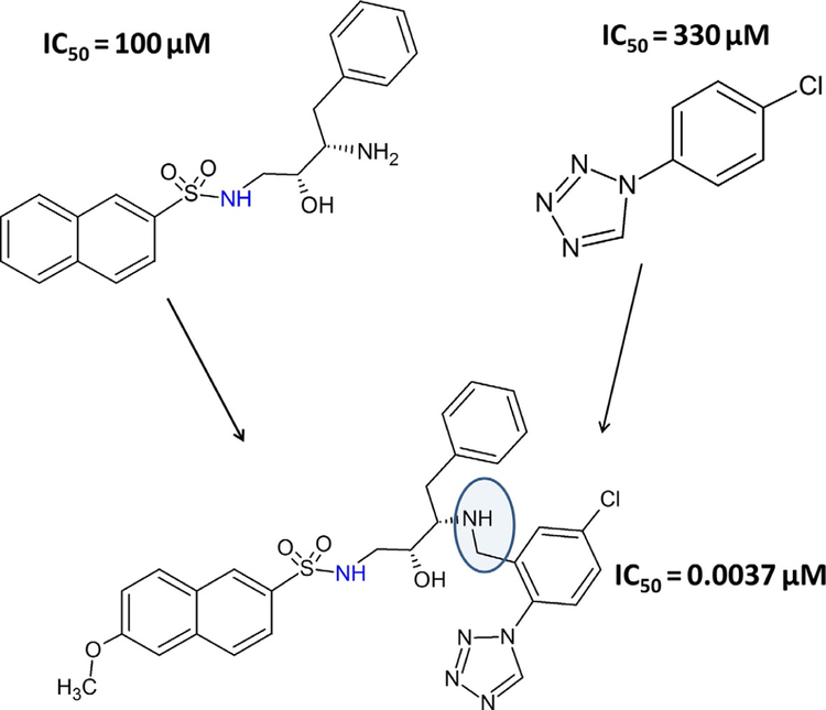
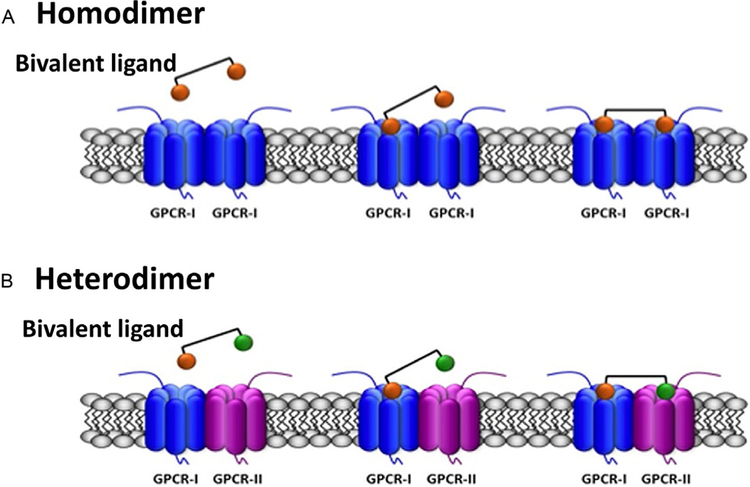
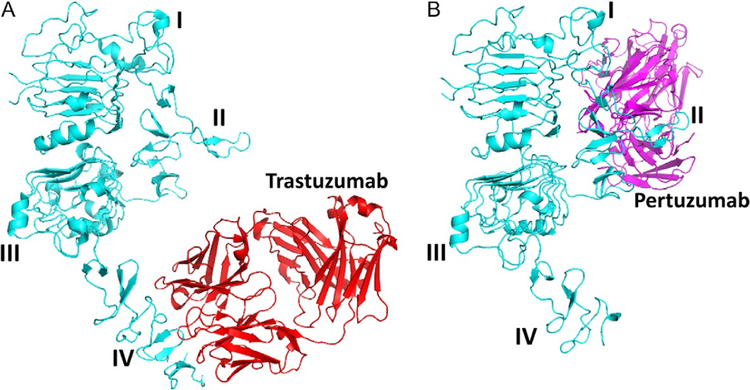
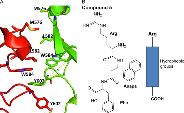
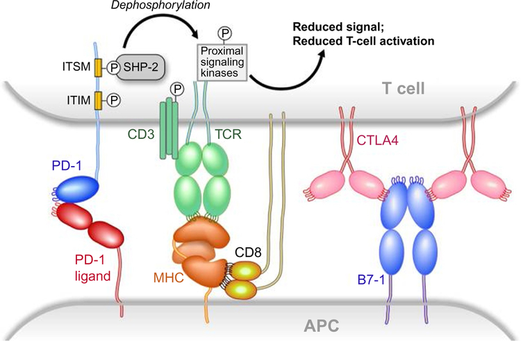
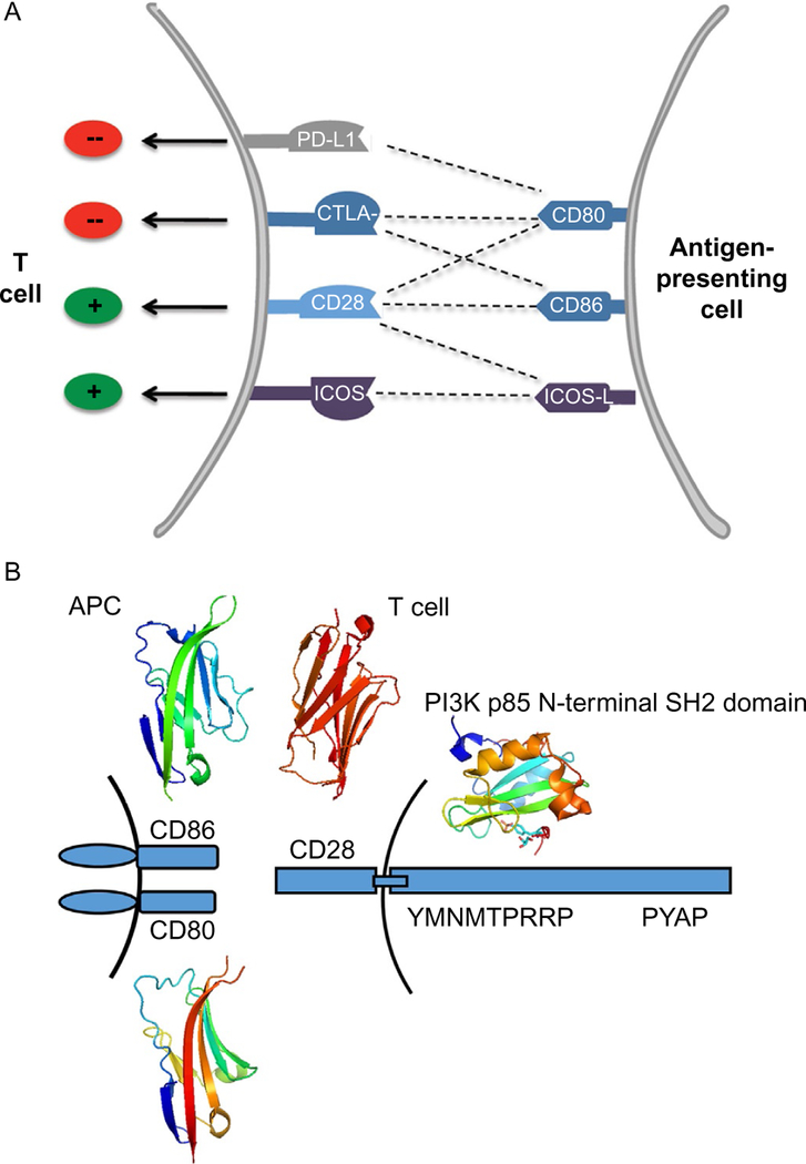
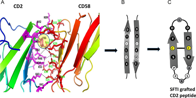
Protein dimerization controls many physiological processes in the body. Proteins form homo-, hetero-, or oligomerization in the cellular environment to regulate the cellular processes. Any deregulation of these processes may result in a disease state. Protein-protein interactions (PPIs) can be inhibited by antibodies, small molecules, or peptides, and inhibition of PPI has therapeutic value. PPI drug discovery research has steadily increased in the last decade, and a few PPI inhibitors have already reached the pharmaceutical market. Several PPI inhibitors are in clinical trials. With advancements in structural and molecular biology methods, several methods are now available to study protein homo- and heterodimerization and their inhibition by drug-like molecules. Recently developed methods to study PPI such as proximity ligation assay and enzyme-fragment complementation assay that detect the PPI in the cellular environment are described with examples. At present, the methods used to design PPI inhibitors can be classified into three major groups: (1) structure-based drug design, (2) high-throughput screening, and (3) fragment-based drug design. In this chapter, we have described some of the experimental methods to study PPIs and their inhibition. Examples of homo- and heterodimers of proteins, their structural and functional aspects, and some of the inhibitors that have clinical importance are discussed. The design of PPI inhibitors of epidermal growth factor receptor heterodimers and CD2-CD58 is discussed in detail.

Keywords: CD2–CD58; EGFR; Heterodimerization; PD-1–PD-L1; PPI inhibition; Protein–protein interaction.


Figures

Fig. 1
Fig. 1
The principle of proximity ligation assay (PLA). (A) Two proteins of interest are targeted by primary antibody from different species. Corresponding secondary antibodies with DNA probes are added. If the two proteins are in proximity the hybridized DNA will be used for rolling circle amplification. (B) Amplified DNA will be detected by a DNA probe. For visualization, a DNA probe with red fluorescence is used. Each dimer of protein in cells is viewed as a red dot with a high-resolution microscope. Adapted from Trifilieff, P., Rives, M. L., Urizar, E., Piskorowski, R. A., Vishwasrao, H. D., Castrillon, J., et al. (2011). Detection of antigen interactions ex vivo by proximity ligation assay: Endogenous dopamine D2-adenosine A2A receptor complexes in the striatum. BioTechniques, 51(2), 111–118. https://doi.org/10.2144/000113719.
Fig. 2
Fig. 2
HER2:HER3 heterodimerization and its inhibition by compound 9 observed by PLA. Concentration-dependent inhibition of HER2:HER3 heterodimers was observed. Control-SKBR-3 cells showing only HER2:HER3 heterodimerization as red spots. CP-A control compound did not inhibit HER2:HER3 heterodimerization. At a sub-optimum dose of compound 9 (0.4μM), HER2:HER3 heterodimerization was inhibited to a lesser extent. At an optimum dose of compound 9 (0.8 μM), HER2:HER3 heterodimerization was significantly inhibited. Reproduced with permission from Kanthala, S., Banappagari, S., Gokhale, A., Liu, Y. Y., Xin, G., Zhao, Y., & Jois, S. (2015). Novel peptidomimetics for inhibition of HER2:HER3 heterodimerization in HER2-positive breast cancer. Chemical Biology & Drug Design, 85, 702–714. https://doi.org/10.1111/cbdd.12453. Copyright (2014) John Wiley and Sons.
Fig. 3
Fig. 3
Inhibition of heterodimerization of HER2:HER3 in HER2, HER3 transfected U2OS cells by compound 9 at different concentrations using enzyme fragment complementation assay (DiscoveRx). Dose–response curve for inhibition of HER2:HER3 heterodimerization by compound 9 in the presence of 0.3μM NRG1 (triangles). Control compound (CP) in the presence of 0.3μM NRG-1 (filled squares). Reproduced with permission from Kanthala, S., Banappagari, S., Gokhale, A., Liu, Y. Y., Xin, G., Zhao, Y., & Jois, S.(2015). Novel peptidomimetics for inhibition of HER2:HER3 heterodimerization in HER2-positive breast cancer. Chemical Biology & Drug Design, 85, 702–714. https://doi.org/10.1111/cbdd.12453. Copyright (2014) John Wiley and Sons.
Fig. 4
Fig. 4
The principle of SPR analysis. (A) SPR chip with analyte flow and SPR angle. (B) SPR sensorgram indicating association and dissociation phases.
Fig. 5
Fig. 5
The structure of PPI domains of (A) CD2 and (B) CD58 with amino acids that form hydrogen bonding, salt bridges, and hydrophobic interaction (PDB ID 1QA9). Mutation studies suggested that the affinity of interaction between the two proteins is reduced when alanine is replaced by some of the residues in the proteins (Kim et al., 2001; Wang et al., 1999). Residues that have the most effect on affinity of binding are represented by large-sized letters for amino acid labels. Residues that partially affect the affinity are represented with medium-sized letters. Residues that do not affect the affinity by replacement with alanine are represented with small-sized letters.
Fig. 6
Fig. 6
A schematic diagram of a decision tree for the design of PPI inhibitors. Adapted from Sable, R., & Jois, S. (2015). Surfing the protein-protein interaction surface using docking methods: Application to the design of PPI inhibitors. Molecules, 20(6), 11569–11603. https://doi.org/10.3390/molecules200611569. MDPI.
Fig. 7
Fig. 7
A schematic representation of the fragment-based drug design principle.(A) Fragments are screened from a database or are built based on PPI hot spots on the protein. Each fragment has a relatively weak dissociation constant. (B) The screened fragments are linked chemically to obtain a high-affinity binding ligand.
Fig. 8
Fig. 8
Example of fragment-based approach. Two molecules with IC50 values of 100 and 330μM, respectively, were linked to obtain a high-affinity ligand that has an IC50 value of 37nM. Adapted from Erlanson, D. A. (2006). Fragment-based lead discovery:A chemical update. Current Opinion in Biotechnology, 17(6), 643–652. https://doi.org/10.1016/j.copbio.2006.10.007. Copyright (2006) Elsevier.
Fig. 9
Fig. 9
Scheme of the simultaneous binding of bivalent ligands with linkers of appropriate length to two receptors in a GPCR dimer. The agonist/antagonist moieties of the bivalent ligand are selective for their respective receptors (orange for GPCR-I; green for GPCR-II) and are linked by a spacer. (A) Binding to two equal GPCRs forming a homo-dimer. (B) Binding to two different GPCRs forming a heterodimer. Reproduced with permission from Franco, R., Martinez-Pinilla, E., Lanciego, J. L., & Navarro, G. (2016). Basic pharmacological and structural evidence for class A g-protein-coupled receptor heteromerization. Frontiers in Pharmacology, 7, 76. https://doi.org/10.3389/fphar.2016.00076. Copyright (2016).
Fig. 10
Fig. 10
Schematic representation of different domains of APP that form homodimers. Arrows indicate the proposed dimerization regions. CAPP, central APP domain; CuBD, copper-binding region; GFLD, growth factor-like domain; ICD, intracellular domain; JM, juxtamembrane region; KPI, Kunitz protease inhibitor domain; TM, transmembrane region. Schematic diagram is drawn based on Khalifa, N. B., Van Hees, J., Tasiaux, B., Huysseune, S., Smith, S. O., Constantinescu, S. N., et al. (2010). What is the role of amyloid precursor protein dimerization? Cell Adhesion & Migration, 4(2), 268–272; Eggert, S., Midthune, B., Cottrell, B., & Koo, E. H. (2009). Induced dimerization of the amyloid precursor protein leads to decreased amyloid-beta protein production. The Journal of Biological Chemistry, 284(42), 28943–28952. https://doi.org/10.1074/jbc.M109.038646.
Fig. 11
Fig. 11
EGFR extracellular domain (ECD). (A) Homodimer (PDB ID 1NJP). Domains II and IV participate in dimerization. (B) and (C) Expanded regions of PPI of domains II and IV. Domain II region has a β-turn structure in the PPI region, forming a hydrophobic core at the interaction site. Molecules that can mimic this region are designed to inhibit PPI.Domain IV also has hydrophobic interactions that can be used to target inhibitors.(D) and (E) EGFR open (PDB ID 1NJP) and closed conformations (1NQL). Domains I–IV are labeled. In the closed conformation, domains II and IV interact, blocking the dimerization arm of domain II and forming a dimer. Notice that in open conformation, the domain IV is moved away from domain II, allowing domain II to interact with other EGFR molecules. Figure was generated using PyMol software (Schrodinger LLC, OR).
Fig. 12
Fig. 12
Hot spots at the interface of PPI in the IL-6/GP130 D1 domain. Modeling of binding hot spots at the IL-6/GP130 D1 domain interface. (A) D1 domain is represented as electrostatic potential surface (red, negatively charged; blue, positively charged; white, hydrophobic). IL-6 is in ribbon representation. The two larger yellow eclipses indicate the two main binding “hot spots,” Leu57-binding site and Trp157-binding site, between IL-6 and GP130. (B) Binding modeling of MDL-A to the GP130 D1 domain. D1 domain is in ribbon representation, and MDL-A is in thick ball-and-stick rendering. Hydrogen bonds are shown as red dotted lines. MDL-A disrupts both binding spots of the GP130 D1 domain. MDL-A forms three hydrogen bonds with Asn92, Cys6, and the carbonyl backbone of Val93 residues of GP130. The modeling indicates that the long butyl tail of MDL-A displaces Leu57 (thin red line), and the indoline moiety partially disrupts Trp157 (thin red line) of the helix D of IL-6. Reproduced with permission from Li, H., Xiao, H., Lin, L., Jou, D., Kumari, V., Lin, J., & Li, C. (2014). Drug design targeting protein-protein interactions (PPIs) using multiple ligand simultaneous docking (MLSD) and drug repositioning: Discovery of raloxifene and bazedoxifene as novel inhibitors of IL-6/GP130 interface. Journal of Medicinal Chemistry, 57(3), 632–641. https://doi.org/10.1021/jm401144z. Copyright (2014) American Chemical Society.
Fig. 13
Fig. 13
Binding of (A) trastuzumab to domain IV of ECD of HER2 protein (PDB ID 1N8Z).(B) Binding of pertuzumab (PDB ID 1S78) to domain II of ECD of HER2 protein. PyMol was used to generate the figures.
Fig. 14
Fig. 14
(A) PPI region of trastuzumab-HER2 domain IV complex (PDB ID 1N8Z). Hydro-phobic region of trastuzumab formed by amino acid residues Tyr52, Pro95,96, Trp99, and Tyr33 interacts with Phe573 and P572 of HER2 protein. On either side of the hydro-phobic region, there is electrostatic interaction formed by Glu558 of HER2 with Arg59 of trastuzumab and from K593 of HER2 to Gly103. (B) This interaction can be used to design a template structure for a PPI inhibitor. Figure was generated using PyMol (Schrodinger LLC, OR).
Fig. 15
Fig. 15
The structure of the designed template compound 5 and a peptidomimetic (B) along with the PPI site (A) of the EGFR homodimer of domain IV (PDB ID 1N8Z and 3NJP). Note that the PPI site is dominated by hydrophobic residues. Peptidomimetic can bind to this PPI site. PyMol (Schrodinger LLC, OR) was used to generate the structure of the protein complexes.
Fig. 16
Fig. 16
Structures of the B7/CD28 family. Structures are modeled on the crystal determinations. Loops have been added to one end of the IgV domains to emphasize the orientation of the CDR-like loops and their interaction with ligand or lack thereof. Reproduced with permission from Freeman, G. J. (2008). Structures of PD-1 with its ligands: Sideways and dancing cheek to cheek. Proceedings of the National Academy of Sciences of the United States of America, 105(30), 10275–10276. https://doi.org/10.1073/pnas.0805459105. Copyright (2008) National Academy of Sciences, USA.
Fig. 17
Fig. 17
(A) Complexities of CD28 costimulatory pathways. The CD28 costimulatory receptor can be ligated by CD80, CD86, and ICOS-L (B7–H2). The CTLA-4 coinhibitor competes with CD28 for binding to CD80 and CD86. However, CD80 can also bind to PD-L1 (B7–H1) and deliver a coinhibitory signal. ICOS competes with CD28 for binding to B7–H2. (B) Schematic representation of PPI interactions of CD28 with CD80/CD86. CD28 cytoplasmic domain motifs YMNM and PYAP that are important in signaling are indicated. Crystal structures of domains of CD28 (1YJD), CD80 (4RWH), CD86 (1NCN), and p53 (5GJI) domain important in binding to CD28 signaling are shown. Panel (A): Reproduced with permission from Ford, M. L., Adams, A. B., & Pearson, T. C. (2014). Targeting co-stimulatory pathways: Transplantation and autoimmunity. Nature Reviews Nephrology, 10(1), 14–24. https://doi.org/10.1038/nrneph.2013.183. Copyright (2013) Nature Publishing Group.
Fig. 18
Fig. 18
(A) PPI interface of CD2–CD58 showing electrostatic and hydrophobic interactions. There are 10 salt bridges, 5 hydrogen bonds, and 1 hydrophobic interaction (PDB ID 1QA9). The design of peptides and multicyclic grafted peptide from CD2 adhesion domain based on the interface residues of CD2 of CD2–CD58 interactions. (B) F and C strands from the CD2 protein that interact with CD58. (C) Grafting of CD2 F and C strand residues onto sunflower trypsin inhibitor peptide to generate stable peptides that inhibit PPI of CD2–CD58 for immunomodulation (Sable et al., 2016 ACS Chemical Biology).

References

    1. Acuner Ozbabacan SE, Engin HB, Gursoy A, & Keskin O (2011). Transient protein-protein interactions. Protein Engineering, Design & Selection, 24(9), 635–648. 10.1093/protein/gzr025. - DOI - PubMed
    1. Adams PD, Seeholzer S, & Ohh M (2002). Identification of associated proteins by coimmunoprecipitation In Protein–protein interactions: A molecular cloning manual (pp. 59–74). New York: Cold Spring Harbor Laboratory Press, Cold Spring Harbor.
    1. Aglan M, Amr K, Ismail S, Ashour A, Otaify GA, Mehrez MA, et al. (2015). Clinical and molecular characterization of seven Egyptian families with autosomal recessive robinow syndrome: Identification of four novel ROR2 gene mutations. American Journal of Medical Genetics. Part A, 167A(12), 3054–3061. 10.1002/ajmg.a.37287. - DOI - PubMed
    1. Arkin MR, Tang Y, & Wells JA (2014). Small-molecule inhibitors of protein-protein interactions: Progressing toward the reality. Chemistry & Biology, 21(9), 1102–1114. 10.1016/j.chembiol.2014.09.001. - DOI - PMC - PubMed
    1. Bakail M, & Ochsenbein F (2016). Targeting protein–protein interactions, a wide open field for drug design. Comptes Rendus Chimie, 19(1), 19–27.