. 2018 May:105:63-72.
doi: 10.1016/j.cyto.2018.02.002. Epub 2018 Feb 17.

Melanoma exosomes promote mixed M1 and M2 macrophage polarization

Affiliations

Melanoma exosomes promote mixed M1 and M2 macrophage polarization

Gina T Bardi et al. Cytokine. 2018 May.

Abstract

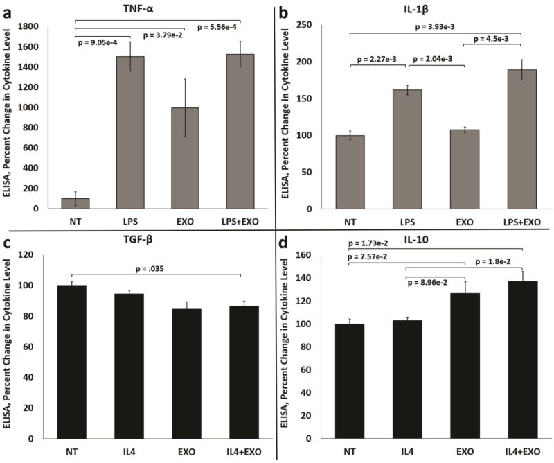
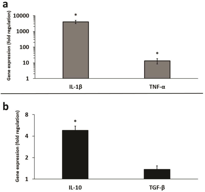
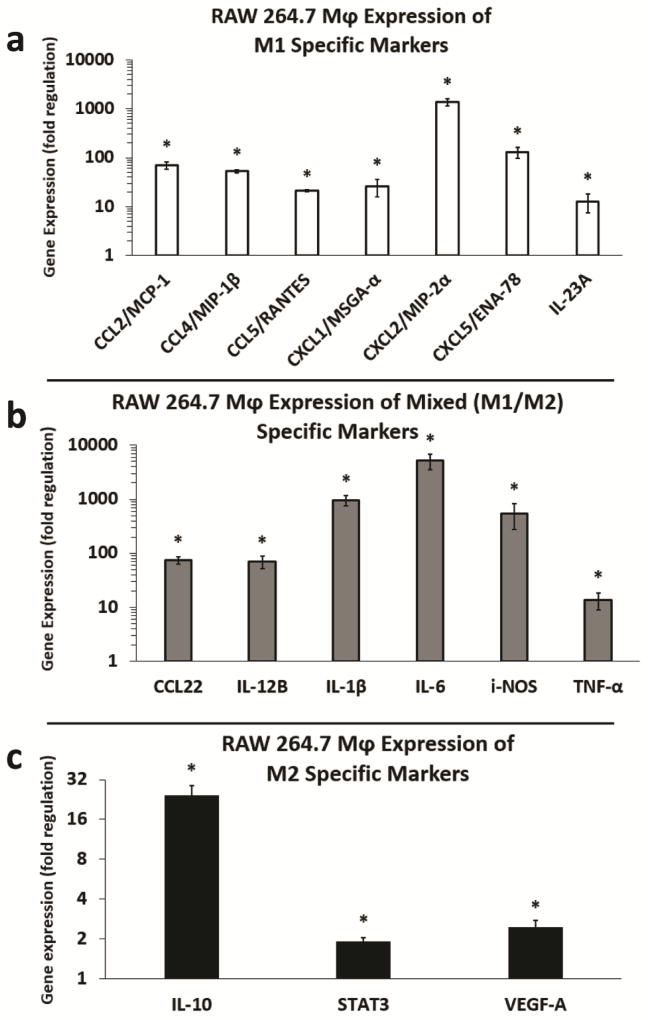
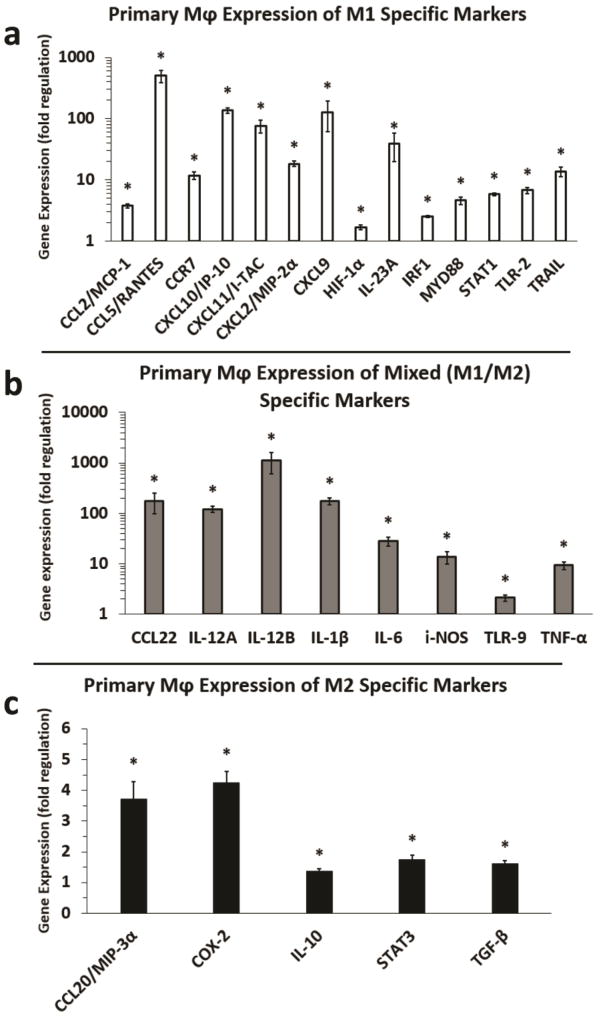
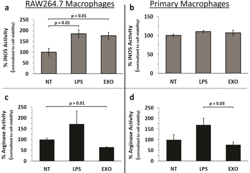
Macrophages are key participants in melanoma growth and survival. In general, macrophages can be classified as M1 or M2 activation phenotypes. Increasing evidence demonstrates that melanoma exosomes also facilitate tumor survival and metastasis. However, the role of melanoma exosomes in directly influencing macrophage function is poorly understood. Herein, we investigated the hypothesis that natural melanoma exosomes might directly influence macrophage polarization. To explore this hypothesis, ELISA, RT-qPCR, and macrophage functional studies were performed in vitro using an established source of melanoma exosomes (B16-F10). ELISA results for melanoma exosome induction of common M1 and M2 cytokines in RAW 264.7 macrophages, revealed that melanoma exosomes do not polarize macrophages exclusively in the M1 or M2 direction. Melanoma exosomes induced the M1 and M2 representative cytokines TNF-α and IL-10 respectively. Further assessment, using an RT-qPCR array with RAW 264.7 and primary macrophages, confirmed and extended the ELISA findings. Upregulation of markers common to both M1 and M2 polarization phenotypes included CCL22, IL-12B, IL-1β, IL-6, i-NOS, and TNF-α. The M2 cytokine TGF-β was upregulated in primary but not RAW 264.7 macrophages. Pro-tumor functions have been attributed to each of these markers. Macrophage functional assays demonstrated a trend toward increased i-NOS (M1) to arginase (M2) activity. Collectively, the results provide the first evidence that melanoma exosomes can induce a mixed M1 and M2 pro-tumor macrophage activation phenotype.

Keywords: Chemokine; Cytokine; Exosomes; M1/M2; Macrophage; Melanoma; Polarization.


Conflict of interest statement

Conflict of Interest Statement: The authors declare no potential conflicts of interest.

Figures

Figure 1
Figure 1
Melanoma exosomes stimulate production of mixed M1 and M2 cytokines by macrophages in cell culture. (a) Induction of the M1 cytokine TNF-α in control, LPS treated, exosome treated and LPS + exosome treated RAW264.7 Mφs. (b) Induction of the M1 cytokine IL-1β in control, LPS treated, exosome treated and LPS + exosome treated RAW264.7 Mφs. (c) Induction of the M2 cytokine TGF-β in control, IL-4 treated, exosome treated and IL-4 + exosome treated RAW264.7 Mφs. (d) Induction of the M2 cytokine IL-10 in control, IL-4 treated, exosome treated and IL-4 + exosome treated RAW264.7 Mφs. n = 3 independent experiments normalized to non-treated (NT) Mφs (percent change = 100%) using three pooled batches of melanoma exosomes. Error bars = S.E.M. Connecting bars denote a statistical comparison between two groups with p values listed above or below the connecting bars, p values < 0.05 were considered statistically significant.
Figure 2
Figure 2
Fold regulation (RT-qPCR) in LPS and IL-4 induced gene expression of common M1 and M2 cytokines in macrophages. (a) LPS induces increased gene expression of IL-1β and TNF-α in RAW264.7 Mφs. (b) IL-4 induces increased gene expression of IL-10 and TGF-β in RAW264.7 Mφs. n = 3 replicate arrays, using three pooled independent batches of melanoma exosomes, and normalized to non-treated Mφs (fold change = 1). Error bars = S.E.M. * = p values < 0.05, and were considered statistically significant compared to non-treated Mφs.
Figure 3
Figure 3
Fold regulation (RT-qPCR) in M1 and M2 cytokine gene expression induced by melanoma exosomes in RAW264.7 macrophages. (a) Induction of M1 markers in RAW264.7 Mφs by melanoma exosomes. (b) Induction of mixed (M1/M2) markers in RAW264.7 Mφs by melanoma exosomes. (c) Induction of M2 markers in RAW264.7 Mφs by melanoma exosomes. n = 3 replicate arrays, using three pooled independent batches of melanoma exosomes, and normalized to non-treated Mφs (fold change = 1). Error bars = S.E.M. * = p values < 0.05, and were considered statistically significant compared to non-treated RAW264.7 Mφs.
Figure 4
Figure 4
Fold regulation (RT-qPCR) in M1 and M2 cytokine gene expression induced by melanoma exosomes in primary macrophages. (a) Induction of M1 markers in primary Mφs by melanoma exosomes. (b) Induction of mixed (M1/M2) markers in primary Mφs. (c) Induction of M2 markers in primary Mφs by melanoma exosomes. n = 3 replicate arrays, using three pooled independent batches of melanoma exosomes, and normalized to non-treated Mφs (fold change = 1). Error bars = S.E.M. * = p values < 0.05, and were considered statistically significant compared to non-treated primary Mφs.
Figure 5
Figure 5
Assessment of macrophage M1 and M2 functional status following treatment with melanoma exosomes. (a) i-NOS activity detected for non-treated, LPS, or melanoma exosome treated RAW 264.7 Mφs (n = 5), (b) i-NOS activity detected for non-treated, LPS, or melanoma exosome treated primary Mφs (n = 9), (c) Arginase activity detected for non-treated, LPS, or melanoma exosome treated RAW264.7 Mφs (n = 3), (d) Arginase activity detected for non-treated, LPS, or melanoma exosome treated primary Mφs (n = 6). Error bars = S.E.M. Connecting bars denote a statistical comparison between two groups with p values listed above the connecting bars, p values < 0.05 were considered statistically significant.

References

    1. Witwer KW, Buzas EI, Bemis LT, Bora A, Lasser C, Lotvall J, Nolte-'t Hoen EN, Piper MG, Sivaraman S, Skog J, Thery C, Wauben MH, Hochberg F. Standardization of sample collection, isolation and analysis methods in extracellular vesicle research. Journal of extracellular vesicles. 2013;2 - PMC - PubMed
    1. Hood JL. The association of exosomes with lymph nodes. Seminars in cell & developmental biology. 2017;67:29–38. - PubMed
    1. Hood JL, Pan H, Lanza GM, Wickline SA. Paracrine induction of endothelium by tumor exosomes. Lab Invest. 2009;89(11):1317–28. - PMC - PubMed
    1. Ekstrom EJ, Bergenfelz C, von Bulow V, Serifler F, Carlemalm E, Jonsson G, Andersson T, Leandersson K. WNT5A induces release of exosomes containing pro-angiogenic and immunosuppressive factors from malignant melanoma cells. Molecular cancer. 2014;13:88. - PMC - PubMed
    1. Peinado H, Aleckovic M, Lavotshkin S, Matei I, Costa-Silva B, Moreno-Bueno G, Hergueta-Redondo M, Williams C, Garcia-Santos G, Ghajar C, Nitadori-Hoshino A, Hoffman C, Badal K, Garcia BA, Callahan MK, Yuan J, Martins VR, Skog J, Kaplan RN, Brady MS, Wolchok JD, Chapman PB, Kang Y, Bromberg J, Lyden D. Melanoma exosomes educate bone marrow progenitor cells toward a pro-metastatic phenotype through MET. Nature medicine. 2012;18(6):883–91. - PMC - PubMed

Publication types

LinkOut - more resources