

Review
doi: 10.1161/STROKEAHA.117.016995.
Epub 2018 Feb 19.
Rodent Models of Cerebral Microinfarct and Microhemorrhage
Affiliations
- PMID: 29459393
- PMCID: PMC5851653
- DOI: 10.1161/STROKEAHA.117.016995
Item in Clipboard
Review
Rodent Models of Cerebral Microinfarct and Microhemorrhage
Stroke.
2018 Mar.
No abstract available
Keywords: arteriole; autopsy; brain; cognitive dysfunction; vascular dementia.
Conflict of interest statement
Figures
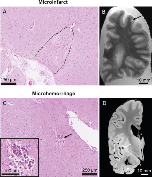
(A) A cortical microinfarct on a Hematoxylin & Eosin-stained
section. (B) Microinfarct (black arrow) on a T2-weighted ex
vivo MRI scan. (C) A cortical microhemorrhage on a
Hematoxylin & Eosin-stained section. (D) Multiple lobar
microbleeds (white arrows) on a GRE ex vivo MRI scan.
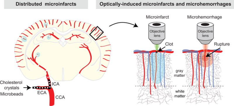
The left hemisphere depicts the production of distributed microinfarcts by
injecting microemboli through the internal carotid artery (ICA). CCA =
common carotid artery, and ECA = external carotid artery. The right
hemisphere shows the selective occlusion of a penetrating arteriole with focal
photothrombosis or rupture of a penetrating arteriole with an amplified laser
during in vivo optical imaging.
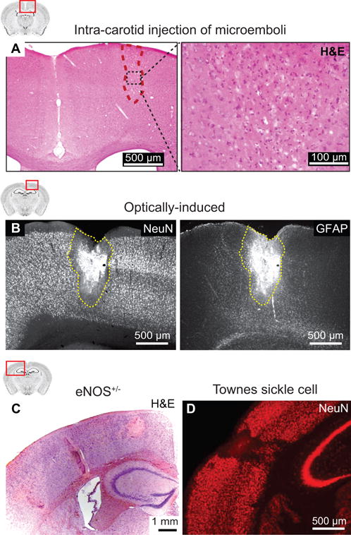
(A) A cortical microinfarct observed in Hematoxylin & Eosin
stained mouse brain sections after injection of cholesterol crystals into the
internal carotid artery. From Wang et al.
(B) A cortical microinfarct observed in NeuN and GFAP immunostained
rat brain sections after occlusion of a single cortical penetrating arteriole by
focal photothrombosis. From Shih et al.
(C) Spontaneous microinfarcts observed in Hematoxylin &
Eosin stained brain sections from an 18 months old eNOS-deficient mouse. From
Tan et al.
(D) Spontaneous microinfarcts observed in NeuN immunostained brain
sections from a 13 months old Townes sickle cell mouse. From Hyacinth et
al.
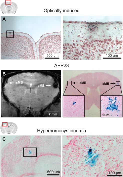
(A) A cortical microhemorrhage observed in Prussian blue stained
mouse brain sections after optically-induced rupture of a single cortical
penetrating arteriole. From Rosidi et al.
(B) Microbleeds detected by T2*-weighted MRI (left) and
corresponding microhemorrhages detected by Prussian blue (right) in an APP23
mouse. From Reuter et al.
(C) Microhemorrhages detected by Prussian blue staining in a mouse
that received a specialized diet to induce hyperhomocysteinemia. From Sudduth
et al.
References
Publication types
MeSH terms
Grants and funding
LinkOut - more resources
Full Text Sources
Other Literature Sources
