. 2018 Mar;13(3):260-266.
doi: 10.1038/s41565-017-0041-7. Epub 2018 Feb 19.

Photoelectrochemical modulation of neuronal activity with free-standing coaxial silicon nanowires

Affiliations

Photoelectrochemical modulation of neuronal activity with free-standing coaxial silicon nanowires

Ramya Parameswaran et al. Nat Nanotechnol. 2018 Mar.

Abstract

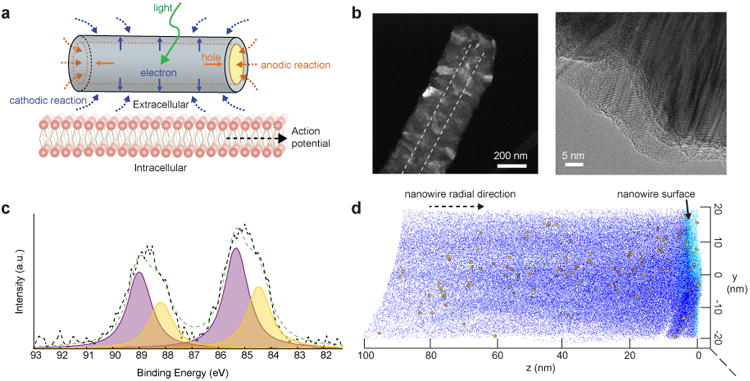
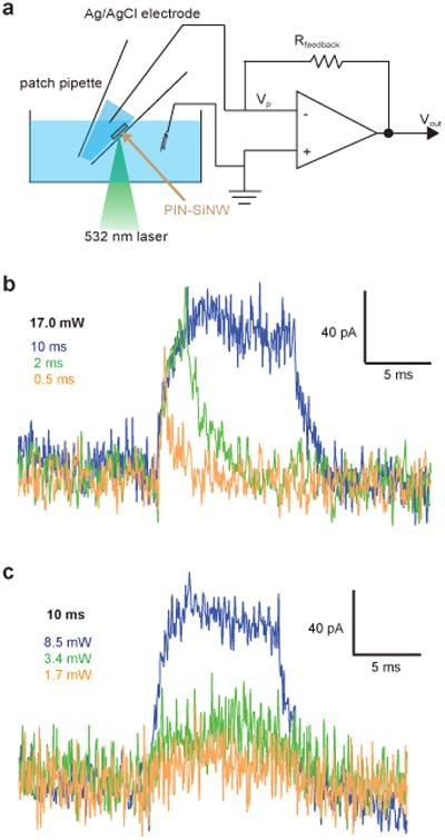
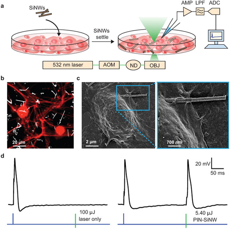
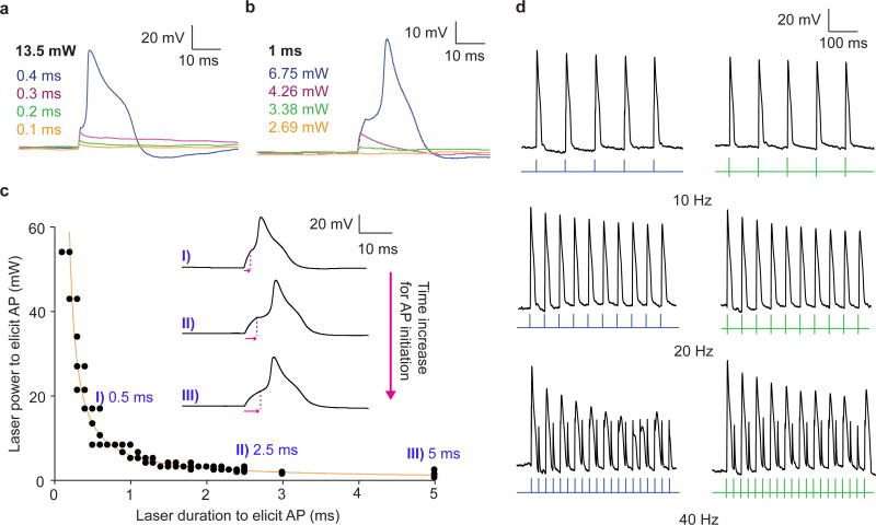
Optical methods for modulating cellular behaviour are promising for both fundamental and clinical applications. However, most available methods are either mechanically invasive, require genetic manipulation of target cells or cannot provide subcellular specificity. Here, we address all these issues by showing optical neuromodulation with free-standing coaxial p-type/intrinsic/n-type silicon nanowires. We reveal the presence of atomic gold on the nanowire surfaces, likely due to gold diffusion during the material growth. To evaluate how surface gold impacts the photoelectrochemical properties of single nanowires, we used modified quartz pipettes from a patch clamp and recorded sustained cathodic photocurrents from single nanowires. We show that these currents can elicit action potentials in primary rat dorsal root ganglion neurons through a primarily atomic gold-enhanced photoelectrochemical process.


Figures

Figure 1
Figure 1. X-ray photoelectron spectroscopy and atom probe tomography reveal the presence of atomic Au on coaxial nanowire surfaces
(a) Schematic of the Faradaic current produced by a PIN-SiNW at the neuronal cell membrane on light stimulation, inducing action potential generation in the neuron via membrane depolarization. Solid blue and orange lines represent movement of electrons and holes towards n-type and p-type Si, respectively, upon light stimulation. Blue and orange dotted lines represent the cathodic and anodic reactions, respectively. (b) HAADF STEM image of PIN-SiNW (left) with p-type core outlined by white dotted line. This is a representative image from one of a total of 64 PIN-SiNWs imaged from 2 independent experiments. TEM image of PIN-SiNW (right). This is a representative image from one of a total of 8 PIN-SiNWs imaged from 3 independent experiments. (c) XPS spectrum (black dotted line) and the fitted curve (green dotted line) of Au 4f signals from the PIN-SiNWs. Deconvoluted peaks of Au 4f 7/2 and Au 4f 5/2 at 84.46 eV and 88.13 eV (yellow) represent nanoclustered Au species. Au 4f 7/2 and Au 4f 5/2 peaks at 85.4 eV and 89 eV (purple) represent atomic-like Au species. (d) 3D chemical reconstruction of local electrode APT data from a single PIN-SiNW displaying Si atoms (dark blue dots, 10% of all Si atoms displayed), O atoms (light blue dots, 100% of all O atoms displayed), and Au atoms (yellow balls, 100% of all Au atoms displayed). This is representative data from one of 3 independent probes that were prepared for APT analysis.
Figure 2
Figure 2. Single nanowire recordings reveal that coaxial Si nanowires are photoelectrochemical current sources
(a) Schematic of photocurrent measurement setup. Vp represents the pipette voltage; Rfeedback is the resistance of a feedback resistor; Vout denotes the output voltage. (b) Photocurrent traces from a single PIN-SiNW illuminated with a 532 nm laser at 17 mW for durations of 0.5 ms (orange), 2 ms (green), or 10 ms (blue). (c) Photocurrent traces from a single PIN-SiNW illuminated with a 532 nm laser at 8.5 mW (blue), 3.4 mW (green), or 1.7 mW (orange) for a duration of 10 ms. The traces in (b) and (c) are representative ones from a total of 40 current traces measured from 5 independent PIN-SiNWs.
Figure 3
Figure 3. Basic silicon nanowire-based neural interfaces
(a) Schematic of the set-up used to record DRG neuron APs in response to a 532 nm laser stimulation at a single neuron/SiNW interface. This set-up includes an ordinary electrophysiology capability for patch clamp experiments, with an amplifier (AMP), used in current clamp mode, a low pass filter (LPF), and an analog to digital converter (ADC). In addition the setup was implemented with a 532 nm laser beam controlled by an acoustic modulator (AOM) and various neutral density (ND) filters (to attenuate the power). The laser beam is aligned to the optical central axis of the objective lens (OBJ) of an inverted microscope. SiNWs were sonicated off of their growth substrate and drop casted onto the primary neuron culture. After 20 min of settling, the experiments were performed. (b) Confocal microscopy image of primary neonatal rat dorsal root ganglion (DRG) neurons stained with anti-β-tubulin III (red) co-cultured with PIN-SiNWs (white). This is a representative image from one of a total of 10 images taken from 2 independent experiments. (c) Scanning electron microscopy images of a single DRG neuron interacting with a single PIN-SiNW (left); zoomed in image displaying neuron-PIN-SiNW interface (right). This is a representative image from one of a total of 63 images taken from 4 independent experiments. (d) Patch clamp electrophysiology current clamp trace of membrane voltage in DRG neuron stimulated (blue pulse) or a laser pulse (green bar) of various energies as labeled at the neuron/SiNW interface. Two conditions are displayed: laser only with no SiNW (left) and PIN-SiNW (right). These are representative traces from one of 173 total action potential traces measured from 30 neurons with PIN-SiNWs, and one of 27 total traces from 2 neurons with PIN-SiNWs.
Figure 4
Figure 4. Photocurrent generated by coaxial nanowires can be harnessed to elicit action potentials in primary rat dorsal root ganglion neurons
(a and b) Patch clamp electrophysiology current clamp traces of membrane voltage in DRG neurons illuminated by a 532 nm laser pulse at the neuron/PIN-SiNW interface at (a) 13.5 mW with durations of 0.1 ms (orange), 0.2 ms (green), 0.3 ms (pink), and 0.4 ms (blue) and (b) 2.69 mW (orange), 3.38 mW (green), 4.26 mW (pink), and 6.75 mW (blue) for 1 ms. These traces in (a) and (b) are representative traces from one of a total of 1398 traces from 30 independent neurons, many of which are sub-threshold depolarizations at various laser powers and durations. 173 traces out of the 1398 represent action potentials. (c) Excitability curve displaying 532 nm laser power and duration combinations that produce APs in neurons (N=6 neurons; total of N=78 replicates) interacting with a single PIN-SiNWs with specific traces and time to peak response for each of those traces (pink arrows) at I) 0.5 ms, II) 2.5 ms, and III) 5 ms durations highlighted. Error bars represent the standard error about the average. Some of the data points are overlaid. (d) AP traces from neurons interacting with single PIN-SiNWs pulsed at 10, 20 and 40 Hz with light (green bars) and injected current from patch amplifier (blue pulses). These are representative traces from a total of 6 pulse train traces for each frequency taken from 3 independent neurons.
Figure 5
Figure 5. The mechanism of coaxial nanowire photocurrent generation and neuronal modulation is primarily photoelectrochemical, aided by surface atomic Au
(a) Schematic of temperature measurement setup for simultaneous measurement of temperature and neuronal APs produced by laser stimulation or injected current through patch amplifier. (b) Patch clamp electrophysiology current clamp trace of membrane voltage (top) in DRG neuron stimulated by injected current (blue pulse) and illuminated by a 532 nm laser pulse at the neuron-PIN-SiNW interface (green bar). Corresponding temperature measurement (bottom) taken 2 μm away from neuron/PIN-SiNW interface produced by calibrating the thermometer pipette resistance with temperature changes. This is a representative temperature measurement from one of 18 total traces from 3 independent neurons. (c) Photocurrent measurement taken from a single p-type SiNW illuminated with 532 nm laser light for 10 ms at a laser illumination power of 85 mW (top). This is a representative trace from one of a total of 71 traces measured from 4 independent p-type SiNWs. Photocurrent measurement taken from a p-type SiNW with diffused Au for 10 ms at laser illumination powers of 8.5 mW (blue) and 6.8 mW (green). These are representative traces from a total of 52 traces measured from 6 independent p-type SiNWs with diffused Au. (d) Patch clamp electrophysiology current clamp trace of membrane voltage (top) in DRG neuron stimulated by injected current (blue pulse) and illuminated by a 1 ms 17.4 μJ 532 nm laser pulse at the neuron-p-type SiNW with diffused Au interface (green bar). This is a representative trace from one of a total of 40 traces measured from 5 independent neurons. (e) Band diagram representing the redox reaction that occurs at the interface between the PIN-SiNW and the electrolyte solution. Kinetic barrier for photoelectrochemical reaction is lowered by the presence of atomic Au. Green arrows represent the light stimulus. Grey arrow shows the excitation of electrons from the valence to the conduction band. Grey dashed lines represent Fermi levels in dark.

Comment in

  • Light touch.
    Lanzani G. Lanzani G. Nat Nanotechnol. 2018 Mar;13(3):181-182. doi: 10.1038/s41565-018-0081-7. Nat Nanotechnol. 2018. PMID: 29459652 No abstract available.

References

    1. Famm K, Litt B, Tracey KJ, Boyden ES, Slaoui M. A jump-start for electroceuticals. Nature. 2013;496:159–161. - PMC - PubMed
    1. Vetter RJ, Williams JC, Hetke JF, Nunamaker EA, Kipke DR. Chronic neural recording using silicon-substrate microelectrode arrays implanted in cerebral cortex. IEEE Transactions on Biomedical Engineering. 2004;51:896–904. - PubMed
    1. Zhou W, Dai XC, Lieber CM. Advances in nanowire bioelectronics. Reports on Progress in Physics. 2017;80 doi: 10.1088/0034-4885/80/1/016701. - DOI - PubMed
    1. Ghezzi D, et al. A hybrid bioorganic interface for neuronal photoactivation. Nature Communications. 2011;2 doi: 10.1038/ncomms1164. - DOI - PubMed
    1. Ghezzi D, et al. A polymer optoelectronic interface restores light sensitivity in blind rat retinas. Nature Photonics. 2013;7:400–406. - PMC - PubMed

Publication types