. 2018 Feb 19;8(1):3310.
doi: 10.1038/s41598-018-21477-9.

c-Jun N-terminal kinase 1 defective CD4+CD25+FoxP3+ cells prolong islet allograft survival in diabetic mice

Affiliations

c-Jun N-terminal kinase 1 defective CD4+CD25+FoxP3+ cells prolong islet allograft survival in diabetic mice

Deepak Tripathi et al. Sci Rep. .

Abstract

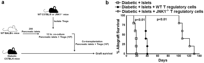
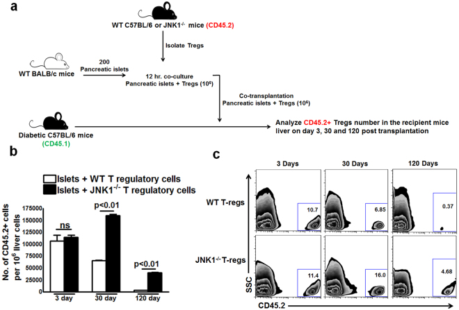
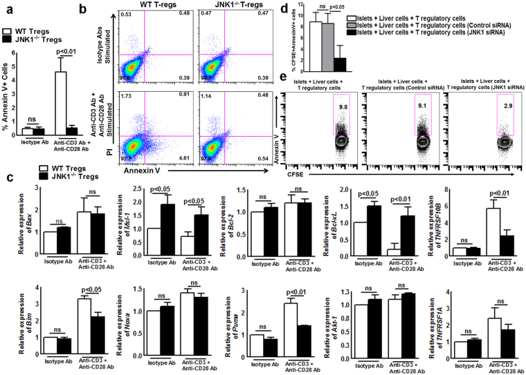
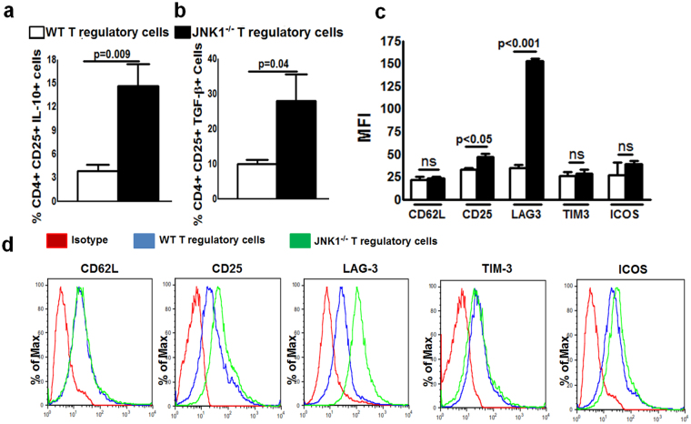
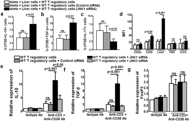
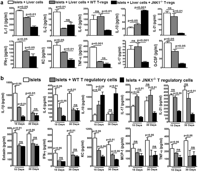
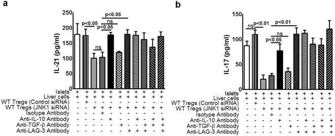
CD4+CD25+FoxP3+ cells (Tregs) inhibit inflammatory immune responses to allografts. Here, we found that co-transplantation of allogeneic pancreatic islets with Tregs that are defective in c-Jun N-terminal kinase 1 (JNK1) signaling prolongs islet allograft survival in the liver parenchyma of chemically induced diabetic mice (CDM). Adoptively transferred JNK1-/- but not wild-type (WT) Tregs survive longer in the liver parenchyma of CDM. JNK1-/- Tregs are resistant to apoptosis and express anti-apoptotic molecules. JNK1-/- Tregs express higher levels of lymphocyte activation gene-3 molecule (LAG-3) on their surface and produce higher amounts of the anti-inflammatory cytokine interleukin (IL)-10 compared with WT Tregs. JNK1-/- Tregs inhibit liver alloimmune responses more efficiently than WT Tregs. JNK1-/- but not WT Tregs are able to inhibit IL-17 and IL-21 production through enhanced LAG-3 expression and IL-10 production. Our study identifies a novel role of JNK1 signaling in Tregs that enhances islet allograft survival in the liver parenchyma of CDM.


Conflict of interest statement

The authors declare no competing interests.

Figures

Figure 1
Figure 1
JNK1 defective Tregs prolong islet allograft survival in liver parenchyma of CDM. A single intraperitoneal injection of streptozotocin (STZ) (180 mg/kg body weight) caused C57BL/6 mice to develop diabetes, as measured by random blood sugar levels after one week. Approximately 200 pancreatic islets obtained from BALB/c mice (donor) were cultured in medium for 12 hours and transplanted into the liver parenchyma of CDM (recipient). Some of the islet allograft recipient mice received CD4+CD25+Foxp3+ cells (106) from WT or JNK−/− mice (both C57BL/6 background) that were isolated and cultured with islets for 12 hours prior to transfer along with islets as mentioned in the Methods section. Blood glucose levels were measured every 72 hours up to 140 days (a). Schematic representation of CD4+CD25+Foxp3+ cell-loaded islets. Blood glucose levels above 300 mg/deciliter were considered to indicate diabetes or failed glucose control (b). Percent allograft survival. The percent graft survival was calculated using the log-rank test. Kaplan–Meier survival curves of the mice are shown. Data presented are representative of five independent experiments. Five mice per group were used.
Figure 2
Figure 2
Lack of JNK1 expression enhances survival of Tregs in recipient mice. Pancreatic islets and Tregs from WT or JNK1−/− mice (CD45.2) were transferred to diabetic mice (CD45.1) as in Fig. 1a. Three, thirty and one hundred twenty days after transplantation, the percentages of donor-derived (CD45.2) cells in recipient mouse liver were determined by flow cytometry. (a) Schematic representation of the experiment. (b) Absolute number of cells. (c) A representative flow cytometry figure. Data presented are representative of five independent experiments. Five mice per group were used. Mean values, p values and SEs are shown.
Figure 3
Figure 3
JNK1−/− Tregs are less apoptotic than WT Tregs. a to c. CD4+CD25+ Tregs from WT or JNK1−/− were stimulated with isotype control antibodies IgG and IgG2 or anti-CD3 (5 μg/ml) and anti-CD28 (1 μg/ml) as described in the Methods section. After six hours, (a) the percentage of Annexin V+ cells was determined by flow cytometry. (b) A representative flow cytometry figure. (c) The mRNA expression of pro- and anti-apoptotic genes (Bax, Mcl-1, Bcl-2, Bcl-Xl, TNFRSF10B, Bim, Noxa, Puma, Akt-1 and TNFRSF1A) was determined by real-time PCR. (d) Liver cells from C57BL/6 mice were isolated and cultured with BALB/c mouse pancreatic islets at a ratio of 10000:1 (1 × 105 liver cells and 10 islets) in the presence or absence of CFSE-labeled JNK1 or control siRNA-transfected Tregs from WT C57BL/6 mice (1 × 104). After 72 hours, the percentages of CFSE+ AnnexinV+ cells were measured by flow cytometry. (e) A representative flow cytometry figure. Mean values, p values and SEs are shown. Data presented are representative of three independent experiments.
Figure 4
Figure 4
JNK1−/− Tregs produce more IL-10 and TGF-β and express more LAG-3 than WT Tregs. CD4+CD25+ Tregs from WT or JNK1−/− were stimulated with anti-CD3 (5 μg/ml) or anti-CD3 anti-CD28 (1 μg/ml) as described in the Methods section. After 72 hours, the percentage of (a) IL-10 and (b) TGF-β producing cells was determined. (c) Mean fluorescence intensity (MFI) of LAG-3, CD62L, ICOS, CD25 and TIM-3 expression by WT and JNK1−/− Tregs. (d) A representative flow cytometry figure. Mean values, p values and SEs are shown. Data presented are representative of three independent experiments.
Figure 5
Figure 5
JNK1 siRNA enhances WT Treg production of IL-10 and TGF-β. a to c. Liver cells from C57BL/6 mice were isolated and cultured with BALB/c mouse pancreatic islets at a ratio of 10000:1 (1 × 105 liver cells and 10 islets) in the presence or absence of freshly isolated CFSE-labeled and JNK1 or control siRNA-transfected Tregs from WT C57BL/6 mice (104). After 72 hours, the percentage of Annexin V + cells expressing (a) IL-10, (b) TGF-β, (c) IL-17, (d) LAG3, CD25, CD62L, ICOS and TIM-3 was measured by flow cytometry. Tregs from WT C57BL/6 mice were isolated and transfected with JNK1 or JNK2 or control siRNA as described in the Methods section. Twelve hours after transfection, the cells were stimulated with anti-CD3 (5 μg/ml) or anti-CD3 and anti-CD28 (1 μg/ml). After 72 hours, real-time PCR analysis was performed to evaluate gene expression. (e) IL-10 (f) TGF-β and (g) Foxp3. Mean values, p values and SEs are shown. Data presented are representative of three independent experiments.
Figure 6
Figure 6
JNK1−/− but not WT Tregs inhibit IL-17 and IL-21-mediated inflammatory responses (a) Liver cells from C57BL/6 mice were isolated and cultured with BALB/c mouse pancreatic islets at a ratio of 10000:1 (1 × 105 liver cells and 10 islets) in the presence or absence of Tregs from WT C57BL/6 or JNK1−/− (1 × 104) mice. After 72 hours, various cytokine levels were measured by multiplex ELISA. Data from three independent experiments are shown. Mean values, p values and SEs are shown (b). Pancreatic islets and Tregs from WT or JNK−/− mice were transferred to CDM as described in Fig. 1. Fifteen and thirty days after transplantation, liver cell homogenates were prepared, and various cytokine levels were measured by multiplex ELISA. Data from five independent experiments are shown. Five mice per group were used. Mean values, p values and SEs are shown.
Figure 7
Figure 7
LAG-3 and IL-10 but not TGF-β are responsible for JNK1−/− Treg-mediated inhibition of IL-21 and IL-17 production. Liver cells from C57BL/6 mice were isolated and cultured with BALB/c mice pancreatic islets at a ratio of 10000:1 (1 × 105 liver cells and 10 islets) in the presence or absence of WT Tregs treated with control or JNK1 siRNA and antibodies against LAG-3 or IL-10 or TGF-β or isotype controls (10 µg/ml). In some wells, a combination of LAG-3, IL-10, TGF-β antibodies (suboptimal dose 2. 5 µg/ml each) was used. After 72 hours, (a) IL-21 and (b) IL-17 levels were measured by multiplex ELISA. Data from three independent experiments are shown. Mean values, p values and SEs are shown.

Similar articles

Cited by

References

    1. Sakaguchi S, Yamaguchi T, Nomura T, Ono M. Regulatory T Cells and Immune Tolerance. Cell. 2008;133:775–787. doi: 10.1016/j.cell.2008.05.009. - DOI - PubMed
    1. Hsieh C-S, Lee H-M, Lio C-WJ. Selection of regulatory T cells in the thymus. Nat. Rev. Immunol. 2012;12:157–167. doi: 10.1038/nri3155. - DOI - PubMed
    1. Workman CJ, Szymczak-Workman AL, Collison LW, Pillai MR, Vignali DAA. The Development and Function of Regulatory T Cells. Cell. Mol. Life Sci. CMLS. 2009;66:2603–2622. doi: 10.1007/s00018-009-0026-2. - DOI - PMC - PubMed
    1. Moon, B.-I., Kim, T. H. & Seoh, J.-Y. Functional Modulation of Regulatory T Cells by IL-2. PLoS ONE10, (2015). - PMC - PubMed
    1. Khan MA. T regulatory Cell–mediated Immunotherapy for Solid Organ Transplantation: A Clinical Perspective. Mol. Med. 2016;22:892–904. doi: 10.2119/molmed.2016.00050. - DOI - PMC - PubMed

Publication types

MeSH terms