. 2018 Feb 19;8(1):3241.
doi: 10.1038/s41598-018-21674-6.

Silibinin alleviates inflammation and induces apoptosis in human rheumatoid arthritis fibroblast-like synoviocytes and has a therapeutic effect on arthritis in rats

Affiliations

Silibinin alleviates inflammation and induces apoptosis in human rheumatoid arthritis fibroblast-like synoviocytes and has a therapeutic effect on arthritis in rats

W W Tong et al. Sci Rep. .

Abstract

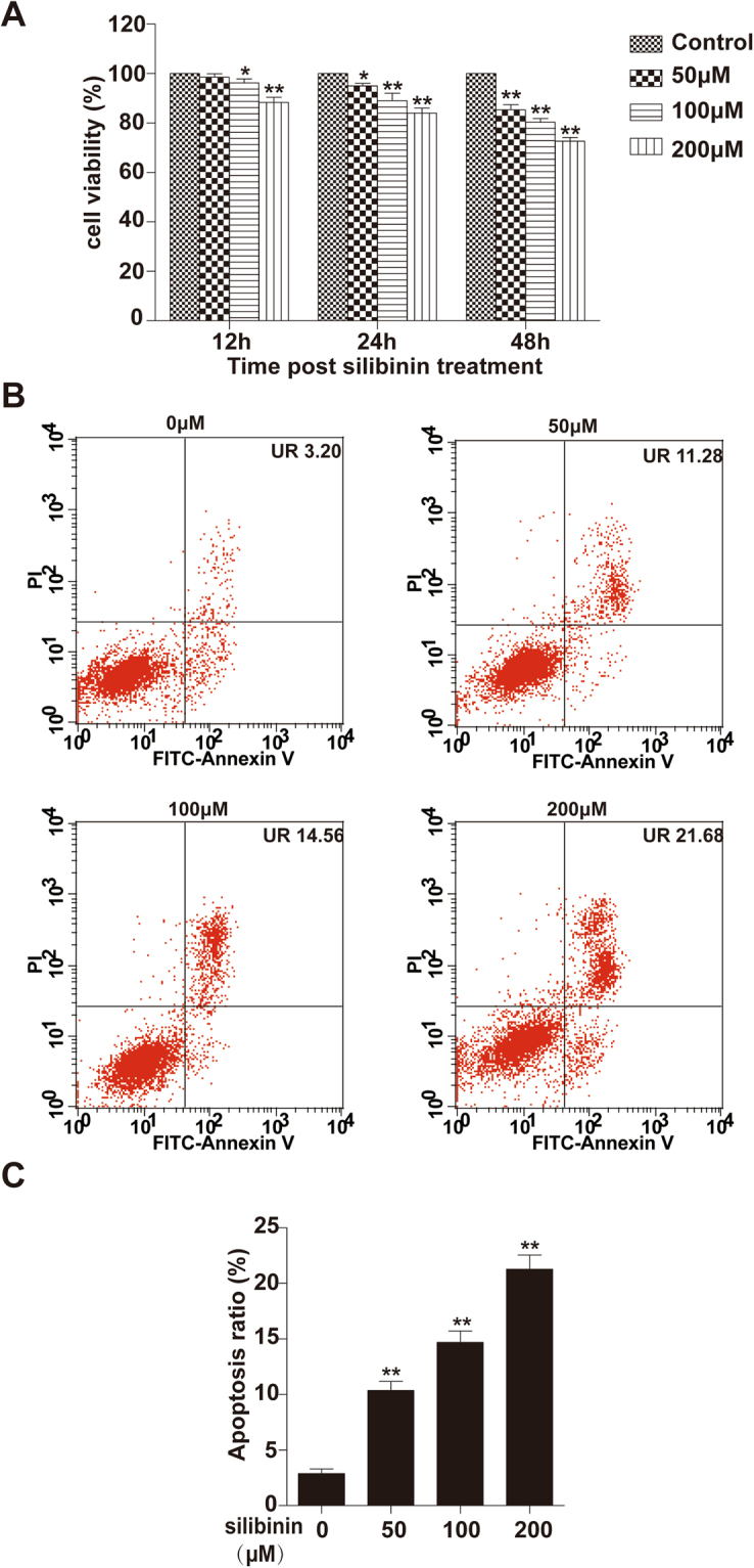
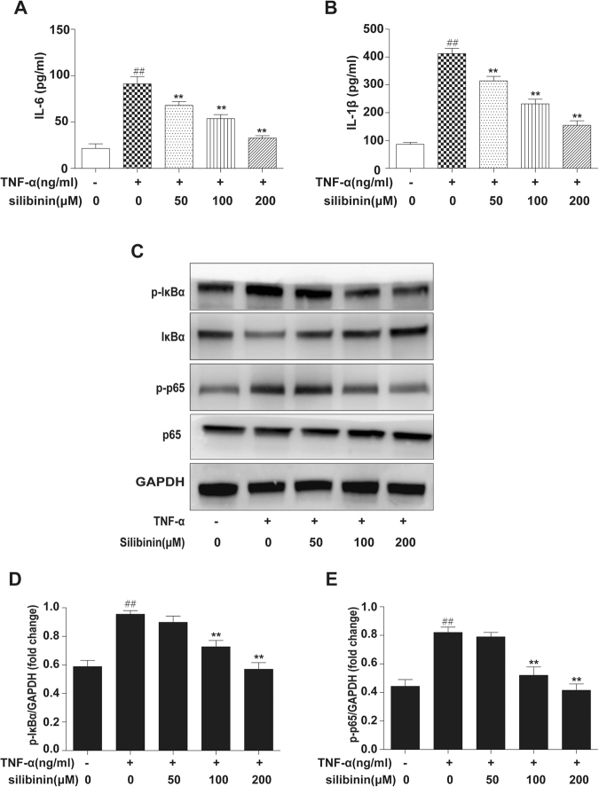
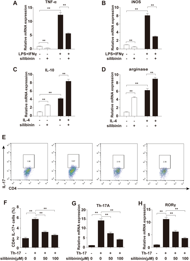
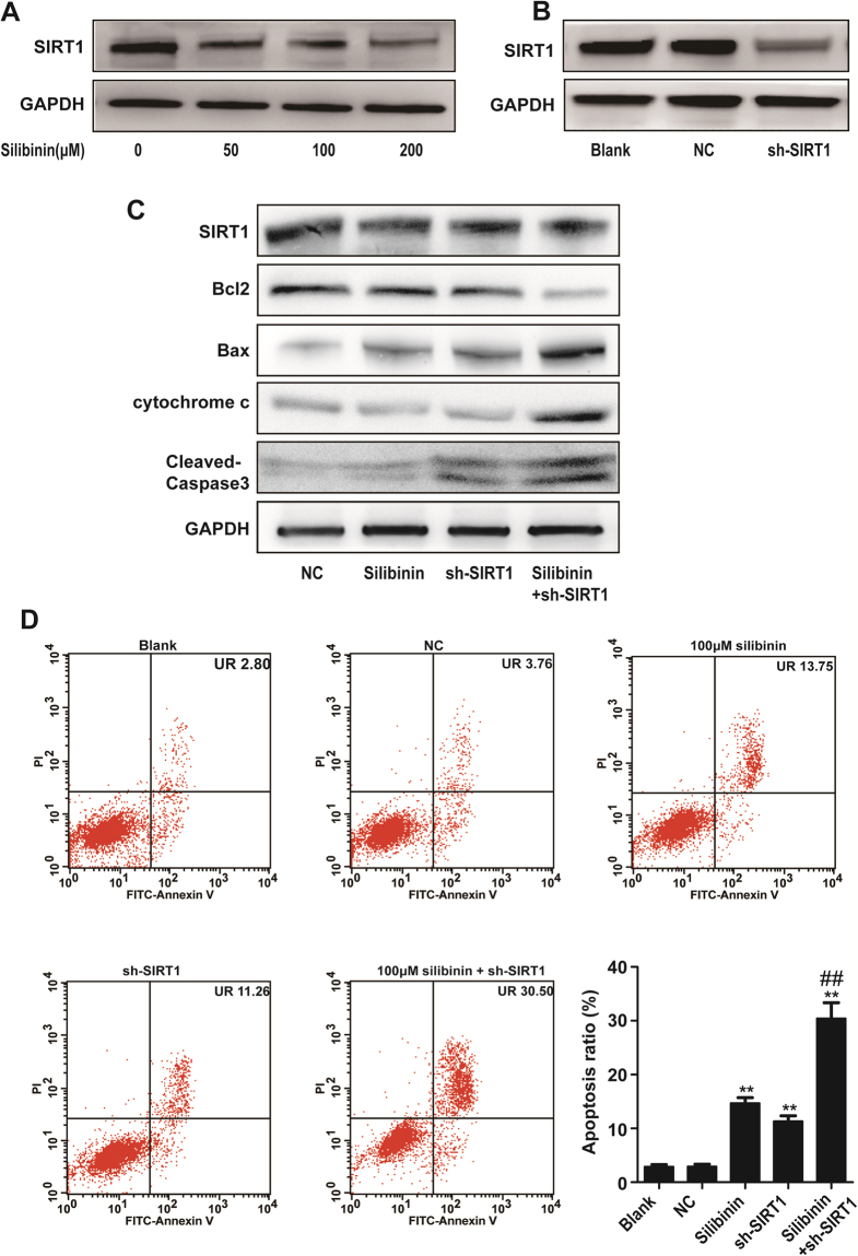
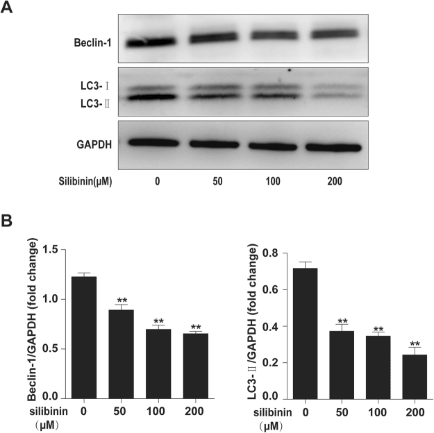
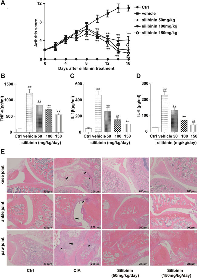
Silibinin, a natural polyphenolic flavonoid, possesses anti-oxidant, anti-inflammation and anti-cancer properties. The present study was designed to investigate the effects of silibinin on rheumatoid arthritis (RA) pathogenesis-related cells and collagen-induced arthritis (CIA) and further explore the potential underlying mechanisms. Our results showed that silibinin suppressed cell viability and increased the percentage of apoptotic RA-fibroblast-like synoviocytes (FLS). Furthermore, the production of inflammatory cytokines in RA-FLS and a CIA rat model was effectively inhibited by silibinin. Silibinin also induced macrophage M2 polarization in RAW264.7 cells. We further demonstrated that silibinin inhibits Th17 cell differentiation in vitro. The nuclear factor kappa B (NF-κB) pathway was suppressed in RA-FLS. In addition, Sirtuin1 (SIRT1) was decreased after silibinin treatment, and RA-FLS transfection with a short hairpin RNA (shRNA) of SIRT1 enhanced silibinin-induced apoptosis. Autophagy was markedly decreased in a dose-dependent manner following silibinin treatment. These findings indicate that silibinin inhibited inflammation by inhibiting the NF-κB pathway, and SIRT1 may participate in silibinin-induced apoptosis. Silibinin also inhibited autophagy in RA-FLS. Thus, silibinin may be a potential therapeutic agent for the treatment of RA.


Conflict of interest statement

The authors declare no competing interests.

Figures

Figure 1
Figure 1
In vitro treatment of human RA-FLS inhibits cell viability and induces apoptosis in a dose- and time-dependent manner. (A) Cells were treated with various concentrations (0, 50, 100, and 200 μM) of silibinin for 12 h, 24 h and 48 h, and then cell viability was determined using the CCK-8 assay as described in the Materials and Methods section. The result is represented as control% compared to the control group. (B) Cells were treated with various concentrations of silibinin (0, 50, 100, and 200 μM) for 48 h and then harvested for apoptosis analysis by staining with Annexin V-FITC and PI. The upper right (UR) quadrant indicates the percentage of late apoptotic cells. (C) Quantitative analysis of the apoptosis rate. Data are presented as the mean ± SD from three independent experiments. *P < 0.05, **P < 0.01 compared to the 0 μM silibinin group.
Figure 2
Figure 2
Effects of silibinin on TNF-α-induced cytokine production and activation of NF-κB in RA-FLS. RA-FLS were pre-treated with or without different concentrations (50, 100, and 200 μM) of silibinin and then exposed to TNF-α (10 ng/ml) for 12 h. (A) IL-6 and IL-1β levels in cultured cell supernatants were measured by ELISA. (B) Phospho-IκBα, IκBα, phospho-p65, p65 and GAPDH were determined by Western blot analysis. The intensity of phospho-IκBα and phospho-p65 was determined by densitometry using ImageJ software and normalized to the control (p-IκBα/GAPDH, p-p65/GAPDH). Data are representative of three independent experiments (mean ± SD). ##P < 0.01 compared to the group without TNF-α. *P < 0.05, **P < 0.01 vs treatment with TNF-α alone.
Figure 3
Figure 3
Silibinin induces Raw264.7 macrophages M2 polarization and inhibits Th17 cell differentiation in vitro. Raw264.7 macrophages were treated with silibinin (100 µM) for 24 h. To induced M1 polarization, the macrophages were incubated with LPS (1 µg/ml) and IFN γ (20 ng/ml) for 12 h. To induced M2 polarization, the macrophages were incubated with IL-4 (20 ng/ml, 12 h). The mRNA expression levels of (A) TNF-α, (B) iNOS, (C) IL-10 and (D) arginase were quantified by qPCR and normalized versus β-actin. Naive CD4+ T cells were purified from the spleens of DBA/1 mice and cultured under Th17-polarizing conditions for 3 days in the presence of silibinin (0, 50, and100 μM), and (E,F) the frequency of IL-17+CD4+ T cells was analyzed by flow cytometry. The flow cytometric profiles are representative of three independent experiments with similar results. The mRNA levels of (G) IL-17 and (H) RORγ were measured by real-time PCR. Data are presented as mean ± SD from three independent experiments. *P < 0.05, **P < 0.01.
Figure 4
Figure 4
Role of SIRT1 in silibinin-induced apoptosis in RA fibroblast-like synoviocytes. (A) RA-FLS was treated with different concentrations (50, 100, and 200 μM) of silibinin for 48 h, and the expression of SIRT1 was analysed in the cell extract by Western blotting. (B) Effect of SIRT1-shRNA (sh-SIRT1) or control-shRNA (NC) transfection on SIRT1 protein levels in RA-FLS cells. SIRT1 expression levels were estimated by Western blotting. (C) Effect of sh-SIRT1 combined with silibinin (100 μM, 48 h) on the expression of SIRT1, Bcl2, Bax, Cytochrome c and Cleaved caspase-3. GAPDH was used as a loading control. The intensity was determined by densitometry using ImageJ software and normalized to the loading control. (D) Silibinin and sh-SIRT1 induced apoptosis in RA-FLS. The apoptosis levels were examined by flow cytometry. Data are presented as the mean ± SD from three independent experiments. *P < 0.05, **P < 0.01 compared to the control group. ##P < 0.01 compared to the silibinin-treated group.
Figure 5
Figure 5
Silibinin inhibits autophagy in RA-FLS. (A) RA-FLS were treated with different concentrations (50, 100, and 200 μM) of silibinin for 48 h, and Beclin-1 and LC3 expression was analysed in the cell extracts by Western blotting. (B) The intensity of Beclin-1 and LC3 was determined by densitometry using ImageJ software and normalized with the loading control (SIRT1/GAPDH). Data are presented as the mean ± SD from three independent experiments. *P < 0.05, **P < 0.01 compared to the 0 μM silibinin group.
Figure 6
Figure 6
Effects of silibinin on CIA. Rats were randomly divided into 5 groups, with six rats in each group: without CIA (Ctrl), CIA+ vehicle, CIA+ silibinin 50 mg/kg/day, CIA+ silibinin 100 mg/kg/day, and CIA+ silibinin 150 mg/kg/day. (A) Treatment with silibinin led to dose-dependent improvements in the clinical arthritis score compared with vehicle. Silibinin also reduced cytokine levels in the sera of CIA rats. (B) TNF-α, (C) IL-1β and (D) IL-6. (E) Pannus formation (arrowheads) and infiltration of inflammatory cells (thin arrows) were attenuated by two weeks treatment of silibinin. Data are expressed as the mean ± SD. ##P < 0.01 compared to the Ctrl group, *P < 0.05, **P < 0.01 represent significant differences when compared with vehicle treatment.

References

    1. Firestein GS. Evolving concepts of rheumatoid arthritis. Nature. 2003;423:356–361. doi: 10.1038/nature01661. - DOI - PubMed
    1. Choy EH, Panayi GS. Cytokine pathways and joint inflammation in rheumatoid arthritis. N. Engl. J. Med. 2001;344:907–916. doi: 10.1056/NEJM200103223441207. - DOI - PubMed
    1. Brennan FM, McInnes IB. Evidence that cytokines play a role in rheumatoid arthritis. J. Clin. Invest. 2008;118:3537–3545. doi: 10.1172/JCI36389. - DOI - PMC - PubMed
    1. Bartok B, Firestein GS. Fibroblast-like synoviocytes: key effector cells in rheumatoid arthritis. Immunol. Rev. 2010;233:233–255. doi: 10.1111/j.0105-2896.2009.00859.x. - DOI - PMC - PubMed
    1. Imboden J. The immunopathogenesis of rheumatoid arthritis. Annu. Rev. Pathol. 2009;4:417–434. doi: 10.1146/annurev.pathol.4.110807.092254. - DOI - PubMed

Publication types

MeSH terms