

A fuzzy feature fusion method for auto-segmentation of gliomas with multi-modality diffusion and perfusion magnetic resonance images in radiotherapy
- PMID: 29459741
- PMCID: PMC5818538
- DOI: 10.1038/s41598-018-21678-2
A fuzzy feature fusion method for auto-segmentation of gliomas with multi-modality diffusion and perfusion magnetic resonance images in radiotherapy
Abstract
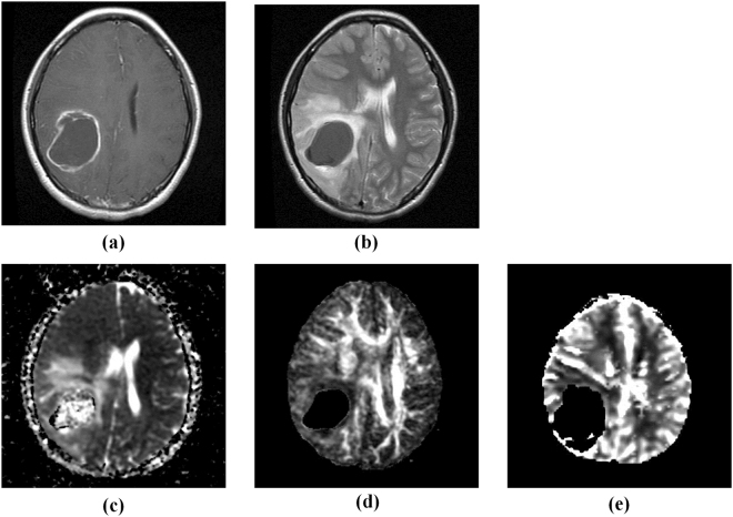
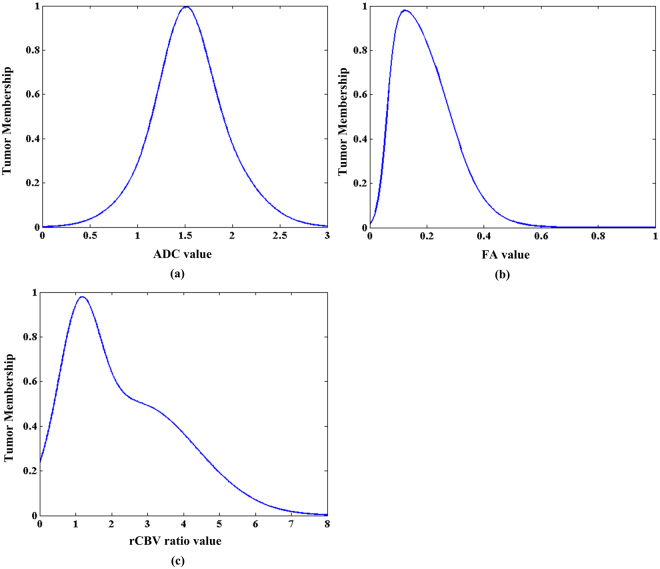
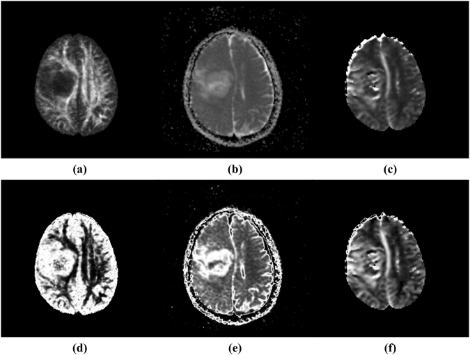
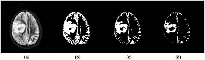
The diffusion and perfusion magnetic resonance (MR) images can provide functional information about tumour and enable more sensitive detection of the tumour extent. We aimed to develop a fuzzy feature fusion method for auto-segmentation of gliomas in radiotherapy planning using multi-parametric functional MR images including apparent diffusion coefficient (ADC), fractional anisotropy (FA) and relative cerebral blood volume (rCBV). For each functional modality, one histogram-based fuzzy model was created to transform image volume into a fuzzy feature space. Based on the fuzzy fusion result of the three fuzzy feature spaces, regions with high possibility belonging to tumour were generated automatically. The auto-segmentations of tumour in structural MR images were added in final auto-segmented gross tumour volume (GTV). For evaluation, one radiation oncologist delineated GTVs for nine patients with all modalities. Comparisons between manually delineated and auto-segmented GTVs showed that, the mean volume difference was 8.69% (±5.62%); the mean Dice's similarity coefficient (DSC) was 0.88 (±0.02); the mean sensitivity and specificity of auto-segmentation was 0.87 (±0.04) and 0.98 (±0.01) respectively. High accuracy and efficiency can be achieved with the new method, which shows potential of utilizing functional multi-parametric MR images for target definition in precision radiation treatment planning for patients with gliomas.
Conflict of interest statement
The authors declare no competing interests.
Figures


References
-
- Weber, M., Giesel, F. & Stieltjes, B. MRI for identification of progression in brain tumors: from morphology to function. Expert Rev. Neurother. 8, 10.1586/14737175.8.10.1507 (2008). - PubMed
Publication types
MeSH terms
LinkOut - more resources
Full Text Sources
Other Literature Sources
Medical
