. 2018 Feb 19;8(1):3252.
doi: 10.1038/s41598-018-21663-9.

Genomics of a pediatric ovarian fibrosarcoma. Association with the DICER1 syndrome

Affiliations

Genomics of a pediatric ovarian fibrosarcoma. Association with the DICER1 syndrome

Jorge Melendez-Zajgla et al. Sci Rep. .

Abstract

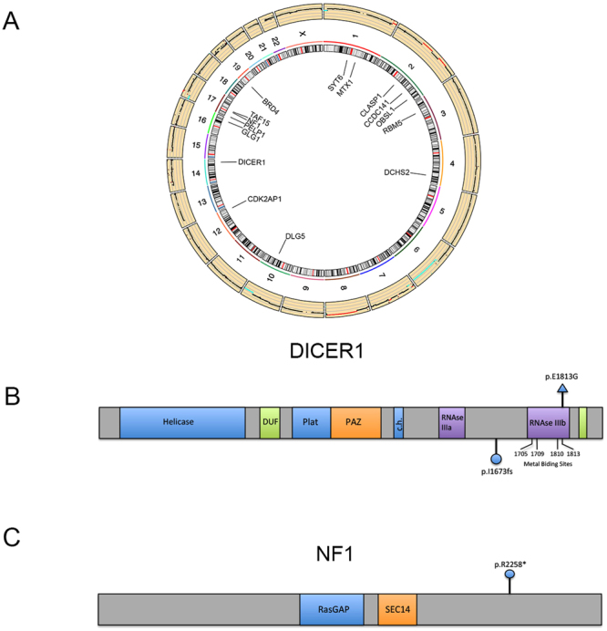
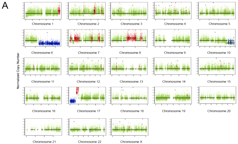
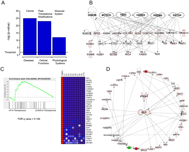
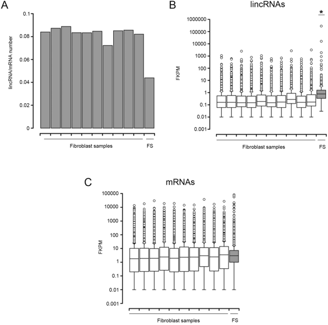
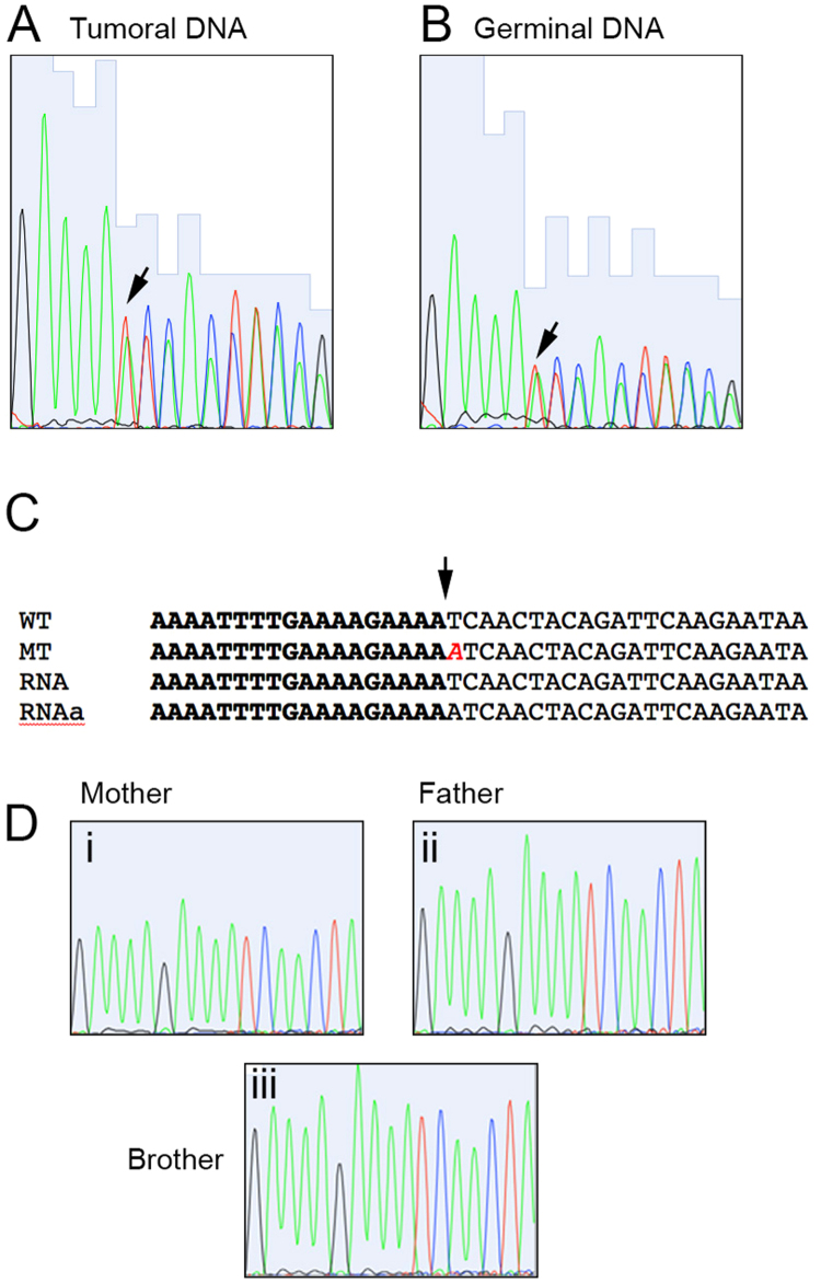
Ovarian fibrosarcomas are extremely rare tumors with little genomic information available to date. In the present report we present the tumoral exome and transcriptome and the germinal exome of an ovarian fibrosarcoma from a 9-years old child. We found a paucity of mutations (0.77/Mb) and CNV alterations. Of these, the most relevant were a point mutation in the metal-binding site of the microRNA-processing DICER1 enzyme and a frame-shift alteration in the tumor suppressor gene NF1. We validated a germinal truncating mutation in DICER1, which was consistent with a DICER1 Syndrome diagnosis, providing the first example of an ovarian fibrosarcoma as the presenting neoplasia in this syndrome. Network and enrichment analyses showed that both a mesenchymal signature and a Hedgehog cascade could be driving the progression of this tumor. We were also able to find a global lincRNA deregulation, as the number of lincRNAs transcripts expressed in the tumor was decreased, with a concomitant upregulation of previously described non-coding transcripts associated with cancer, such as MALAT1, MIR181A1HG, CASC1, XIST and FENDRR. DICER1 Syndrome should be considered as a possible diagnosis in children ovarian fibrosarcoma. The role of lncRNAs in neoplasias associated with DICER1 alterations need to be studied in more detail.


Conflict of interest statement

The authors declare no competing interests.

Figures

Figure 1
Figure 1
Ovarian fibrosarcoma pathology. (A) Hematoxylin-Eosin staining at 40x, (B) 100x and (C) 200x. (D) Immunohistochemistry of Vimentin. (E) Immunohistochemistry of Inhibin. Scale bars are shown at the right bottom of each micrograph.
Figure 2
Figure 2
Mutational landscape of the ovarian fibrosarcoma. (A) Circos plot showing, at the outermost ring, the copy-number alterations found in the tumor. Mutations (SNV and InDels) in codifying genes are shown at the center. (B) Diagram of the DICER1 gene, showing its main domains. The mutation found in the tumor is marked at the upper side of the diagram, whereas the mutation found in the germinal DNA is depicted below it. (C) Diagram of the NF1 gene, showing its main domains. The mutation found in the tumor is shown.
Figure 3
Figure 3
Copy number alterations in the ovarian fibrosarcoma sample. (A) Copy-number profile in the tumor. The normalized copy number is depicted against the length of each chromosome. The mean copy number is marked with a line. Deletions are depicted below and amplifications above the mean copy number line.
Figure 4
Figure 4
Network analysis of expression data. (A) The top enriched function in each category is plotted with the log p-value. The significance threshold is marked. (B) Mesenchymal network derived from the upstream/master regulators found in the expression dataset. (C) Gene Set Enrichment Analysis (GSEA) of the mesenchymal hallmark signature. Left Panel: The enrichment score is compared with the up-regulated (left) and down-regulated (right) genes. The False Discovery Rate is also shown. Right Panel: The top regulated genes found in the analysis. Lighter gray, higher expression. Darker gray, lower expression. (D) The Hedgehog pathway is enriched in the ovarian fibrosarcoma sample. Gray tones represent expressional changes.
Figure 5
Figure 5
Long-intergenic non-coding RNAs are deregulated in the fibrosarcoma sample. (A) Plot showing the ratio of lincRNAs versus mRNAs in the fibrosarcoma (FS) and control fibroblasts samples. (B) Boxplots showing the lincRNAs expression, in Fragments Per Kilobase of Exon per Million Fragments Mapped (FPKM), of each analyzed sample. The median, upper and lower quartiles and outliers are depicted for the fibrosarcoma (FS) and control fibroblasts samples. (C) Boxplots showing the mRNAs expression in FPKM, of each analyzed sample. The median, upper and lower quartiles and outliers are depicted for the fibrosarcoma (FS) and control fibroblasts samples.
Figure 6
Figure 6
DICER1 mutations. Sequence traces from Sanger sequencing of the (A) Tumoral and (B) Germinal DNA. The arrow shows the insertion point, where a duplicate signal starts. (C) Nucleotide alignment of wild type (WT) and mutated (MT) sequences derived from the exome data, with the sequences obtained from RNA (RNA and RNAa). The inserted adenine is shown in cursive. (D) Sequence traces from the mother (i), father (ii) and brother (iii) of the affected individual.

References

    1. Christman JE, Ballon SC. Ovarian fibrosarcoma associated with Maffucci’s syndrome. Gynecol Oncol. 1990;37:290–291. doi: 10.1016/0090-8258(90)90350-T. - DOI - PubMed
    1. Kraemer BB, Silva EG, Sneige N. Fibrosarcoma of ovary. A new component in the nevoid basal-cell carcinoma syndrome. Am J Surg Pathol. 1984;8:231–236. doi: 10.1097/00000478-198403000-00010. - DOI - PubMed
    1. Kruger S, Schmidt H, Kupker W, Rath FW, Feller AC. Fibrosarcoma associated with a benign cystic teratoma of the ovary. Gynecol Oncol. 2002;84:150–154. doi: 10.1006/gyno.2001.6408. - DOI - PubMed
    1. Nieminen U, Numers CV, Purola E. Primary sarcoma of the ovary. Acta Obstet Gynecol Scand. 1969;48:423–432. doi: 10.3109/00016346909156657. - DOI - PubMed
    1. Ghosh AK. Bilateral fibrosarcoma of the ovary following hysterectomy. Am J Obstet Gynecol. 1972;112:1136–1138. doi: 10.1016/0002-9378(72)90196-2. - DOI - PubMed

Publication types