. 2018 Mar;25(3):208-216.
doi: 10.1038/s41594-018-0030-z. Epub 2018 Feb 19.

2'-O-methylation in mRNA disrupts tRNA decoding during translation elongation

Affiliations

2'-O-methylation in mRNA disrupts tRNA decoding during translation elongation

Junhong Choi et al. Nat Struct Mol Biol. 2018 Mar.

Abstract

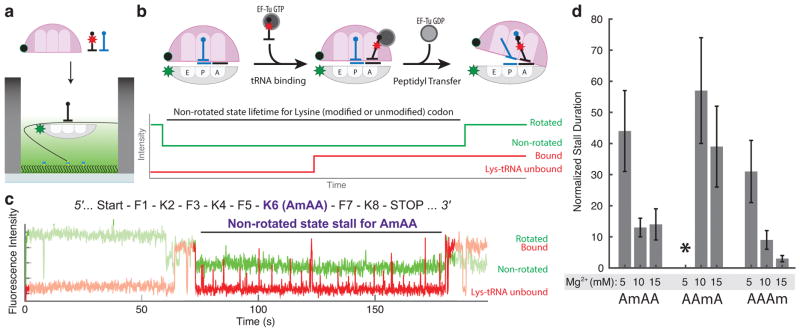
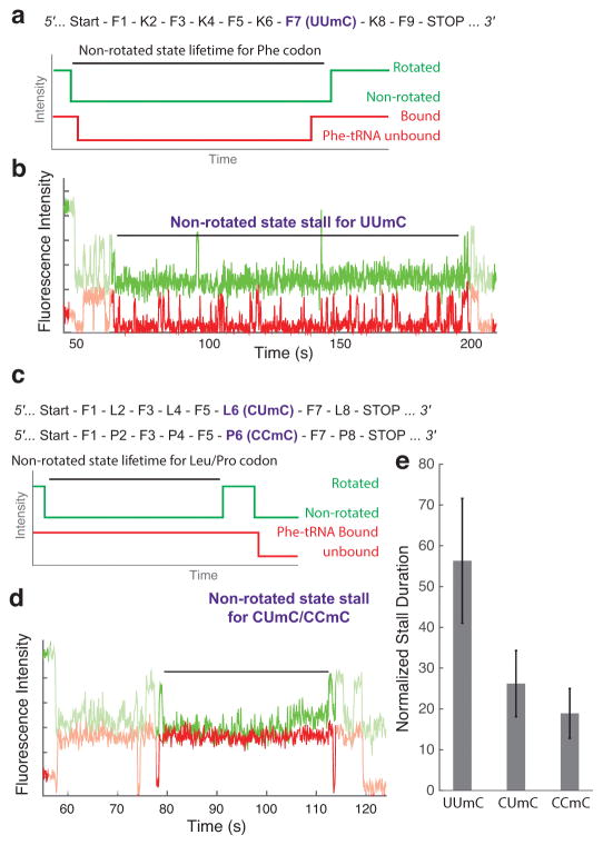
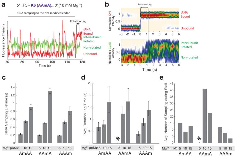
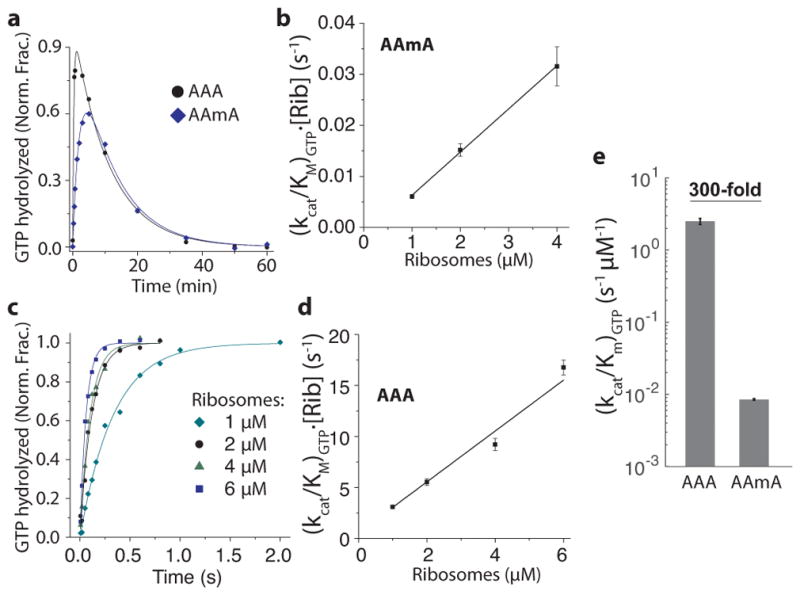
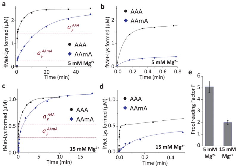
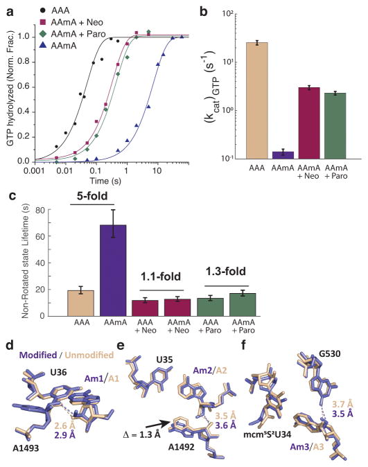
Chemical modifications of mRNA may regulate many aspects of mRNA processing and protein synthesis. Recently, 2'-O-methylation of nucleotides was identified as a frequent modification in translated regions of human mRNA, showing enrichment in codons for certain amino acids. Here, using single-molecule, bulk kinetics and structural methods, we show that 2'-O-methylation within coding regions of mRNA disrupts key steps in codon reading during cognate tRNA selection. Our results suggest that 2'-O-methylation sterically perturbs interactions of ribosomal-monitoring bases (G530, A1492 and A1493) with cognate codon-anticodon helices, thereby inhibiting downstream GTP hydrolysis by elongation factor Tu (EF-Tu) and A-site tRNA accommodation, leading to excessive rejection of cognate aminoacylated tRNAs in initial selection and proofreading. Our current and prior findings highlight how chemical modifications of mRNA tune the dynamics of protein synthesis at different steps of translation elongation.


Conflict of interest statement

Competing Financial Interests Statement

The authors declare no competing financial interests.

Figures

Figure 1
Figure 1. Multiple cognate tRNAs are rejected during decoding of the 2′-O-methylated codon
(a) Single-molecule experimental schematics. Fluorescently labeled pre-initiation complex is tethered to the surface of the ZMW well via biotinylated mRNA, and necessary translation factors are delivered in the beginning of the signal acquisition. (b) Expected sequence of events between translocation from the previous codon and the peptidyl transfer reaction. Both translocation and peptidyl transfer events are detected from the ribosomal inter-subunit conformation change from the rotated state to the non-rotated state via change in the dye-quencher signal. Independently, tRNA binding events are detected from co-localized fluorescence from fluorescently-labeled tRNA. (c) Representative experimental trace at 5 mM Mg2+ condition with the first-base modified codon (AmAA) shows a long stall between the translocation to the modified codon and its peptidyl transfer event. (d) Quantification of stall on the different modified Lys codon at different Mg2+ condition. The stall duration measured as in (c) was normalized to the non-rotated state lifetimes for the unmodified codon, as specified in the Supplementary Note 1 (n = 147, 154, 77, 111, 94, 111, 120, and 108 from left to right; error bars were calculated from propagating s.e. from fitting the single-exponential distributions; * on AAmA codon at 5 mM Mg2+ condition indicates that out of all traces that showed translation leading to the modified codon, less than 5% showed a tRNA-binding/ribosome-rotation event on the modified codon, hindering calculation of the normalized stall duration).
Figure 2
Figure 2. Codon-context alters the magnitude of 2′-O-methylation-induced stall
(a) mRNA sequence used to test the effect of 2′-O-methylation on the Phe codon, and the expected sequence of events between translocation from the previous codon and the peptidyl transfer reaction on Phe codon using Phe-(Cy5)-tRNAPhe. (b) Representative experimental trace shows a long stall between the translocation to the modified codon and its peptidyl transfer event on the UUmC codon. (c) mRNA sequence used to test the effect of 2′-O-methylation on the Leu and Pro codons, and the expected sequence of events between translocation from the previous codon and the peptidyl transfer reaction on the Leu or Pro codon using Phe-(Cy5)-tRNAPhe. (d) Representative experimental trace shows a long stall between the translocation to the modified codon and its peptidyl transfer event on the CUmC and CCmC codon. (e) Quantification of stall on the different modified codons at 5 mM Mg2+ condition (n = 131, 108 and 104 from left to right; error bars were calculated from propagating s.e. from fitting the single-exponential distributions).
Figure 3
Figure 3. 2′-O-methylation prolongs time between the initial tRNA binding and its full accommodation event
(a) A representative trace from the 10 mM total Mg2+ experimental condition (AAmA codon) shows a time-lag between the initial tRNA binding and the ribosomal intersubunit rotation, which follows the peptidyl transfer reaction. tRNA samplings and rotation lags on the modified codon were observed in all 3 2′-O-methylated codons. (b) Contour plots of the normalized Cy3B and Cy5 intensity trajectories, generated by post-synchronizing to the initial tRNA binding event (red; unbound to bound state) that leads to the ribosomal rotation (green; non-rotated to rotated state; n = 81). (c) Lifetimes of futile tRNA samplings on the modified codon observed in AmAA, AAmA and AAAm codons. (n = 1008, 1188, 759, 1961, 3294, 1452, 731, 862 and 316 from left to right; error bars represent s.e. from fitting the single-exponential distributions) (d) Average time between the initial tRNA binding and the ribosomal intersubunit rotational event on the modified codon observed in AmAA, AAmA and AAAm codons. (n = 65, 123, 42, 81, 64, 64, 124 and 92 from left to right; error bars represent s.e. from fitting the single-exponential distributions; * on AAmA codon at 5 mM Mg2+ condition indicates that out of all traces that showed translation leading to the modified codon, less than 5% showed a tRNA-binding/ribosome-rotation event on the modified codon, same as in Figure 1d). (e) Average number of futile sampling events prior to the successful tRNA accommodation on the 2′-O-methylated codons (n = 147, 154, 77, 111, 94, 111, 120 and 108 from left to right).
Figure 4
Figure 4. Bulk kinetics measurements of (kcat/Km)GTP with modified (AAmA) and unmodified (AAA) codons at 5 mM Mg2+
(a) A representative time course of GTP hydrolysis on AAmA codon. 0.7 μM Lys-tRNALys ternary complexes were reacted to 1 μM ribosomes initiated with AAA or AAmA codons in the A site. AAmA reaction was run in parallel with AAA reaction and the curves fitted with shared parameters to increase precision (see Online Methods). The decrease of the fraction of hydrolyzed [3H]GTP was due to its regeneration by pyruvate kinase. (b) Ribosome titration to determine kcat/Km value of GTP hydrolysis on EF-Tu with AAmA codon in the A site (error bars represent s.d. from n = 3 technical replicates). (c) Time courses of GTP hydrolysis on AAA codon at different ribosome concentrations. Lys-tRNALys ternary complexes (0.3 μM) were reacted to 70S programed with AAA codon in the A site. (d) Ribosome titration to determine (kcat/Km)GTP with AAA codon in the A site (error bars represent s.d. from n = 3 technical replicates). (e) Estimates of (kcat/Km)GTP from b and d (2.5 ± 0.2 and 0.0085 ± 0.0002 s-1 μM−1 for AAA and AAmA, respectively). Error bars represent s.d. from fitting procedure.
Figure 5
Figure 5. The effect of 2′-O-methylation on proofreading
(a) Time courses of fMet-Lys dipeptide formation at 5 mM total Mg2+ (1.3 mM free). Parallel reactions with Lys-tRNALys ternary complexes (2 μM) were performed with ribosomes (4.2 μM) containing AAA or AAmA codon in the A site. The curves were fitted to a model with two exponentials (see Online Methods). The proofreading factor F is expressed as the ratio of the fast phase amplitudes (aF) on AAA(aFAAA) and AAmA(aFAAmA)codons:=aFAAA/aFAAmA. The aF values obtained from the fit are shown as red dotted lines. The lower aF for AAmA codon indicates the rejection of Lys-tRNALys after GTP hydrolysis in the presence of the modification. (b) The first 0.8 min of the experiment in a expanded to highlight the fast phase of the reaction. (c) The same reaction as in a, but at 15 mM total Mg2+ (7.5 mM free) with 1.5 μM ribosomes and 1 μM ternary complexes. (d) The first 0.5 min of c, expanded to highlight the fast phase of the reaction. (e) Proofreading factor measured at 5 mM Mg2+ condition (F = 5.1 ± 0.4), and at 15 mM Mg2+ condition (F = 2 ± 0.2; error bars represent s.d. from n = 3 technical replicates).
Figure 6
Figure 6. The delay in GTP-hydrolysis by 2′-O-methylation is due to the inactivation of the monitoring bases
(a) Kinetics of GTP hydrolysis at 15 mM Mg2+ condition. Lys-tRNALys ternary complexes (0.4 μM) were reacted to 70S ribosomes (1 μM) programed with AAA or AAmA codon in the A site. 10 μM neomycin (Neo) or paromomycin (Paro) were added where indicated. (b) Estimates of (kcat)GTP from a. Error bars represent s.d. from n = 3 technical replicates. (c) Non-rotated state lifetimes measured on either AAA or AAmA codon in the A site with or without 10 μM neomycin (Neo) or paromomycin (Paro) using single-molecule assay (n = 194, 172, 153, 201, 182 and 243 instances; error bars represent s.e. from fitting the single-exponential distribution) (d–f) X-ray crystallography structures of the decoding center (UUU anticodon and A1492, A1493 and G530 monitoring bases) while decoding (d) the first base modified (AmAA), (e) the second base modified (AAmA), and (f) the third base modified Lys codon (AAAm), soaked in the 30S subunit crystals in the presence of paromomycin. Displayed distances indicate interatomic distance (dashed lines) for possible hydrogen bonds between the monitoring bases and 2′-O-moiety on the decoded codon (purple for the modified base and tan for the unmodified base). 1.3 Å shift in A1492 base position is indicated by black arrow in panel e.
Figure 7
Figure 7
Mechanism of epitranscriptomic-induced translation elongation stall. (Top) The schematic of translation decoding, starting from aa-tRNA TC binding event to codon recognition event, monitoring base activation, GTP-hydrolysis, two-step proofreading and full accommodation/peptidyl-transfer reaction (left to right). While 2′-O-methylation does not interrupt codon-anticodon RNA duplex formation (the faded-out step), it sterically hinders dynamics of monitoring bases, delaying GTPase activation and causing rejections of multiple tRNA molecules during proofreading. (Bottom) m6A modification disrupts codon-anticodon RNA duplex stability during initial selection and for proofreading.

References

    1. Zhao BS, Roundtree IA, He C. Post-transcriptional gene regulation by mRNA modifications. Nat Rev Mol Cell Biol. 2016;18:31–42. - PMC - PubMed
    1. Liu N, et al. N6-methyladenosine-dependent RNA structural switches regulate RNA–protein interactions. Nature. 2015;518:560–564. - PMC - PubMed
    1. Fu Y, Dominissini D, Rechavi G, He C. Gene expression regulation mediated through reversible m6A RNA methylation. Nat Rev Genet. 2014;15:293–306. - PubMed
    1. Choi J, et al. N6-methyladenosine in mRNA disrupts tRNA selection and translation-elongation dynamics. Nat Struct Mol Biol. 2016;23:110–115. - PMC - PubMed
    1. Dai Q, et al. Nm-seq maps 2′-O-methylation sites in human mRNA with base precision. Nat Methods. 2017 doi: 10.1038/nmeth.4294. - DOI - PMC - PubMed

Publication types