. 2018 Mar;25(3):217-224.
doi: 10.1038/s41594-018-0031-y. Epub 2018 Feb 19.

GlcNAc-1-P-transferase-tunicamycin complex structure reveals basis for inhibition of N-glycosylation

Affiliations

GlcNAc-1-P-transferase-tunicamycin complex structure reveals basis for inhibition of N-glycosylation

Jiho Yoo et al. Nat Struct Mol Biol. 2018 Mar.

Abstract

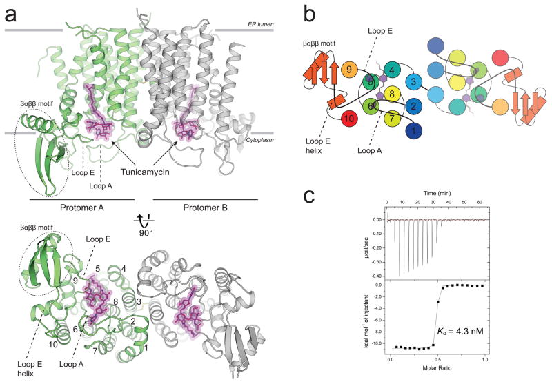
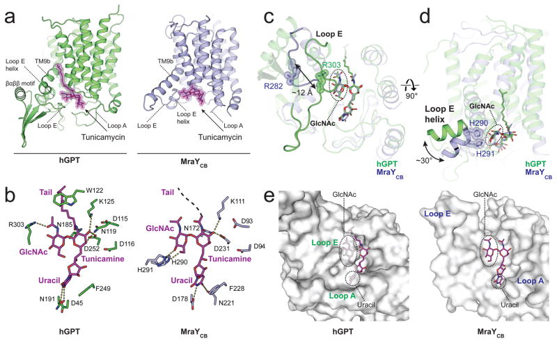
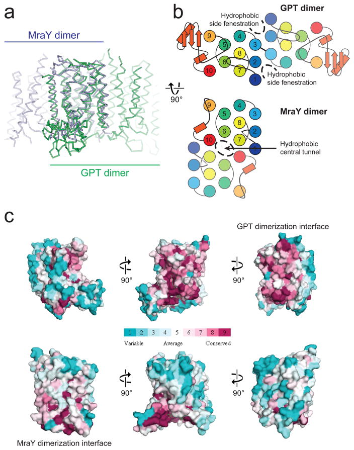
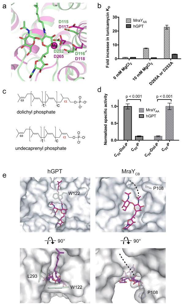
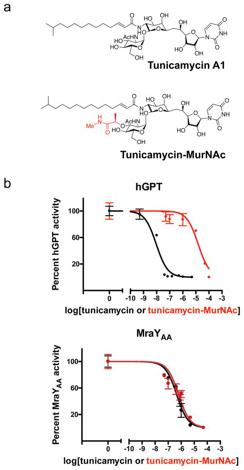
N-linked glycosylation is a predominant post-translational modification of protein in eukaryotes, and its dysregulation is the etiology of several human disorders. The enzyme UDP-N-acetylglucosamine:dolichyl-phosphate N-acetylglucosaminephosphotransferase (GlcNAc-1-P-transferase or GPT) catalyzes the first and committed step of N-linked glycosylation in the endoplasmic reticulum membrane, and it is the target of the natural product tunicamycin. Tunicamycin has potent antibacterial activity, inhibiting the bacterial cell wall synthesis enzyme MraY, but its usefulness as an antibiotic is limited by off-target inhibition of human GPT. Our understanding of how tunicamycin inhibits N-linked glycosylation and efforts to selectively target MraY are hampered by a lack of structural information. Here we present crystal structures of human GPT in complex with tunicamycin. Structural and functional analyses reveal the difference between GPT and MraY in their mechanisms of inhibition by tunicamycin. We demonstrate that this difference could be exploited to design MraY-specific inhibitors as potential antibiotics.


Conflict of interest statement

Competing financial interests The authors declare no competing financial interests.

Figures

Figure 1
Figure 1. The crystal structure of human GPT in complex with tunicamycin reveals the structural basis for high affinity binding
a, hGPT crystallized as a homodimer with one tunicamycin molecule (magenta sticks) bound to the cytosolic active site cavity of each protomer, tightly enclosed by loops A and E. The two protomers are related by pseudo-twofold rotational symmetry and are covalently linked by a disulfide bond between TM3 of each protomer. b, Cartoon representation of the cytoplasmic view, rainbow colored from N-terminus (blue) to C-terminus (red). c, Representative ITC raw data (top) and binding isotherm (bottom) for tunicamycin interacting with wild-type hGPT; Kd = 4.3 nM, ΔH° = −10.8 kcal/mol. This ITC experiment was performed in triplicate (technical replicates (5.6+/−1.3 nM, mean +/− S.E.M.)).
Figure 2
Figure 2. Tunicamycin binding to GPT is analogous but distinct from that to MraY
a, Tunicamycin (magenta sticks) binds to the active site of both hGPT (green) and bacterial MraYCB (blue, PDB 5JNQ) near the cytoplasmic side of the membrane. The aliphatic tail is not resolved in the MraY-tunicamycin structure. b, Interactions between tunicamycin and either hGPT or MraYCB. c, Loop E of hGPT is more extensive and closer to tunicamycin than it is the MraYCB structure, bringing the invariant residue Arg303 into contact with the GlcNAc moiety of tunicamycin. The corresponding residue in MraYCB (Arg282) does not interact with tunicamycin. d, The Loop E helix of hGPT is swiveled by 30 degrees towards the horizontal plane as compared to MraYCB. e, Tunicamycin is almost completely enclosed in the binding pocket of hGPT, as compared to the solvent-exposed groove of MraY. In hGPT, the uracil and GlcNAc moieties of tunicamycin are completely covered by loop A and loop E respectively, while are both solvent-exposed in MraY.
Figure 3
Figure 3. Divergence between GPT and MraY is underpinned by their different dimerization interfaces
a, Superposition of hGPT (green) with MraYCB (blue) reveals strikingly different dimerization interfaces, despite the conserved fold of each protomer. b, The different dimerization interfaces of GPT and MraY result in altered membrane accessibility. Hydrophobic side fenestrations penetrate the GPT dimer, while a central hydrophobic tunnel occupies the MraY dimer. c, The interface for GPT dimerization is conserved only among GPT orthologue sequences but not among MraY sequences, and vice versa is true for MraY. Surfaces are colored in increasing conservation from cyan (least) to magenta (most). Sequence conservation was mapped onto the hGPT and MraYCB structures using 30 orthologue sequences for each alignment.
Figure 4
Figure 4. MraY and GPT bind tunicamycin and magnesium differently and they are selective for distinct lipid substrates
a, Structural overlay of hGPT-tunicamycin (green) and Mg2+-bound MraYAA (magenta, PDB ID: 4J72). Labeled residues in magenta are known to be critical for MraYAA activity and Asp265 coordinates the magnesium cofactor (magenta sphere). b, Fold increase in Kd of tunicamycin with either hGPT or MraY as measured by ITC. The Kd of tunicamycin for hGPT without added MgCl2 is 5.6 ± 1.3 nM (triplicate, technical replicates). The Kd of tunicamycin for MraYAA without added MgCl2 is 37 ± 1 nM (triplicate, technical replicates). Data are shown as the mean of three technical replicates ± s.e.m. c, Chemical structures of dolichyl phosphate and undecaprenyl phosphate. d, TLC-based specific activity assay for hGPT and MraYAA in the presence of either dolichyl phosphate (C55-dol-P) or undecaprenyl phosphate (C55-P). hGPT is selective for the saturated α-isoprenyl position in its substrate dolichyl-phosphate, while MraY is selective for the unsaturated α-isoprenyl position in its substrate undecaprenyl-phosphate. Measurements were performed in triplicate (technical replicates). Specific activity measurements were normalized relative to the C55-Dol-P condition for hGPT and to the C55-P condition for MraY. Data are shown as the mean of normalized specific activity ± s.e.m. Significance was determined with P < 0.001, two-tailed Student’s t-test. e, Cytoplasmic view of the tunicamycin lipid tail binding sites in hGPT and MraYCB. In GPT, the aliphatic tail of tunicamycin packs against the conserved Trp122, which forms a lipid-binding tunnel. By contrast, the corresponding residue in MraYCB (Pro108) is unable lock the lipid tail into place, leaving the hydrophobic groove exposed.
Figure 5
Figure 5. Chemically modifying tunicamycin can introduce ligand selectivity between hGPT and MraYAA
a, Chemical structures of tunicamycin and its MurNAc derivative. The substructure highlighted in red differs from tunicamycin, being a MurNAc-like moiety rather than a GlcNAc moiety as in tunicamycin. b, IC50 measurements of tunicamycin and its MurNAc analog (tunicamycin-MurNac) with hGPT and MraYAA, respectively. hGPT is much less inhibited by tunicamycin-MurNAc than tunicamycin, while MraY appears to be similarly inhibited by both tunicamycin and tunicamycin-MurNAc. The IC50 for hGPT with tunicamycin is ~9 nM; for hGPT with tunicamycin-MurNAc is ~15 μM; for MraYAA with tunicamycin is ~450 nM; for MraYAA with tunicamycin-MurNAc is ~640 nM. Each IC50 value was determined using the TLC-based phosphoglycosyl transferase assay. Data are shown as the mean of triplicate measurements (technical replicates) ± s.e.m.

Similar articles

Cited by

References

    1. Lehrman MA. Biosynthesis of N-acetylglucosamine-P-P-dolichol, the committed step of asparagine-linked oligosaccharide assembly. Glycobiology. 1991;1:553–562. - PubMed
    1. Belaya K, et al. Mutations in DPAGT1 cause a limb-girdle congenital myasthenic syndrome with tubular aggregates. Am J Hum Genet. 2012;91:193–201. doi: 10.1016/j.ajhg.2012.05.022. - DOI - PMC - PubMed
    1. Wu X, et al. Deficiency of UDP-GlcNAc:Dolichol Phosphate N-Acetylglucosamine-1 Phosphate Transferase (DPAGT1) causes a novel congenital disorder of Glycosylation Type Ij. Hum Mutat. 2003;22:144–150. doi: 10.1002/humu.10239. - DOI - PubMed
    1. Wurde AE, et al. Congenital disorder of glycosylation type Ij (CDG-Ij, DPAGT1-CDG): extending the clinical and molecular spectrum of a rare disease. Mol Genet Metab. 2012;105:634–641. doi: 10.1016/j.ymgme.2012.01.001. - DOI - PubMed
    1. Liwosz A, Lei T, Kukuruzinska MA. N-glycosylation affects the molecular organization and stability of E-cadherin junctions. J Biol Chem. 2006;281:23138–23149. doi: 10.1074/jbc.M512621200. - DOI - PubMed

Publication types

MeSH terms

Substances