. 2018 Feb 5:9:47.
doi: 10.3389/fneur.2018.00047. eCollection 2018.

Novel Approaches to the Diagnosis of Chronic Disorders of Consciousness: Detecting Peripersonal Space by Using Ultrasonics

Affiliations

Novel Approaches to the Diagnosis of Chronic Disorders of Consciousness: Detecting Peripersonal Space by Using Ultrasonics

Antonino Naro et al. Front Neurol. .

Abstract

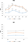
The assessment of behavioral responsiveness in patients suffering from chronic disorders of consciousness (DoC), including Unresponsive Wakefulness Syndrome (UWS) and Minimally Conscious State (MCS), is challenging. Even if a patient is unresponsive, he/she may be covertly aware in reason of a cognitive-motor dissociation, i.e., a preservation of cognitive functions despite a solely reflexive behavioral responsiveness. The approach of an external stimulus to the peripersonal space (PPS) modifies some biological measures (e.g., hand-blink reflex amplitude) to the purpose of defensive responses from threats. Such modulation depends on a top-down control of subcortical neural circuits, which can be explored through changes in cerebral blood flow velocity (CBFV), using functional transcranial Doppler (fTCD) and, thus, gaining useful, indirect information on brain connectivity. These data may be used for the DoC differential diagnosis. We evaluated the changes in CBFV by measuring the pulsatility index (PI) in 21 patients with DoC (10 patients with MCS and 11 with UWS) and 25 healthy controls (HC) during a passive movement and motor imagery (MI) task in which the hand of the subject approached and, then, moved away from the subject's face. In the passive movement task, the PI increased progressively in the HCs when the hand was moved toward the face and, then, it decreased when the hand was removed from the face. The PI increased when the hand was moved toward the face in patients with DoC, but then, it remained high when the hand was removed from the face and up to 30 s after the end of the movement in the patients with MCS (both MCS+ and MCS-) and 1 min in those with UWS, thus differentiating between patients with MCS and UWS. In the MI task, all the HCs, three out of four patients with MCS+, and one out of six patients with MCS- showed an increase-decrease PI change, whereas the remaining patients with MCS and all the patients with UWS showed no PI changes. Even though there is the possibility that our findings will not be replicated in all patients with DoC, we propose fTCD as a rapid and very easy tool to differentiate between patients with MCS and UWS, by identifying residual top-down modulation processes from higher-order cortical areas to sensory-motor integration networks related to the PPS, when using passive movement tasks.

Keywords: cerebral blood flow; chronic disorders of consciousness; motor imagery; peripersonal space; transcranial ultrasound.


Figures
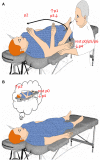
Figure 1
Figure 1
Summarizes the experimental paradigm. Concerning passive movement task (A), the subject was verbally instructed to keep the eyes open, to relax, and to be prepared to be moved the right upper limb from trunk toward the face and vice versa. The speed of passive movement was kept as constant as possible. When the hand reached a predetermined position (p), the examiner kept hold of the participant’s forearm up to the next movement onset, which occurred every 15 heartbeats. Concerning motor imagery task (B), the subject was provided with two sequential instructions, provided every 15 heartbeats: (i) move your right hand toward the face and keep hold of it, and (ii) came back where you started. All instructions were 2 s in length and presented by loudspeakers.
Figure 2
Figure 2
Shows mean (±SD; vertical error bar) pulsatility index (PI) values during passive movement (PM) and motor imagery (MI) tasks for each group [healthy controls (HC), minimally conscious state (MCS), and unresponsive wakefulness syndrome (UWS)] across the hand positions (p) explored. There was a significant difference at p0 (baseline) between the PI values of HCs and patients with disorders of consciousness (p < 0.001), and between patients with MCS and UWS (p = 0.01). PM induced a significantly different PI modulation at each p among the groups (p < 0.001). On the contrary, MI induced a significant increase of PI at p2 and decrease at p4 only in the HCs. *Refer to the significance of intragroup PI change at each p as compared to the previous one (Bonferroni corrected p-value, ***p < 0.001, **p = 0.001, *p < 0.0125).
Figure 3
Figure 3
Shows mean (±SD; vertical error bar) heart rate values [in beats per minute (bpm)] during passive movement (PM) and motor imagery (MI) tasks for each group [healthy controls (HC), minimally conscious state (MCS), and unresponsive wakefulness syndrome (UWS)] across the hand positions (p) explored. There was a significant difference at p0 (baseline) between the bpm values of HCs and patients with disorders of consciousness (p < 0.001), and between patients with MCS and UWS (p = 0.01). PM induced a significantly different bpm modulation at each p among the groups (p < 0.001). On the contrary, MI did not induce any significant bpm change. *Refer to the significance of intragroup pulsatility index change at each p as compared to the previous one (Bonferroni corrected p-value, ***p < 0.001, **p = 0.001, *p < 0.0125).
Figure 4
Figure 4
Shows the transcranial Doppler waveforms (on the right) from left middle (MCA), anterior (ACA), and posterior (PCA) cerebral arteries at rest in a representative subject for each group (HC, MCS, and UWS). Flow velocity (in centimeter per second, right vertical bar) are provided.
Figure 5
Figure 5
Shows the transcranial Doppler waveforms (on the right) with color Doppler (on the left) from left middle cerebral artery during passive mobilization across the six hand positions (p) explored in a representative subject for each group [healthy controls, minimally conscious state (MCS), and unresponsive wakefulness syndrome (UWS)]. Both insonation depth (left vertical bar) and flow velocity (centimeter per second, right vertical bar) are provided.
Figure 6
Figure 6
Shows the transcranial Doppler waveforms (on the right) with color Doppler (on the left) from left middle cerebral artery during motor imagery across the two hand positions (p) explored in a representative subject for each group [HC, minimally conscious state (MCS), and unresponsive wakefulness syndrome (UWS)]. Both insonation depth (left vertical bar) and flow velocity (cm/s, right vertical bar) are provided.
Figure 7
Figure 7
The power of motor task-induced cerebral blood flow velocity modulation across the hand-position employed in differentiating patients with disorders of consciousness is shown by the area under the receiver operating characteristic curve (AUC) for motor imagery (MI) and passive mobilization tasks.

Similar articles

Cited by

References

    1. Multi-Society Task Force on PVS. Medical aspects of the persistent vegetative state. N Engl J Med (1994) 330:1499–508.10.1056/NEJM199405263302107 - DOI - PubMed
    1. Giacino JT, Ashwal S, Childs N, Cranford R, Jennett B, Katz DI, et al. The minimally conscious state: definition and diagnostic criteria. Neurology (2002) 58:349–53.10.1212/WNL.58.3.349 - DOI - PubMed
    1. Giacino JT, Kalmar K, Whyte J. The JFK Coma Recovery Scale-Revised: measurement characteristics and diagnostic utility. Arch Phys Med Rehabil (2004) 85:2020–9.10.1016/j.apmr.2004.02.033 - DOI - PubMed
    1. Laureys S, Owen AM, Schiff ND. Brain function in coma, vegetative state, and related disorders. Lancet Neurol (2004) 3:537–46.10.1016/S1474-4422(04)00852-X - DOI - PubMed
    1. Bruno MA, Vanhaudenhuyse A, Thibaut A, Moonen G, Laureys S. From unresponsive wakefulness to minimally conscious PLUS and functional locked-in syndromes: recent advances in our understanding of disorders of consciousness. J Neurol (2011) 258(7):1373–84.10.1007/s00415-011-6114-x - DOI - PubMed