. 2018 Feb 5:9:61.
doi: 10.3389/fimmu.2018.00061. eCollection 2018.

Genome-Wide Bimolecular Fluorescence Complementation-Based Proteomic Analysis of Toxoplasma gondii ROP18's Human Interactome Shows Its Key Role in Regulation of Cell Immunity and Apoptosis

Affiliations

Genome-Wide Bimolecular Fluorescence Complementation-Based Proteomic Analysis of Toxoplasma gondii ROP18's Human Interactome Shows Its Key Role in Regulation of Cell Immunity and Apoptosis

Jing Xia et al. Front Immunol. .

Abstract

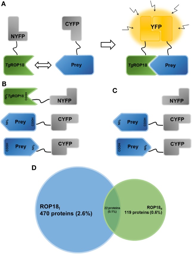
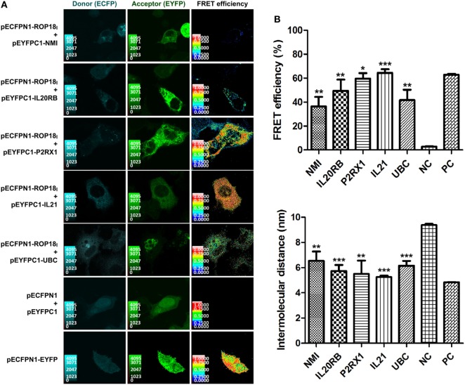
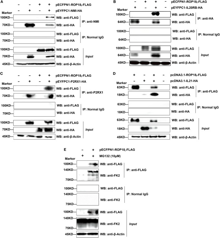
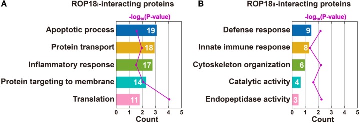
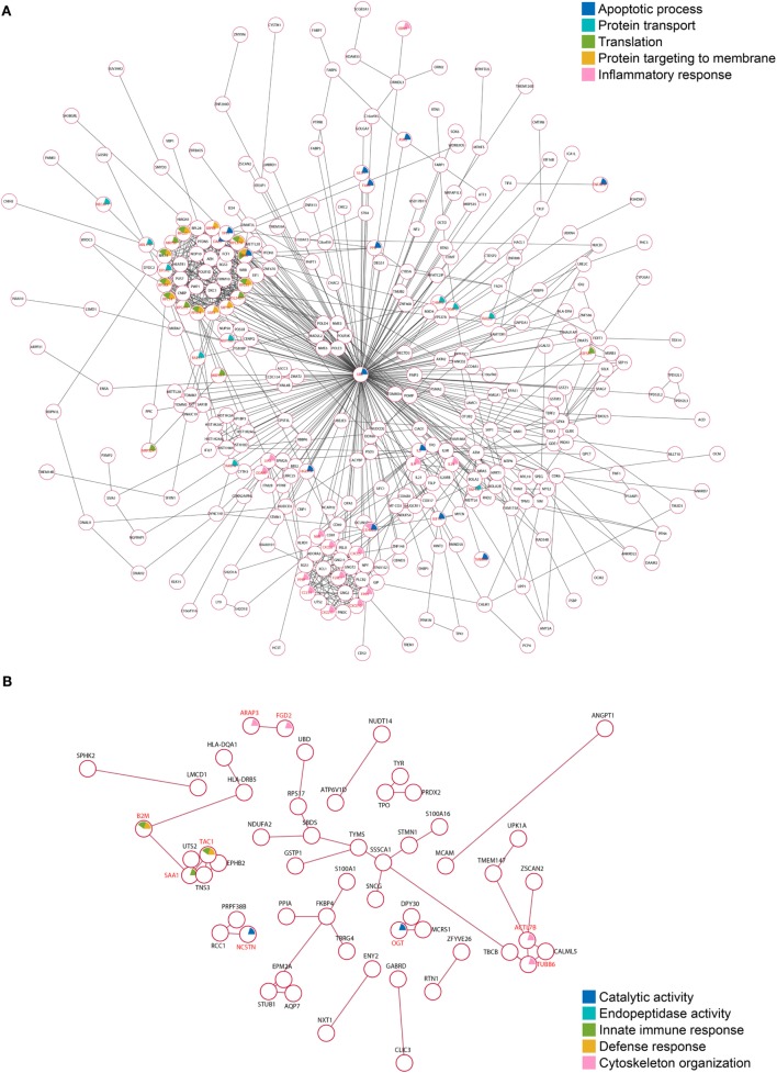
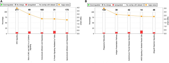
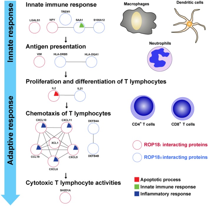
Toxoplasma gondii rhoptry protein ROP18 (TgROP18) is a key virulence factor secreted into the host cell during invasion, where it modulates the host cell response by interacting with its host targets. However, only a few TgROP18 targets have been identified. In this study, we applied a high-throughput protein-protein interaction (PPI) screening in human cells using bimolecular fluorescence complementation (BiFC) to identify the targets of Type I strain ROP18 (ROP18I) and Type II strain ROP18 (ROP18II). From a pool of more than 18,000 human proteins, 492 and 141 proteins were identified as the targets of ROP18I and ROP18II, respectively. Gene ontology, search tool for the retrieval of interacting genes/proteins PPI network, and Ingenuity pathway analyses revealed that the majority of these proteins were associated with immune response and apoptosis. This indicates a key role of TgROP18 in manipulating host's immunity and cell apoptosis, which might contribute to the immune escape and successful parasitism of the parasite. Among the proteins identified, the immunity-related proteins N-myc and STAT interactor, IL20RB, IL21, ubiquitin C, and vimentin and the apoptosis-related protein P2RX1 were further verified as ROP18I targets by sensitized emission-fluorescence resonance energy transfer (SE-FRET) and co-immunoprecipitation. Our study substantially contributes to the current limited knowledge on human targets of TgROP18 and provides a novel tool to investigate the function of parasite effectors in human cells.

Keywords: ROP18; Toxoplasma gondii; bimolecular fluorescence complementation; genome-wide; human interactome.


Figures

Figure 1
Figure 1
Establishment of the HT-BiFC screening system and TgROP18-interacting proteins. (A) Principle of the bimolecular fluorescence complementation (BiFC) assay. The non-fluorescent fragments of a fluorescent reporter protein are fused with the proteins of interest and expressed in human cells. If the interaction between the proteins of interest takes place, the split fragments will be pulled close enough to refold together and reconstitute the functional fluorescent entity. (B) Schematic representation illustrating the TgROP18/prey BiFC constructs generated in the present study. TgROP18 is fused with the N-terminal fragment of YFP (NYFP) at the C-terminus, and the prey protein is tethered with the C-terminal fragment of YFP (CYFP) at either the N- or C-terminus. (C) Schematic representation illustrating the control screening. Non-fused NYFP is mated with each CYFP-prey/prey-CYFP constructs. (D) Venn diagram depicting the number (percentage) of ROP18I-specific targets (blue), ROP18II-specific targets (green), and ROP18I/ROP18II targets (in the middle).
Figure 2
Figure 2
Validation of the TgROP18-interacting proteins by SE-FRET assay. (A) Co-localization and FRET interaction of ROP18I with NMI, IL20RB, P2RX1, IL21, and UBC. Localization and co-localization of ROP18I and the five indicated candidates are shown in the donor channel (column 1) and the acceptor channel (column 2), respectively. The FRET efficiency is shown in column 3, in which a thermal pseudo color-matched FRET signal intensity scale is indicated for each image. (B) Quantitative analysis of FRET efficiency and intermolecular distance between ROP18I and the five indicated candidates. Error bars represent the means ± SD of triplicates. Student’s t-tests results are between the six experimental groups and NC, *p < 0.05; **p < 0.01; ***p < 0.001. Abbreviations: NC, negative control; PC, positive control.
Figure 3
Figure 3
Validation of the TgROP18-interacting proteins by co-immunoprecipitation (Co-IP) assay. (A–D) Lysates of COS-7 cells co-overexpressing ROP18I and the indicated interacting proteins were immunoprecipitated with the indicated antibodies. Rabbit, mouse, or goat normal control IgG were used as negative controls. The immunoprecipitates were detected by SDS-PAGE and western blotting using the antibodies indicated. (E) Lysates of COS-7 cells overexpressing ROP18I in the presence of MG132 (10 μM) for 12 h were immunoprecipitated with the anti-FLAG antibody. Endogenous UBC (a smear of bands) was detected in the immunoprecipitates through western blotting with anti-FK2 antibody, which recognizes mono- and polyubiquitinylated conjugates.
Figure 4
Figure 4
Top five enriched biological processes for ROP18I (A) and ROP18II-interacting proteins (B) identified by GO analysis. (A) ROP18I-interacting proteins were significantly overrepresented in the biological processes of apoptotic process, protein transport, inflammatory response, protein targeting to membrane, and translation. (B) ROP18II-interacting proteins were significantly overrepresented in the biological processes of defense response, innate immune response, cytoskeleton organization, catalytic activity, and endopeptidase activity.
Figure 5
Figure 5
Protein–protein interaction (PPI) networks of the TgROP18-interacting proteins. (A) Among the ROP18I-interacting proteins, 353 (71.7%) are tied to a single large network with 785 edges. (B) Among the ROP18II-interacting proteins, 55 (39.0%) are enriched in a PPI network with 44 edges.
Figure 6
Figure 6
Top five enriched pathways for ROP18I (A) and ROP18II (B) interacting proteins. The stacked bar chart indicates the number of proteins overlapped with the database, and the connected orange points represent the logarithm of the p-values.
Figure 7
Figure 7
TgROP18-interacting proteins involved in the immune response. Numerous ROP18I (red circles) and ROP18II (blue circles) identified interacting proteins related to the processes of immune response. During T. gondii infection, innate immune response acts rapidly to provide the first line of defense and activate the adaptive immune response, with release of proinflammatory cytokines by macrophages, dendritic cells, and neutrophils. In the milieu of proinflammatory cytokines, T cell-mediated immune response is initiated when naïve CD4+ or CD8+ T cells encounter the parasite antigens presented by antigen presenting cells (APCs). Once the antigen-specific T lymphocytes are activated, they proliferate, differentiate, and traffic to the sites of infection, protecting the host by exhibiting cytotoxic T lymphocyte (CTL) activities toward infected cells.

Similar articles

Cited by

References

    1. Moncada PA, Montoya JG. Toxoplasmosis in the fetus and newborn: an update on prevalence, diagnosis and treatment. Expert Rev Anti Infect Ther (2012) 10:815–28. 10.1586/eri.12.58 - DOI - PubMed
    1. Morrison DA. Evolution of the apicomplexa: where are we now? Trends Parasitol (2009) 25:375–82. 10.1016/j.pt.2009.05.010 - DOI - PubMed
    1. Nichols BA, Chiappino ML, O’Connor GR. Secretion from the rhoptries of Toxoplasma gondii during host-cell invasion. J Ultrastruct Res (1983) 83:85–98. 10.1016/S0022-5320(83)90067-9 - DOI - PubMed
    1. Boothroyd JC, Dubremetz JF. Kiss and spit: the dual roles of Toxoplasma rhoptries. Nat Rev Microbiol (2008) 6:79–88. 10.1038/nrmicro1800 - DOI - PubMed
    1. Howe DK, Sibley LD. Toxoplasma gondii comprises three clonal lineages: correlation of parasite genotype with human disease. J Infect Dis (1995) 172:1561–6. 10.1093/infdis/172.6.1561 - DOI - PubMed

Publication types