. 2018 Feb 5:9:79.
doi: 10.3389/fimmu.2018.00079. eCollection 2018.

Type I Interferon Signaling Is Required for CpG-Oligodesoxynucleotide-Induced Control of Leishmania major, but Not for Spontaneous Cure of Subcutaneous Primary or Secondary L. major Infection

Affiliations

Type I Interferon Signaling Is Required for CpG-Oligodesoxynucleotide-Induced Control of Leishmania major, but Not for Spontaneous Cure of Subcutaneous Primary or Secondary L. major Infection

Ulrike Schleicher et al. Front Immunol. .

Abstract

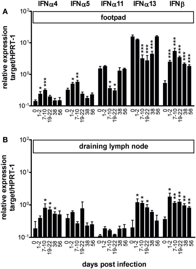
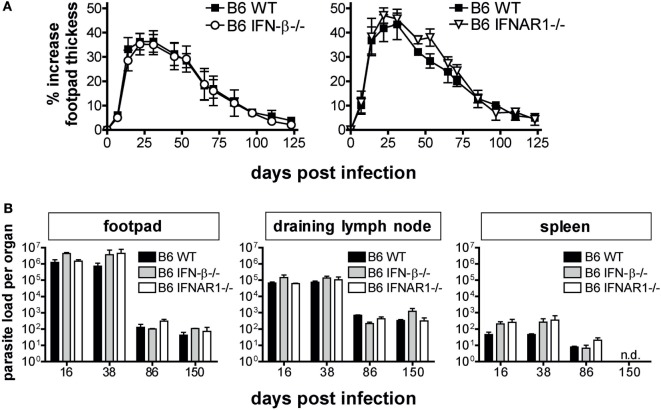
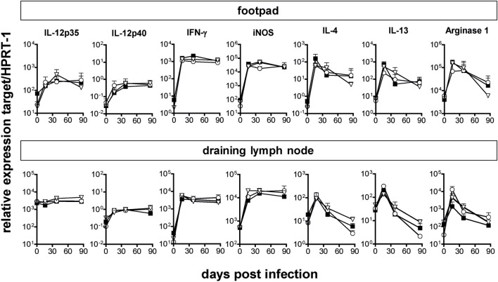
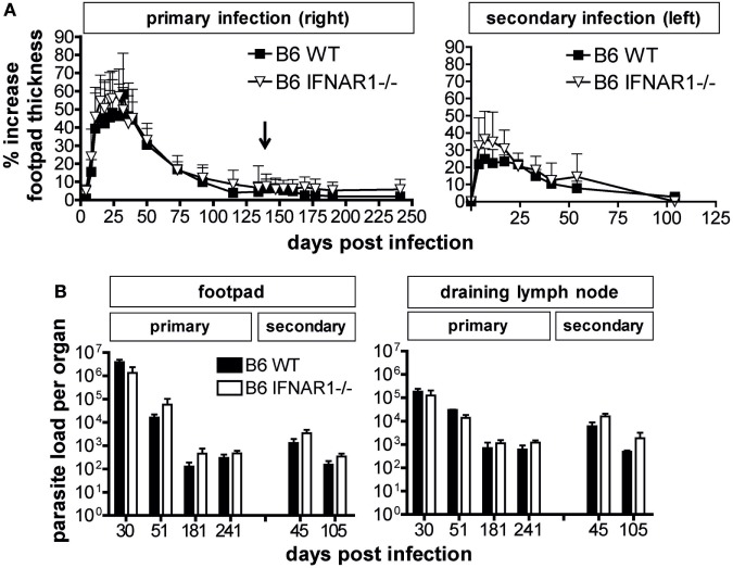
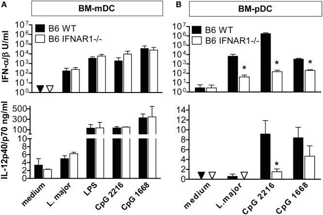
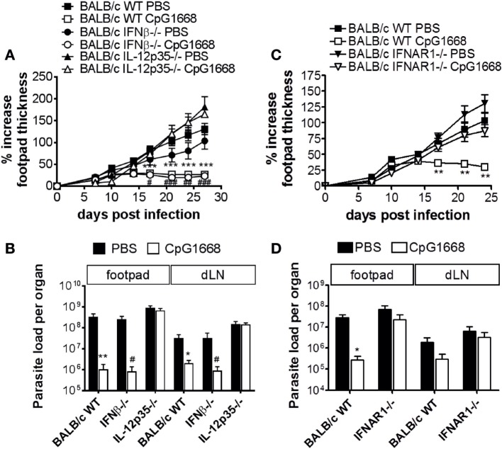
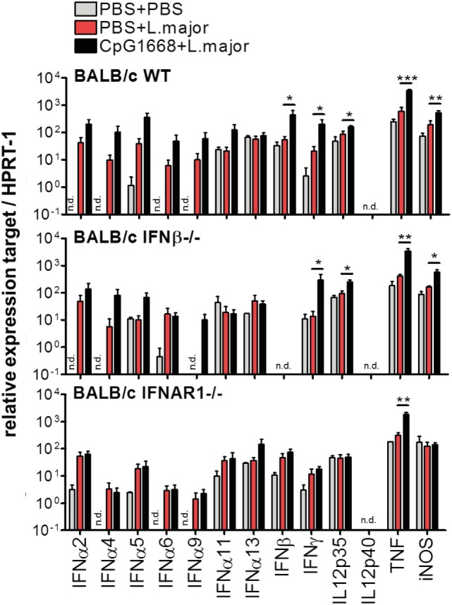
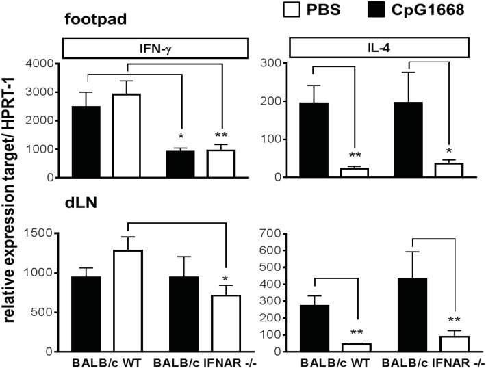
We previously showed that in mice infected with Leishmania major type I interferons (IFNs) initiate the innate immune response to the parasite at day 1 and 2 of infection. Here, we investigated which type I IFN subtypes are expressed during the first 8 weeks of L. major infection and whether type I IFNs are essential for a protective immune response and clinical cure of the disease. In self-healing C57BL/6 mice infected with a high dose of L. major, IFN-α4, IFN-α5, IFN-α11, IFN-α13, and IFN-β mRNA were most prominently regulated during the course of infection. In C57BL/6 mice deficient for IFN-β or the IFN-α/β-receptor chain 1 (IFNAR1), development of skin lesions and parasite loads in skin, draining lymph node, and spleen was indistinguishable from wild-type (WT) mice. In line with the clinical findings, C57BL/6 IFN-β-/-, IFNAR1-/-, and WT mice exhibited similar mRNA expression levels of IFN-γ, interleukin (IL)-4, IL-12, IL-13, inducible nitric oxide synthase, and arginase 1 during the acute and late phase of the infection. Also, myeloid dendritic cells from WT and IFNAR1-/- mice produced comparable amounts of IL-12p40/p70 protein upon exposure to L. major in vitro. In non-healing BALB/c WT mice, the mRNAs of IFN-α subtypes (α2, α4, α5, α6, and α9) were rapidly induced after high-dose L. major infection. However, genetic deletion of IFNAR1 or IFN-β did not alter the progressive course of infection seen in WT BALB/c mice. Finally, we tested whether type I IFNs and/or IL-12 are required for the prophylactic effect of CpG-oligodesoxynucleotides (ODN) in BALB/c mice. Local and systemic administration of CpG-ODN 1668 protected WT and IFN-β-/- mice equally well from progressive leishmaniasis. By contrast, the protective effect of CpG-ODN 1668 was lost in BALB/c IFNAR1-/- (despite a sustained suppression of IL-4) and in BALB/c IL-12p35-/- mice. From these data, we conclude that IFN-β and IFNAR1 signaling are dispensable for a curative immune response to L. major in C57BL/6 mice and irrelevant for disease development in BALB/c mice, whereas IL-12 and IFN-α subtypes are essential for the disease prevention by CpG-ODNs in this mouse strain.

Keywords: Leishmania major; cutaneous leishmaniasis; innate immunity; interferon-alpha/beta; type I interferon.


Figures

Figure 1
Figure 1
Expression of type I interferon (IFN) subtypes in Leishmania major-infected C57BL/6 mice. C57BL/6 wild-type mice were infected with 3 × 106 stationary phase L. major promastigotes into the hind footpads or treated with PBS. The mRNA expression of type I IFN subtypes was analyzed in the footpad tissues (A) and in the draining lymph nodes (B) at different time-points after infection using quantitative RT-PCR (day 0 are naïve mice). Results are shown as mRNA levels (mean ± SEM) normalized to the endogenous control HPRT-1 from four independent experiments (three mice per group and time point, respectively). Asterisks represent the respective significance values compared with uninfected mice (*p < 0.05; **p < 0.01; and ***p < 0.001; Mann–Whitney test).
Figure 2
Figure 2
Interferon (IFN)-β and IFNAR signaling are dispensable for the control of primary infection with Leishmania major in C57BL/6 mice. (A) Development of footpad lesions in C57BL/6 wild-type (WT) vs. IFN-β−/− vs. IFNAR1−/− mice after infection with 3 × 106 stationary phase L. major promastigotes into both hind footpads. The mean (±SEM) of the relative footpad thickness increase of 4 independent experiments with 9–12 mice per group is shown. (B) Parasite burden in the footpads, draining lymph nodes, and the spleens of C57BL/6 WT vs. IFN-β−/− vs. IFNAR1−/− mice. At the indicated time-points, three mice per group were analyzed for their parasite load in different organs by limiting dilution assays. The mean results (±SEM) of one representative out of four independent experiments (A) are presented. n.d., Not detectable.
Figure 3
Figure 3
Comparable mRNA expression of cytokines and effector pathways in C57BL/6 wild-type (WT), IFN-β−/−, and IFNAR1−/− mice during Leishmania major infection. Total RNA was isolated from footpad tissue or draining popliteal lymph nodes of WT (solid squares), IFN-β−/− (open circles), and IFNAR1−/− mice (open triangles) and reverse transcribed. Gene expression levels were determined by quantitative RT-PCR analysis using assays for the respective genes. Expression levels were calculated relative to the expression level of the endogenous control gene (HPRT-1). Results are mean expression levels from three mice per group and time point with error bars indicating SDs. The results of one of two experiments are shown.
Figure 4
Figure 4
Comparable control of secondary Leishmania major infection in C57BL/6 wild-type (WT) and IFNAR1−/− mice. For primary infection, mice were injected subcutaneously with 3 × 106 stationary phase L. major promastigotes into the right hind footpad. At day 136 (indicated by ↓), i.e., after healing of the primary skin lesion, mice were reinfected with an identical parasite inoculum into the left hind footpad. (A) Clinical course of infection in C57BL/6 WT vs. IFNAR1−/− mice. The mean (±SD) of the relative footpad thickness increase during primary (right footpad) and secondary infection (left footpad) is shown. One of three independent experiments with 12–18 mice per group is presented. In panel (B), the tissue parasite burden in the right and left footpad and draining lymph node at various time points after primary (left) and secondary infection (right) is depicted (please note the different time scale of the abscissas). At the indicated time points, three mice per group were analyzed by limiting dilution assays. The mean results (±SEM) of one representative out of three independent experiments are shown.
Figure 5
Figure 5
IFNAR signaling affects the Leishmania major-induced production of interferon (IFN)-α/β and interleukin (IL)-12 in BM-plasmacytoid dendritic cells (pDCs), but not bone marrow-derived dendritic cells (BM-mDCs). CD11b+CD11c+CD86 sorted BM-mDCs and B220+CD11bintCD11c+ sorted BM-pDCs were cultured for 48 h with L. major promastigotes (multiplicity of infection = 3), 200 ng/ml LPS, 1 µM CpG-oligodesoxynucleotides (ODN) 2216 or CpG-ODN 1668. Culture supernatants were analyzed for IFN-α/β (vesicular stomatitis virus bioassay with L929 fibroblasts) and IL-12p40/p70 content (ELISA). Triangles depict values below the detection limit of the assays. (A) BM-mDCs, (B) BM-pDCs. Mean results (±SEM) of three to four independent experiments are shown. Asterisks depict significant differences between wild-type (WT) and IFNAR1−/− cells (*p < 0.05; Mann–Whitney test).
Figure 6
Figure 6
Course of Leishmania major infection in untreated or CpG-oligodesoxynucleotides (ODN) 1668-treated BALB/c wild-type (WT), interferon (IFN)-β−/−, IFNAR1−/−, and interleukin (IL)-12p35−/− mice. Mice were infected with 3 × 106 stationary phase L. major promastigotes into both hind footpads. 2 h before infection, 5 nmol CpG-ODN 1668 was administered subcutaneously at the site of infection, and 10 h postinfection additional 5 nmol CpG-ODN 1668 was injected intraperitoneally. (A,C) Development of footpad lesions in untreated vs. CpG-ODN 1668-treated BALB/c WT vs. IFN-β−/− vs. IL-12p35−/− or BALB/c WT vs. IFNAR1−/− mice. The relative footpad thickness increase (mean ± SEM) of two to three independent experiments with three to five mice per group is shown. (B,D) Parasite burden in the footpads and draining lymph nodes of untreated vs. CpG-ODN 1668-treated BALB/c WT vs. IFN-β−/− vs. IL-12p35−/− or BALB/c WT vs. IFNAR1−/− mice at day 27 (B) or day 24 (D) after infection. Mean results (±SEM) of two to three independent experiments with three to five mice per group are shown. Significant differences by Mann–Whitney test between PBS- and CpG-treated BALB/c WT mice (*p < 0.05; **p < 0.01; and ***p < 0.001) or PBS- and CpG-treated IFN-β−/− mice (#p < 0.05; ##p < 0.01; and ###p < 0.001) are indicated.
Figure 7
Figure 7
Effect of CpG-oligodesoxynucleotides (ODN) 1668 treatment on the cytokine mRNA expression in the footpad of Leishmania major-infected mice. BALB/c wild-type (WT), interferon (IFN)-β−/− and IFNAR1−/− mice were infected with 3 × 106 stationary phase L. major promastigotes into both hind footpads or injected with PBS as control. 2 h before infection, 5 nmol CpG-ODN 1668 was administered subcutaneously at the site of infection, and 10 h postinfection additional 5 nmol CpG-ODN 1668 was injected intraperitoneally. Control mice received PBS. Total RNA was isolated from footpad tissue 36 h p.i. and reverse transcribed. Gene expression levels were determined as described in legend of Figure 3. Results are mean expression levels (±SEM) from three independent experiments with two to three mice per group. Significant differences by Mann–Whitney test between PBS- and CpG-treated infected BALB/c WT, IFN-β−/−, or IFNAR1−/− mice (*p < 0.05; **p < 0.01; and **p < 0.001) are indicated. n.d., Not detectable.
Figure 8
Figure 8
Interferon (IFN)-γ and interleukin (IL)-4 mRNA expression in the footpads and draining lymph nodes (dLN) of Leishmania major-infected BALB/c wild-type (WT) and IFNAR1−/− mice treated with CpG-oligodesoxynucleotides (ODN) 1668. BALB/c WT and IFNAR1−/− mice were infected with 3 × 106 stationary phase L. major promastigotes into both hind footpads. 2 h before infection, 5 nmol CpG-ODN 1668 was administered subcutaneously at the site of infection, and 10 h postinfection additional 5 nmol CpG-ODN 1668 was injected intraperitoneally. Control mice received PBS (same volume). Total RNA was isolated from footpad and dLN tissue at day 23 or day 24 of infection and reverse transcribed. Gene expression levels of IFN-γ and IL-4 were determined as described in legend of Figure 3. Results are mean expression levels (±SEM) from two independent experiments with three mice per group. Significant differences by Mann–Whitney test (*p < 0.05 and **p < 0.01) are indicated.

References

    1. van Pesch V, Lanaya H, Renauld J-C, Michiels T. Characterization of the murine alpha interferon gene family. J Virol (2004) 78:8219–28.10.1128/JVI.78.15.8219-8228.2004 - DOI - PMC - PubMed
    1. Uze G, Schreiber G, Piehler J, Pellegrini S. The receptor of the type I interferon family. Curr Top Microbiol Immunol (2007) 316:71–95. - PubMed
    1. Sadler AJ, Williams BR. Interferon-inducible antiviral effectors. Nat Rev Immunol (2008) 8(7):559–68.10.1038/nri2314 - DOI - PMC - PubMed
    1. Haller O, Weber F. The interferon response circuit in antiviral host defense. Verh K Acad Geneeskd Belg (2009) 71(1–2):73–86. - PubMed
    1. Snell LM, McGaha TL, Brooks DG. Type I interferon in chronic virus infection and cancer. Trends Immunol (2017) 38(8):542–57.10.1016/j.it.2017.05.005 - DOI - PMC - PubMed

Publication types

MeSH terms