. 2018 Feb 1;68(1):63-73.

Model of Traumatic Spinal Cord Injury for Evaluating Pharmacologic Treatments in Cynomolgus Macaques (Macaca fasicularis)

Affiliations

Model of Traumatic Spinal Cord Injury for Evaluating Pharmacologic Treatments in Cynomolgus Macaques (Macaca fasicularis)

Nitin Seth et al. Comp Med. .

Abstract

Here we present the results of experiments involving cynomolgus macaques, in which a model of traumatic spinal cord injury (TSCI) was created by using a balloon catheter inserted into the epidural space. Prior to the creation of the lesion, we inserted an EMG recording device to facilitate measurement of tail movement and muscle activity before and after TSCI. This model is unique in that the impairment is limited to the tail: the subjects do not experience limb weakness, bladder impairment, or bowel dysfunction. In addition, 4 of the 6 subjects received a combination treatment comprising thyrotropin releasing hormone, selenium, and vitamin E after induction of experimental TSCI. The subjects tolerated the implantation of the recording device and did not experience adverse effects due the medications administered. The EMG data were transformed into a metric of volitional tail moment, which appeared to be valid measure of initial impairment and subsequent natural or treatment-related recovery. The histopathologic assessment demonstrated widespread axon loss at the site of injury and areas cephalad and caudad. Histopathology revealed evidence of continuing inflammation, with macrophage activation. The EMG data did not demonstrate evidence of a statistically significant treatment effect.


Figures
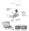
Figure 1.
Figure 1.
The transmitter to collect EMG data is surgically implanted in the macaque's lower back and connected to recording wire electrodes in the flexor cauda longus and brevis of the tail. The EMG data are transmitted by a radiofrequency link to a computer. Adapted and modified from reference.
Figure 2.
Figure 2.
Standard radiograph of catheter inserted into the epidural space via laminotomy in a cadaveric subject. The balloon is not inflated in this image.
Figure 3.
Figure 3.
CT image of the inflated balloon in the epidural space of the thoracic spine. The balloon is inflated with air, which appears black in the spinal column (red arrow). Note that 60% of the column is occupied by the balloon, and the spinal cord is displaced. This image is from a cadaveric subject.
Figure 4.
Figure 4.
(A) Photomicrograph of the spinal cord at the epicenter of the lesion. The gray and white parenchyma are disrupted by dilated myelin sheaths (spongiosis; arrows), which are widespread. These findings are noted caudal and cephalad to the lesion, in both treated and untreated subjects. Hematoxylin and eosin stain; magnification, 40×. (B) Photomicrograph of the spinal cord (white matter funiculi) at epicenter of experimental injury. There are many dilated myelin sheaths (spongiosis; black arrows), some of which contain degenerating or swollen axons (red arrows). These findings are noted caudal and cephalad to the lesion, in both treated and untreated subjects. Haematoxylin and eosin stain; magnification, 400×.
Figure 5.
Figure 5.
Photomicrograph of the spinal cord cranial to epicenter of the spinal cord lesion. Dilated myelin sheaths (black arrows) and swollen axons (spheroids; red arrows) are present. Luxol fast blue stain; magnification, 400×.
Figure 6.
Figure 6.
(A) Photomicrograph of the spinal cord at the epicenter of experimental lesion. Reactivity to Iba1 is present within the white matter adjacent to the gray matter with spongiosis of the parenchyma; magnification, 40×. (B) Increased magnification of region denoted by the box in A. These findings suggest that substantial inflammation continues at the 90-d time point. Iba1 immunohistochemistry with DAB staining and counterstained with hematoxylin; 400x magnification.
Figure 7.
Figure 7.
Photomicrograph of the spinal cord gray matter at the epicenter of experimental lesion. This figure shows Iba1-positive cells at the level of the lesion. These microglia show the hypertrophic phenotype typical of activated microglia Iba1 immunohistochemistry with DAB staining and counterstained with hematoxylin. Magnification, 400×.
Figure 8.
Figure 8.
Photomicrograph of the spinal cord cephalad to epicenter of spinal cord lesion. Immunohistochemistry using Iba1 antibody highlights microglial cells. Microglial cells (black arrows) demonstrate an activated phenotype (that is, thickened processes, enlarged cell body). Iba1 immunohistochemistry with DAB staining and counterstained with hematoxylin. Magnification, 200×.
Figure 9.
Figure 9.
The aggregate fitted Q-values, representing tail movement, are plotted over 90 d for the left (top) and right (bottom) sides. Values are normalized to range between 0 to 1, with higher values indicating greater muscle activity. Of note, in both the left and right side, Q values are decreased immediately after creation of the lesion. On the left side, the effect of the lesion in the treatment group (orange) was attenuated, compared with the nontreatment group (grey). On both the left and right sides, Q scores improved over time.

Similar articles

Cited by

References

    1. Algotar AM, Stratton MS, Ahmann FR, Ranger-Moore J, Nagle RB, Thompson PA, Slate E, Hsu CH, Dalkin BL, Sindhwani P, Holmes MA, Tuckey JA, Graham DL, Parnes HL, Clark LC, Stratton SP. 2012. Phase 3 clinical trial investigating the effect of selenium supplementation in men at high-risk for prostate cancer. Prostate 75:328–335. - PMC - PubMed
    1. Allen AR. 1911. Surgery of experimental lesion of spinal cord equivalent to crush injury of fracture dislocation of spinal column. JAMA LVII:878–880.
    1. Anderson DK, Saunders RD, Demediuk P, Dugan LL, Braughler JM, Hall ED, Means ED, Horrocks LA. 1985. Lipid hydrolysis and peroxidation in injured spinal cord: partial protection with methylprednisolone or vitamin E and selenium. Cent Nerv Syst Trauma 2:257–267. - PubMed
    1. Beattie MS, Bresnahan JC, Komon J, Tovar CA, VanMeter M, Anderson DK, Faden AL, Hsu CY, Noble LJ, Salzman S, Young W. 1997. Endogenous repair after spinal cord contusion injuries in the rat. Exp Neurol 148:453–463. - PubMed
    1. Brown TG. 1911. The intrinsic factors in the act of progression in the mammal. Proc R Soc Lond B Biol Sci 84:308–319.

Publication types